Cervical lymph nodes Carotid body tumor Solid neck

Cervical lymph nodes Carotid body tumor Solid neck swellings Cervical rib Sternomastoid tumor

• Metastases in cervical lymph nodes are common in head and neck cancers. • common sites of involvement in lymphoma. • Tuberculous lymphadenitis is common in South East Asia.

ANATOMY

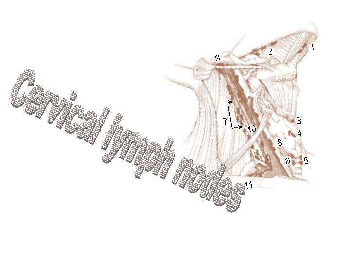
• Cervical lymph nodes are composed of lymphoid tissue and are located along the lymphatic vessels in the neck. • about 300 lymph nodes in the neck • the lymph nodes are embedded in the soft tissues of the neck and are either partly or completely surrounded by fat

• Classification • Level 1: Submandibular, submental. • Level 2: Internal jugular from skull base to carotid bifurcation. • Level 3: Internal jugular below carotid bifurcation to omohyoid. • Level 4: Internal jugular below omohyoid. • Level 5: Posterior triangle. (also known as accessory chain) • Level 6: Adjacent to thyroid. • Level 7: Tracheal esophageal groove and superior mediastinum.

NODAL PATHOLOGY • Malignant Adenopathy Is associated with a 50% reduction in long term survival. Abnormal (malignant) Nodes • Size: Greater than 1 centimeters • Hard, irregular, fixed nodes • Necrosis: Regardless of size. • Extracapsular spread: Regardless of size.

• Staging information is necessary for selection for most appropriate treatment option. • 30% of malignant nodes are clinically undetected on physical examination due to deep location especially in retropharyngeal and high internal jugular chains. • Accuracy of nodal staging CT: 90 -95% Physical exam- 75%

Clinical Nodal Staging • NX: Not assessable. • N 0: No clinically positive nodes. • N 1: Single clinically positive ipsilateral node less than or equal to 3 cm • N 2: Greater than 3 cm, less than 6 cm • N 2 A: Single, ipsilateral. • N 2 B: Multiple ipsilateral. • N 3: Greater than 6 cm • N 3 A: Ipsilateral. • N 3 B: Bilateral. • N 3 C: Contralateral

• Extracapsular spread carries a grave prognosis and may be the best indicator of treatment failure. Signs of Extracapsular Spread • Spiculated margins. • Fatty invasion. • Encasement of vessels.

FNAC under local anesthesia in op or cytology clinic. Useful if malignancy is suspected. Advantage • Accurate histological diagnosis • No spread of tumor • If not palpable image guided aspiration under USG or CT

Accuracy Comparison CT versus MRI • CT = MR for detecting and sizing nodes. • CT better than MR for demonstratin necrosis. • CT better than MR for detecting extracapsular spread.

Nodes from Unknown Primary • Approximately 10% of patients with abnormal cervical nodes present without obvious primary. Most common sites for unknown primary: • Nasal Pharynx. • Pyriform Sinus. • Tongue Base. • Tonsillar Crypts. • Thyroid. • Lung. Knowledge of drainage patterns may help in search for primary.

Probable Source of Nodal Metastasis • Level 1: Oral cavity, submandibular gland. • Level 2: Nasal pharynx, oral pharynx, parotid, superglottic larynx. • Level 3: Oral pharynx, hypopharynx, superglottic larynx. • Level 4: Subglottic larynx, hypopharynx, esophagus, thyroid. • Level 5: Nasal pharynx, oral pharynx. • Level 6 & 7: Thyroid, larynx, lung. • Note: Bilateral nodes are common with cancers of soft pallet, tongue, epiglottis, and nasal pharynx.

Lymphoma • Usually non-Hodgkin's lymphoma: Large nodes, enlargement of Waldeyer's ring, extra lymphatic enlargement of particular glands such as the thyroid. • Hodgkin's lymphoma may be present (25%of head & neck lymphoma) particularly if there is also a mediastinal involvement.

• Lymphomas can cross fascial planes easily. • Can undergo rapid enlargement. • Differential diagnosis: squamous cell cancer infectious mononucleosis.

Non-Malignant Adenopathy • Granulomatous disease: TB, sarcoid. • Fungal infections: . • Cat scratch fever. • Castleman's. • AIDS. • Post radiation changes.

Tuberculosis • Painless posterior neck mass. • Necrotic nodes particularly in level 5. • Multi-loculated with thick rim enhancement. • May calcify following treatment. • Brightly enhanced with contrast.

Infectious Mononucleosis • Multiple large non-necrotic nodes. • Enlargement of Waldeyer's ring. • Appears similar to AIDS, sarcoid, leukemia, lymphoma.

Cat Scratch Fever • Bilateral large nodes including intraparotid nodes. • Uncertain etiology ? viral or ricketsial

AIDS • Multiple small nodes. • Non-necrotic. • Enlargement of Waldeyer's ring. • Associated lympho epithelial cyst.

Sonographic features for diagnosis of cervical lymphadenopathy • Distribution Commonly involved nodal groups Mets from oropharynx, hypopharynx, larynx Internal jugular chain Mets from oral cavity Submandibular Upper cervical Mets from nasopharyngeal carcinoma Upper cervical Posterior triangle

• Metastases from papillary carcinoma of the thyroid. Internal jugular chain • Metastases from non-head and neck carcinoma Supraclavicular fossa Posterior triangle • Lymphoma - • Tuberculosis- Submandibular, Upper cervical Posterior triangle Supraclavicular fossa Posterior triangle

Size Shape Intranodal necrosis Echogenic hilus Echogenicity Calcification Nodal border

• radical neck dissection, originally described by Crile • popularized by Martin et al. , has been modified in various ways, giving rise to several types of cervical lymph node dissections that are currently used for the surgical treatment of the neck. • these modifications were classified according to a random system of terminology depending on the author at the time.

• In 1991 the Academy’s Committee for Head and Neck Surgery and Oncology classified four major types of neck dissections:
 Radical neck dissection; 2) Modified radical neck dissection; 3) Selective neck dissection supraomohyoid,")
1) Radical neck dissection; 2) Modified radical neck dissection; 3) Selective neck dissection supraomohyoid, posterolateral, Lateral anterior 4) Extended radical neck dissection.

Radical neck dissection • defined as removing all of the lymphatic tissue in regions I-V including removal of the spinal accessory nerve(SAN), sternocleidomastoid muscle (SCM) internal jugular vein (IJV).

Indications Ø in patients with Ø extensive cervical lymph node metastasis and/or extension beyond the capsule with invasion into the spinal accessory nerve, IJV, and SCM. Ø Many surgeons will elect to perform a RND if there is extensive disease surrounding the spinal accessory nerve without gross evidence of invasion.
 is defined as excision of all lymph nodes routinely")
Modified radical neck dissection (MRND) is defined as excision of all lymph nodes routinely removed by radical neck dissection with preservation of one or more nonlymphatic structures, i. e. , SAN, IJV, SCM. 4 Medina subclassifies the MRND into types I-III; type. I preserves the SAN, • type. II preserves the SAN and IJV, • type. III preserves the SAN, IJV, &SCM. • type III referred to as the "functional neck dissection" (Bocca), *in his classic description the submandibular gland is not excised.

Indications • in patients with gross nodal metastasis to the neck that does not directly infiltrate or adhere to the non-lymphatic structures • Bilateral MRND is indicated when there is contralateral nodal involvement

Selective neck dissection • is defined as any type of cervical lymphadenectomy where there is preservation of one or more lymph node groups removed by the radical neck dissection. • There are four common subtypes,

Supraomohyoid neck dissection • This removes lymph tissue contained in regions I-III.

Indications • patients with primary tumors arising from the oral cavity without clinical or radiologic evidence of cervical metastasis but who have a high probability of occult lymphatic disease. • in patients with staged T 2 -T 4 N 0 or TXN 1 when the palpable node is less than 3 cm, clearly mobile, and located in levels I or II. • Bilateral SOHND is indicated in patients who have carcinomas of the anterior tongue or oral tongue and floor of mouth.

• Posterolateral neck dissection, refers to the removal of the suboccipital lymph nodes, retroauricular lymph nodes, levels II-IV, and level V Medina suggests subclassification of the posteriolateral neck dissection to types I-III to mirror preservation of SAN, IJV, and SCM as in MRND.

Indications • This type of neck dissection is primarily used to treat the neck in patients with cutaneous malignancies and soft tissue sarcomas.

Lateral neck dissection removes lymph tissue in levels II-IV. Indications removal of nodal disease associated with carcinomas arising in the oropharynx, hypopharynx, and larynx.

Anterior neck dissection • is the last subtype of selective neck dissection and refers to the removal of lymph nodes surrounding the visceral structures of the anterior aspect of the neck
 selected cases of differentiated thyroid carcinoma, • (2) parathyroid carcinoma •")
Indications • (1) selected cases of differentiated thyroid carcinoma, • (2) parathyroid carcinoma • (3) subglottic carcinoma • (4) glottic carcinomas with subglottic extension,

Extended neck dissection • defined as removal of one or more additional lymph node groups and/or nonlymphatic structures not encompassed by radical neck dissection, such as parapharyngeal, superior mediastinal, and paratracheal.

Indications • Extended neck dissections are usually performed when MRND or RND is planned for N+ necks. • • The decision to extend the neck dissection may either be made preoperatively based on findings on CT or MR or intraoperatively based on findings of tumor invasion of surrounding structures. • The most significant example is when cervical disease involves the carotid artery.

Radiotherapy Along with RND when : • • lymph node more than 3 cm multiple extra capsular invasion. occult primary
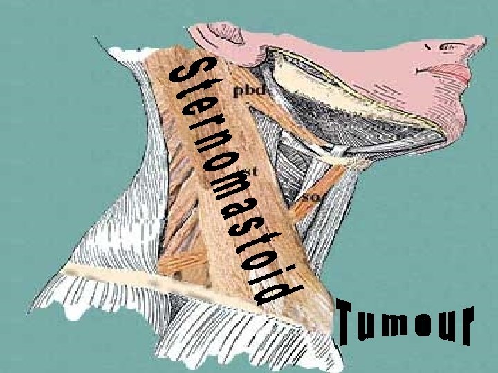

CLINICAL FEATURES Not present at birth Appears at 20 -28 days of life lump of size 1 -2 cm tender in the 1 st few wks of life firm in consistency smooth surface mobile sideways but not along the length of muscle
")
• Sternomastoid shortened and tight • Restricts head movement (turned to opposite side) • No signs of inflammation • Infant otherwise well • May be associated with plagiocephaly

TREATMENT If noticed at birth daily physiotherapy may help to prevent torticollis If noticed only after the torticollis has developed; A brace may be used to correct the torticillis. If this fails surgery is the only option. Best technique is to divide the muscle at its proximal or distal attachment care should be taken not to injure the accessory nerve

Before surgery Tumour removed

CAROTID BODY TUMOUR

• Carotid body tumours are derived from both, the mesoderm of the second branchial arch and the ectoderm of the neural crest. • also called paragangliomas because they arise from the paraganglionic cells • malignant potential in 2. 6 - 5% of the cases. • Lymphatics are the most favoured route of spread.
Sporadic type - more common 2)")
On the basis of incidence two types : 1)Sporadic type - more common 2) Familial type - autosomal dominant.

CLINICAL FEATURES • SYMPTOMS • most common complaint is of a lump in the neck • HIGHEST INCIDENCE IS BETWEEN AGE OF 40 AND 60 YEARS 32 incidence of bilaterality % • Expansion of the tumour may lead to cranial nerve paresis (VII, IX, X, XI, and XII) resulting in such symptoms as dysphagia, choking, or hoarseness. Horners syndrome also may be present. .

• SIGNS • non tender round swelling size 2 -10 cm hard in consistency • it can be moved vertically but not sideways. • Symptoms -10 per cent of patients

• The investigation of choice is a bilateral cerebral angiography tumour appears as a hypervascular oval mass. • angiography also helps to identify the sources of blood supply.

• Open biopsy is also confirmatory. • CT scan may also be useful

TREAMENT • If tumours are benign, their natural course is of slow enlargement with eventual compression of local structures resulting in symptoms such as nerve palsies. • In the elderly patient without symptoms it may be appropriate to do nothing. • BUT in Untreated, 75 per cent -develop symptoms • 30 per cent will die from invasion of local structures or metastatic disease.

The operative treatment • consists of excision of the tumour and maintenance of blood supply to the brain • . The dissection generally proceeds along the subadventitial plane but when deeper involvement of the wall is present, full thickness excision of the base of the tumour is done • When the carotid artery requires clamping, the cerebral function is monitored an indwelling carotid shunt (Javed Shunt) is used to maintain cerebral perfusion

• When the tumour is very large, the carotid artery may have to be sacrificed and the circulation is restored with an arterial graft. • If the pressure in the carotid stump is more than 65 mm of Hg, then the carotid artery may be ligated • the complication of this procedure is contralateral hemiplegia.

• When the tumour is unresectable, radiotherapy may be tried. CAROTID BODY TUMOR MUST ALWAYS BE KEPT IN MIND WHENEVER A SOLID SWELLING OF NECK IS MET WITH BECAUSE OF CHANCES OF SEVERE BLEEDING DURING BIOPSY

CERVICAL RIB
 arises from C")
Most common neurovascular compression at neck base ØCervical rib (extra rib) arises from C 7 vertebra ØRib narrows interval between scalenes ØHigher barrier for nerves and vessels to pass over ØCompression Worsened by ØShoulder sag Øin elderly or Muscle Weakness Øb. Carrying heavy object in hand ØLevels C 8 to T 1 most commonly affected
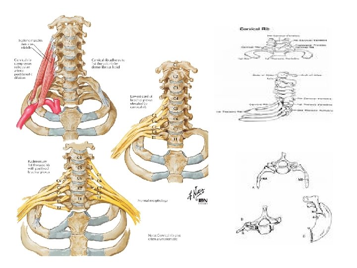

Symptoms Affects hand inner forearm Pain and Paresthesia along Ulnar Nerve course Hand weakness, numbness, and clumsiness Associated symptoms Hand cold sensation Raynaud's Phenomenon Gangrene

Sign – – – Palpable cervical rib Tender brachial Plexus distribution Muscle Weakness and atrophy (lower trunk) • • – Sensation decreased • • • – Ulnar forearm Arm Ulnar 1. 5 fingers Circulatory insufficiency • • – Interosseus muscles Hypothenar muscles Swelling Cold sensation Distal cyanosis Trophic skin change Adson's Test
")
Diagnostics Chest XRay Extra rib at C 7 vertebra (Often bilateral)

Electromyogram

TREATMENT • In mild cases exercises to strengthen the muscles of shoulder girdle. • In advanced cases excision of cervical rib or dividing the scalenus anterior muscle (scalenotomy)is the choice • Its important that periosteum also should be removed so that there are no chances of regeneration. CERVICAL RIB AFTER SURGERY

Thoracic outlet syndrome • rare condition caused by cervical rib • there is compression of vessels and nerves in the area of the clavicle.

summary • CERVICAL LYMPH NODE bcos secondary carcinomatous infiltration of cervical node is a common occurrence from primary sites like nasopharynx, tonsil , tongue, pyriform fossa, supraglottic larynx etc. • FEATURES OF MALIGNANT NODES • >1 CM • Irregular , hard, Fixed • INVESTIGATIONS FNAC, USG, CT/ MRI • • • NECK DISSECTIONS-TYPES Radical modified radical selective extended

STERNOMASTOID TUMOR • FEATURES • Arising from the sternomastoid muscle at 20 -28 days of life • 1 -2 cm, firm , smooth, mobile sideways • Associated with torticollis • TREATMENT • DEFINITIVE-excision of tumor

CAROTID BODY TUMOR Arising from chemoreceptor cells of the carotid bulb 40 -60 yrs of life FEATURES 2 -10 cm, hard, mobile vertically, non tender TREATMENT Surgery best avoided in elderly if needed full thickness excision of base of tumor

CERVICAL RIB • Most common cause for NV compression at neck base • FEATURES • Pain and parasthesia along ulnar N course • Weakness and numbness of hand • Circulatory insufficiency • TREATMENT • DEFINITIVE-Excision of rib along with periosteum or scalenotomy

- Slides: 73