Cerebrum It is divided into two cerebral hemispheres


Cerebrum

It is divided into two cerebral hemispheres, separated by longitudinal fissure 2 cerebral hemispheres longitudinal fissure

falx cerebri corpus callosum The fissure contains the sickle-shaped fold of dura mater, the falx cerebri Two hemispheres connected together by CC

The cerebral hemispheres are separated from the cerebellum by a horizontal fold of dura mater called the tentorium cerebelli Cerebral hemisphere Posteriorly cerebellum Tentorium cerebelli (cut)

2 - Medial Surface 1 - Superolateral Surface CC 3 - Inferior 3 A- Orbital Surface 3 B-Tentorial part Each cerebral hemisphere has 3 surfaces

Surfaces of the Cerebral Hemisphere 1 - Superolateral surface: the widest surface of the hemisphere. - This is a convex surface which is directed upward and laterally. 2 - Medial surface: is a flat surface which is separated from the opposite side by the longitudinal fissure which lodges the falx cerebri. It contains the corpus callosum which connects the two cerebral hemispheres. 3 - Inferior surface: is directed inferiorly and is divided by the stem of the lateral sulcus into two parts: a- Anterior (orbital surface) rests on the roof of the orbit. b- Posterior (tentorial surface) rests on the tentorium cerebelli.

Occipital pole Frontal pole Temporal pole Each cerebral hemisphere has 3 poles

Main Sulci and Lobes of the cerebral hemisphere
 a deep sulcus")
central sulcus Superolateral Surface 1 - Central sulcus (Fissure of Rolando) a deep sulcus about 1/2 inch behind the midpoint between frontal and occipital poles. - It extends obliquely downwards and forwards and ends slightly above the lateral sulcus. - It extends a little on the medial surface

1 -central sulcus The central sulcus is the only sulcus of any length on the superolateral surface of the hemisphere that indents the superomedial border to the medial surface

The stem arises on the inferior surface Long posterior ramus anterior ascending ramus Short horizontal ramus 2 - Lateral sulcus (fissure of Sylvius) consists of a short stem that divides into three rami.

parieto-occipital sulcus 5 cm occipital pole 3 - Parieto-occipital sulcus begins on the superior medial margin of the hemisphere about 2 inches (5 cm) anterior to the occipital pole, extends downward & forward

parieto-occipital sulcus calcarine sulcus 4 - Calcarine sulcus; begins below the splenium of the corpus callosum to the occipital pole. - It is divided by parieto-occipital sulcus into precalcarine and postcalcarine sulcus.

frontal lobe Parietal lobe temporal lobe occipital lobe Each cerebral hemisphere has 4 lobes

Frontal Parietal Occipital Corpus callosum Temporal Lobes on medial Surface

Sulci & Gyri of the superolateral surface

12 Important Sulci on the supero-lateral surface
: 2")
Sulci on the Supero-lateral surface 1 - Lateral sulcus (fissure of sylvius): 2 - Central sulcus (Fissure of Rolando): 3 - Precentral sulcus: about 1 cm (finger’s breadth) infront the central sulcus. 4 & 5 - Superior and inferior frontal sulci: begin close to the precentral sulcus and extend forwards. 6 - Postcentral sulcus: about 1 cm (finger's breadth) behind the central sulcus. 7 - Intraparietal sulcus: extends backwards from the middle of the postcentral sulcus. 8 & 9 - Superior and inferior temporal sulci: on the temporal lobe parallel to the lateral sulcus. 10 - Parieto-occipital sulcus: 2 inches infront the occipital pole. 11 - Calcarine sulcus: its posterior end reaches to the occipital pole. 12 - Lunate sulcus (Simian) at the occipital lobe

Important gyri on Superolateral Surface
 Supramarginal gyrus 40 39 Angular gyrus")
Long posterior ramus of lateral sulcus (area 40) Supramarginal gyrus 40 39 Angular gyrus (area 39) Wernicke's area Superior temporal sulcus Supramarginal gyrus (area 40) is gyrus around the posterior end of the lateral sulcus into the parietal region Angular gyrus (area 39): is gyrus around the posterior end of the superior temporal sulcus into the parietal region

Gyri On the supero-lateral surface A- Frontal lobe; I- Precentral gyrus between the central and precentral sulci. 2 - Superior frontal gyrus; lies above the superior frontal sulcus. 3 - Middle frontal gyrus lies between the superior and inferior frontal sulci. 4 - Inferior frontal gyrus; below inferior frontal sulcus, from anterior to posterior: a- Orbital part below the horizontal ramus. b- Triangular between the horizontal, and ascending rami. c- Opercular part behind the ascending ramus. B- Parietal lobe; 1 - Postcentral gyrus: between the central and postcentral sulci. 2 - Superior parietal gyrus (lobule) above the intraparietal sulcus. 3 - Inferior parietal gyrus (lobule) below the intraparietal sulcus. 4 - Supramarginal gyrus around the posterior end of the lateral sulcus. C- Temporal lobe; 1 - Superior temporal gyrus between lateral sulcus and superior temporal sulcus. 2 - Middle temporal gyrus lies between the superior and inferior temporal sulci. 3 - Inferior temporal gyri: lies below the inferior temporal sulcus. 4 - Angular gyrus around the posterior end of the superior temporal sulcus.

Functional areas of the Superolateral surface

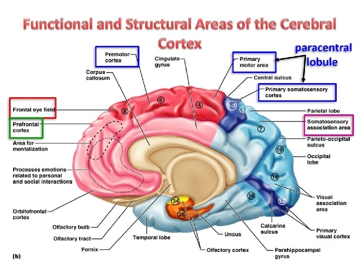
Functional and Structural Areas of the Cerebral Cortex
 52 structurally distinct")
Functional areas of the supero-lateral surface (Brodmann areas in all surfaces) 52 structurally distinct areas of cerebral cortex
,")
Motor area 4 • Primary motor cortex corresponds to the precentral gyrus (area 4), anterior part of the paracentral lobule Controls motor functions

A body represented in upside down. Head represented in lower part of precentral gyrus, leg and foot, perineum represented on medial surface of hemisphere in paracentral lobule, size depends on skill

Premotor area 6 • • Located anterior to the precentral gyrus It is the origin of extrapyramidal fibers Controls more complex movements Involved in the planning of movements

Frontal eye field area 8 6 6 4 - It lies anterior to the premotor cortex. - It controls movements of the eyes when eyes follow a moving target.
 44, 45 8 6 6 4 • Motor speech (Broca's) area (areas")
(Broca’s area) 44, 45 8 6 6 4 • Motor speech (Broca's) area (areas 44, 45) is located in inferior frontal gyrus between the anterior and ascending rami of the lateral sulcus of the dominant hemisphere (95%). • It brings about the formation of words by its connections with the adjacent primary motor areas; the muscles of the speech. • Lesion in this area produces motor aphasia (loss of speech).
 8 6 6 4 44 45 • Writing area (Exner's area); -")
(Writing area) 8 6 6 4 44 45 • Writing area (Exner's area); - It lies in the middle frontal gyrus. - The person able to express himself in written words - Lesion leading to Agraphia (loss of ability to write)

Prefrontal areas 8 6 9 6 4 10 W 11 W 44 45 12 Prefrontal area (areas 9, 10, 11, & 12) - It lies in the most anterior part of the frontal lobe. - It is responsible for: A- Planning, thinking, remember and problem solving B- Motivating, emotions, good & sinful behavior, mood, psychological activities. C- Telling of lies and truth
 cortex corresponds")
Somatosensory area 1, 2, 3 2 1 3 - Somatosensory (Pry sensory) cortex corresponds to postcentral gyrus (area 3, 1, 2), posterior part of paracentral lobule - It receives sensations from opposite side of body. - The body represented upside down

8 1 6 9 6 4 10 W 3 11 W 44 45 12 2 Secondary sensory 5&7 Secondary (Association) sensory area (area 5, 7); - It occupies the superior parietal gyrus. - Function, stereognosis (ability to identify the familiar objective manually) shape, roughness, size of objects

Supramarginal gyrus 40 Superior temporal sulcus 39 Angular gyrus Wernicke's area Sensory speech area (Wernicke’s- area 39, 40). - It is responsible for understanding spoken and written language - Lesion produces sensory aphasia.

Pry auditory area 41&42 Secod auditory area 22 • Primary auditory area (areas 41, 42) - It is present in the floor of the posterior ramus of the lateral sulcus and the middle part of the superior temporal gyrus (Heschl's gyrus). • Auditory association area (Secondary) ( area 22): - It lies behind the auditory area. - It is responsible for recognition and interpretation of the sounds.

Gustatory area 43 • Involved in the conscious awareness of taste stimuli • Located on the “roof” of the lateral sulcus

the lips of the lateral sulcus are separated Insula - Insula lies at the bottom of the deep lateral sulcus and cannot be seen from the surface unless the lips of the sulcus are separated. - It is believed that it contains the area for taste -

Sulci & Gyri of the medial surface

Anterior ramus Central sulcus posterior ramus parietooccipital sulcus cingulate sulcus callosal sulcus Sulci of the medial surface calcarine sulcus

• Sulci on the Medial Surface 1 - Callosal sulcus: close to the upper surface of the corpus callosum. 2 - Cingulate sulcus; about finger's breadth above and parallel to the callosal sulcus. - It ends by dividing into two rami in front and behind the central sulcus. 3 - Central sulcus: between the two branches of the cingulate sulcus. 4 - Parieto-occipital sulcus. 5 - Calcarine sulcus.

cingulate gyrus medial frontal gyrus paracentral lobule precuneus parietooccipital sulcus cingulate sulcus cuneus callosal sulcus corpus callosum Lingual gyrus Post-calcarine sulcus
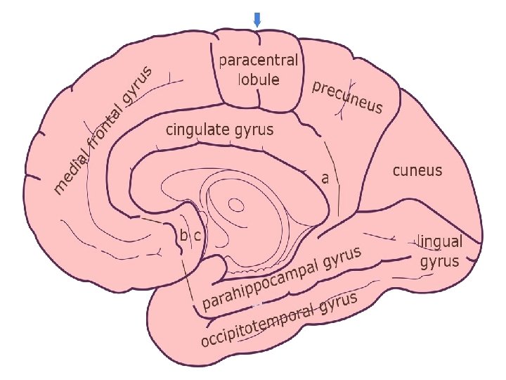

Gyri on the Medial Surface 1 - Cingulate gyrus: between the callosal and cingulate sulci. - The lower part of the posterior end curves downward behind the splenium of corpus callosum and forms a narrow area (isthmus) that connects it with the para-hippocampal gyrus. 2 - Medial frontal gyrus: between the superomedial border and cingulate sulcus. 3 - Paracentral lobule: surrounds the central sulcus between the two rami of the cingulate sulcus. 4 - Precuneus; between the posterior branch of the cingulate sulcus and the parieto-occipital sulcus. 5 - Cuneus: the triangular gyrus between the parieto-occipital and postcalcarine sulci (between the two branches of the Y). 6 - Lingual gyrus: the elongated, tongue-like gyrus extending below the postcalcarine sulcus to the occipital pole.

Functional areas of the medial surface
")
Paracentral lobule Secondary visual areas 18&19 Primary Visual area (area 17)

Cortical Centers of the medial surface 1 - Paracentral lobule; - It is continues with the motor and sensory areas in the lateral surface. - It is the center to the leg, foot and perineum of the opposite side. - It controls the micturition and defecation reflexes. 2 - primary Visual area (area 17); - It lies on the depth of calcarine sulcus - It receives visual sensation. 3 - secondary Visual (association) area (area 18, 19): - It lies in the occipital lobe surrounding the primary visual area. - Damage of this area causes visual agnosia (people can not identify the objects).

Sulci & Gyri of the inferior surface

gyrus rectus Olfactory Sulcus H- shaped orbital sulcus olfactory bulb, tract orbital gyri The inferior surface of the frontal lobe

Stem of lateral sulcus Rhinal sulcus L. Occipitotemporal gyrus Occipitotemporal sulcus M. Occipitotemporal gyrus Collateral sulcus The tentorial surface

A- On the orbital surface: 1 - Olfactory sulcus; on the orbital surface close and parallel to the medial orbital border. It contains olfactory bulb and tract. • Gyrus rectus: between medial orbital border and olfactory sulcus. 2 - Orbital sulcus: is H shaped sulcus lateral to the olfactory sulcus. 2 - Anterior, posterior, lateral and medial orbital gyri: on the orbital surface. B- On the tentorial surface: 1 - Stem of the lateral sulcus between the frontal and temporal lobes. 2 - Occipito-temporal sulcus: from occipital pole to temporal pole. 3 - Rhinal su. Icus: extends from the temporal pole. 4 - Collateral sulcus: begins close to the posterior end of the rhinal sulcus to the occipital pole. 3 - Medial and Lateral occipitotemporal gyrus: medial and lateral to occipitotemporal sulcus.

uncus rhinal sulcus parahippocampal gyrus hippocampal sulcus calcarine sulcus collateral sulcus lingual gyrus The tentorial surface
 Anterior part of parahippocampal gyrus")
Olfactory area uncus (area 34, 28) Anterior part of parahippocampal gyrus

On the tentorial surface: - Lingual gyrus between collateral sulcus and calcarine sulcus - Para hippocampal gyrus anterior to the lingual gyrus - Uncus anterior to Para hippocampal gyrus, a hook-shaped convolution close to the temporal pole medial to the rhinal sulcus. Center of the olfactory

Th ank Qu you est ion s I/Azzam - 2004
- Slides: 56