CEREBRUM 2 BRAIN EYEBALL MOTOR HOMUNCULUS MOTOR HOMUNCULUS

CEREBRUM - 2 BRAIN & EYEBALL
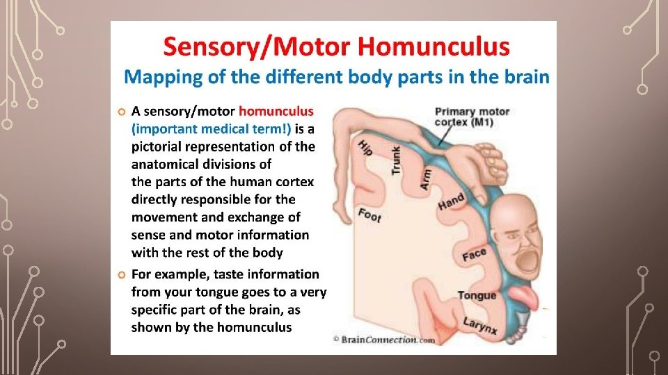
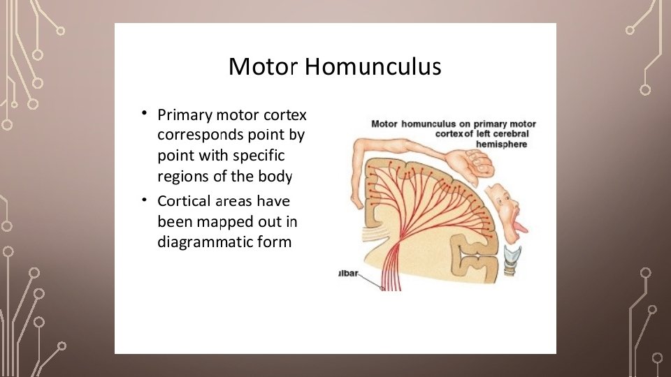

MOTOR HOMUNCULUS

MOTOR HOMUNCULUS
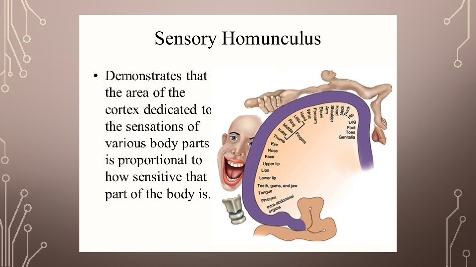

SENSORY HOMUNCULUS

SENSORY HOMUNCULUS

BOTH MOTOR & SENSORY HOMUNCULUS

EASY FIGURE TO UNDERSTAND

EASY FIGURE TO UNDERSTAND THE GYRUS

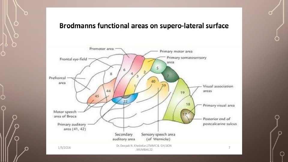
FUNCTIONAL AREA

FUNCTIONAL AREA

FUNCTIONAL AREA

FUNCTIONAL AREA

FUNCTIONAL AREA

FUNCTIONAL AREA
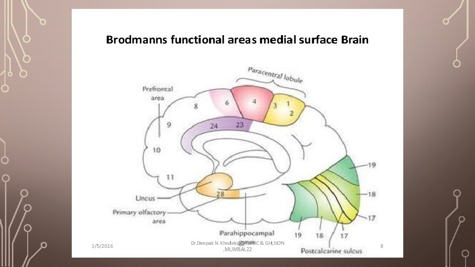

FUNCTIONAL AREA – AT A GLANCE

HISTOLOGICAL STRUCTURE OF CEREBRUM

HISTOLOGICAL STRUCTURE OF CEREBRUM

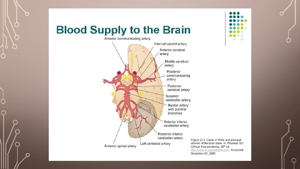
BLOOD SUPPLY OF CEREBRUM Arteries of the Brain • The brain is supplied by the two internal carotid and the two vertebral arteries. The four arteries lie within the subarachnoid space, and their branches anastomose on the inferior surface of the brain to form the circle of Willis. Internal Carotid Artery • The internal carotid artery begins at the bifurcation of the common carotid artery, where it usually possesses a localized dilatation, called the carotid sinus. It ascends the neck and perforates the base of the skull by passing through the carotid canal of the temporal bone. The artery then runs horizontally forward through the cavernous sinus and emerges on the medial side of the anterior clinoid process by perforating the dura mater. It now enters the subarachnoid space by piercing the arachnoid mater and turns posteriorly to the region of the medial end of the lateral cerebral sulcus.

BLOOD SUPPLY OF CEREBRUM Branches of the Cerebral Portion • 1. The ophthalmic artery arises as the internal carotid artery emerges from the cavernous sinus. It enters the orbit through the optic canal below and lateral to the optic nerve. It supplies the eye and other orbital structures, and its terminal branches supply the frontal area of the scalp, the ethmoid and frontal sinuses, and the dorsum of the nose. • 2. The posterior communicating artery is a small vessel that originates from the internal carotid artery close to its terminal bifurcation. The posterior communicating artery runs posteriorly above the oculomotor nerve to join the posterior cerebral artery, thus forming part of the circle of Willis. • 3. The choroidal artery, a small branch, also originates from the internal carotid artery close to its terminal bifurcation. The choroidal artery passes posteriorly close to the optic tract, enters the inferior horn of the lateral ventricle, and ends in the choroid plexus. It gives off numerous small branches to surrounding structures, including the crus cerebri, the lateral geniculate body, the optic tract, and the internal capsule.

BLOOD SUPPLY OF CEREBRUM • 4. The anterior cerebral artery is the smaller terminal branch of the internal carotid artery. It runs forward and medially superior to the optic nerve and enters the longitudinal fissure of the cerebrum. Here, it is joined to the anterior cerebral artery of the opposite side by the anterior communicating artery. It curves backward over the corpus callosum and, finally, anastomoses with the posterior cerebral artery. The cortical branches supply all the medial surface of the cerebral cortex as far back as the parieto-occipital sulcus. They also supply a strip of cortex about 1 inch (2. 5 cm) wide on the adjoining lateral surface. The anterior cerebral artery thus supplies the “leg area” of the precentral gyrus. A group of central branches pierces the anterior perforated substance and helps to supply parts of the lentiform and caudate nuclei and the internal capsule.

BLOOD SUPPLY OF CEREBRUM • 5. The middle cerebral artery, the largest branch of the internal carotid, runs laterally in the lateral cerebral sulcus. Cortical branches supply the entire lateral surface of the hemisphere, except for the narrow strip supplied by the anterior cerebral artery, the occipital pole, and the inferolateral surface of the hemisphere, which are supplied by the posterior cerebral artery. This artery thus supplies all the motor area except the “leg area. ” Central branches enter the anterior perforated substance and supply the lentiform and caudate nuclei and the internal capsule.

ARTERY SUPPLY OF CEREBRUM

ARTERY SUPPLY OF CEREBRUM

ARTERY SUPPLY OF CEREBRUM
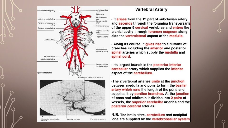

BASILAR ARTERY

CIRCLE OF WILLIS

CIRCLE OF WILLIS
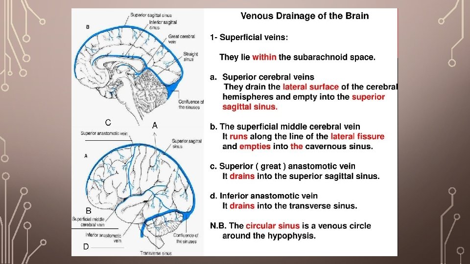

VENOUS DRAINAGE

VENOUS DRAINAGE

VENOUS DRAINAGE

VENOUS DRAINAGE

THANK YOU
- Slides: 40