Cerebrovascular Diseases 1 Pathophysiological Facts For maintained normal

Cerebrovascular Diseases 1

Pathophysiological Facts : • For maintained normal brain function : – Continuous blood flow-15% of cardiac output – High oxygen requirement- 20% of total body O 2 consumption – Maintenance of glucose • ↓ BP ≤ 50 mm. Hg is critical Hypoxia & ischemia • BP ≥ 200 mm. Hg critical! risk of Hemorrhage

• STROKE : The clinical term of acute neurological dysfunction occurring as a result of hemorrhagic or obstructive vascular lesions, causing irreversible damage or death.

Types of Cerebrovascular Disease : • A- Impairment of blood supply & oxygenation of CNS tissue : – Global Cerebral Ischemia due to generalized cerebral blood flow – Localized vascular obstruction due to thrombosis or embolism (Infarction)

• B- Rupture of CNS blood vessel : – Intracerebral hemorrhage – Subarachnoid hemorrhage – Subdural hemorrhage – Epidural hemorrhage

GLOBAL Cerebral Ischemia Hypotenstion, Hypoperfusion, and low flow states

Definition : • Generalized reduction of cerebral perfusion • Symptoms vary from transient mild confusion to irreversible damage

• Causes include : – Sever Hypotension – Cardiac arrest – Shock

Features of global ischemia : • Cells: neurons > glia, but all if severe and prolonged • Selective Vulnerability of neurons in certain locations : – Pyramidal cells of hippocampus – Purkinje cells of the cerebellum – Cortical pyramidal neurons

• MORPHOLOGY : – Brain swelling – Wide gyri and narrow sulci – Poor demarcation between grey & white matter

• Microscopic changes : - Acute : 12 -24 hrs - RED NEURONS - Infiltration by neutrophils - Subacute changes: 24 hrs – 2 weeks - tissue necrosis, influx of macrophages, vascular proliferation & gliosis - Repair: after 2 weeks - Removal of necrotic tissue - Gliosis
 infarcts (Regions of brain at the most distal reaches")
• Watershed (Borderzone ) infarcts (Regions of brain at the most distal reaches of arterial blood supply, border zones between arterial territories) include : - Boundary between anterior & middle cerebral arteries (Cerebral Convexities)

Watershed Vascular Territories

Watershed Infarcts

Outcome : • Depends on: – Duration of ischemia – Magnitude and rapidity of reduction of flow • Result varies from persistent neurological deficit to brain death.

Focal Cerebral Ischemia • Focal brain ………… necrosis due to complete and prolonged ischemia

Mechanisms leading to infarction : • Embolic occlusion : hemorrhagic/ red - Source: heart or atherosclerosis in carotid - Middle Cerebral artery most affected • Thrombotic occlusion, mainly due to Atherosclerosis : Ischemic/ pale Carotid bifurcation Origin of middle cerebral artery Basilar artery at either end

Types of infarction Acute white infarct in the distribution of the Acute red infarct in the distribution of the right middle cerebral artery. right anterior cerebral artery.

• Macroscopic appearance : - No gross change before 48 hrs - Soft swollen pale or hemorrhagic WEDGE SHAPED infarct involving grey & white matter. - Red infarcts: surrounding hemorrhage due to reperfusion of damaged vessels and tissue - After 10 days-3 weeks : liquefaction - Cavity within 1 - 6 months

• Microscopic appearance : – Very similar to global ischemia but more regional.
 EDEMA B) “RED” NEURONS C) NEUTROPHILS D) MACROPHAGES E) GLIOSIS Histopathologic progression of")
A) EDEMA B) “RED” NEURONS C) NEUTROPHILS D) MACROPHAGES E) GLIOSIS Histopathologic progression of CNS infarcts

Old infarction

REMEMBER THE GENERAL RULES FOR DATING INFARCTS : • RED Neurons 12 -18 hours • Neutrophils 24 -48 hours • Macrophages peak at 48 hours – 3 weeks, may persist for months to years • Reactive astrocytes can be seen at week 1 & remain for several months

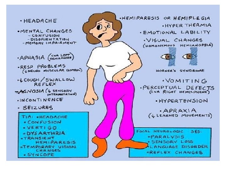
• Clinical picture : linked to site of infarction - Contralateral hemiparesis - Loss of sensation - Visual field abnormalities - Aphasia …… etc

 Subarachnoid")
INTRACRANIAL HEMORRHAGE • According to location, divided into : • • Intracerebral (intraparenchymal) Subarachnoid Epidural Subdural
 - Mostly")
I- Primary Intracerebral Hemorrhage: Causes : • Commonest cause is hypertension (50%) - Mostly due to rupture of small intraparenchymal vessel. - 15% of death in hypertensive patients. • Other causes : Cerebral Amyloid angiopathy Vasculitis Coagulation disorders Neoplasms
, thalamus, pons, cerebellar hemispheres and others.")
§ Intracerebral hemorrhage in HT - Putamen (60%), thalamus, pons, cerebellar hemispheres and others. - Acute: Extravasation of blood with compression of adjacent parenchyma - Old: Cavitary destruction of brain with a rim of brown discoloration.

Intracerebral hemorrhage into lateral ventricle

Other Vascular lesions in hypertension include : Atherosclerosis in larger arteries • Hyaline arteriolosclerosis in smaller vessels →hemorrhage • Arteriolosclerosis in vessels ≤ 150 µm → lacunar infarcts • Charcot Bouchard Microaneurysms in vessels ≤ 300 µm in basal ganglia → rupture •

§ Lacunar Infarcts : -Tiny cystic infarcts in hemispheric white matter, basal ganglia & brainstem due to hypertensive arteriolosclerosis of deep pentrating artries and arterioles. - Multiple small cavitary infarcts (Lake-like spaces, less than 15 mm wide). - Asymptomatic or severe neurologic impairment

§ Slit Hemorrhages : - Rupture of smaller penetrating vessels - Resorb, leaving behind a slit-like cavity

§ Acute Hypertensive Encephalopathy : - Diffuse cerebral dysfunction: – Headaches, Confusion, Vomiting, and Convulsions – Coma
 is deposited in wall")
2 - Cerebral Amyloid Angiopathy : - Amyloid (Aβ 40) is deposited in wall of medium & small meningeal & cortical vessels → Weak wall → hemorrhage.


- Slides: 37