Cerebrovascular Accident CVA Cerebrovascular Accident v v Results

Cerebrovascular Accident CVA

Cerebrovascular Accident v v Results from ischemia to a part of the brain or hemorrhage into the brain that results in death of brain cells. Approximately 750, 000 in USA annually v v v Third most common cause of death #1 leading cause of disability 25% with initial stroke die within 1 year 50 -75% will be functionally independent 25% will live with permanent disability Physical, cognitive, emotional, & financial impact

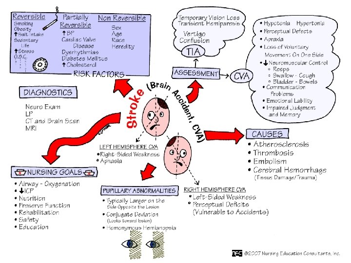
Cerebrovascular Accident Risk Factors v Nonmodifiable: v v Age – Occurrence doubles each decade >55 years Gender – Equal for men & women; women die more frequently than men Race – African Americans, Hispanics, Native Americans, Asian Americans -- higher incidence Heredity – family history, prior transient ischemic attack, or prior stroke increases risk

Cerebrovascular Accident Risk Factors Controllable Risks with Medical Treatment & Lifestyle Changes: High blood pressure Cigarette smoking High blood cholesterol Heart Disease Oral contraceptive use Sickle cell disease Hypercoagulability Diabetes TIA (Aspirin) Obesity Atrial fibrillation Physical inactivity Asymptomatic carotid stenosis

CVA – Risk Factors

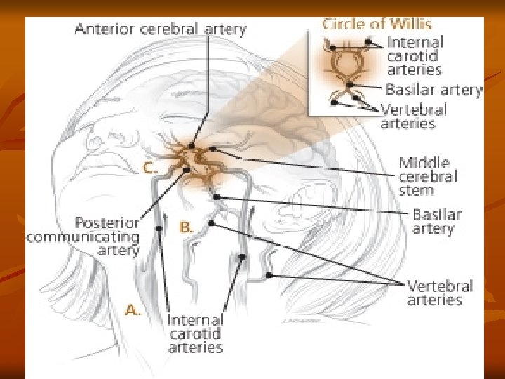
Cerebrovascular Accident Anatomy of Cerebral Circulation v Blood Supply v Anterior: Carotid Arteries – middle & anterior cerebral arteries v v Posterior: Vertebral Arteries – basilar artery v v frontal, parietal, temporal lobes; basal ganglion; part of the diencephalon (thalamus & hypothalamus) Mid and lower temporary & occipital lobes, cerebellum, brainstem, & part of the diencephalon Circle of Willis – connects the anterior & posterior cerebral circulation

Cerebrovascular Accident Anatomy of Cerebral Circulation v Blood Supply v 20% of cardiac output— 750 -1000 ml/min v >30 second interruption– neurologic metabolism is altered; metabolism stops in 2 minutes; brain cell death < 5 mins.

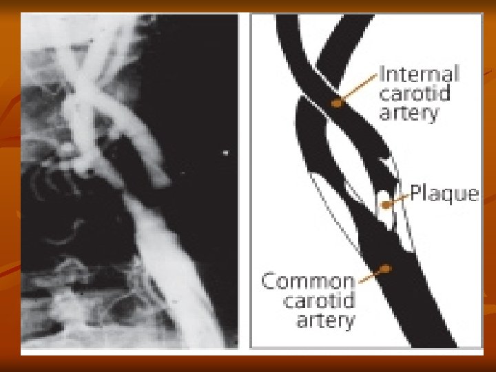
Cerebrovascular Accident Pathophysiology v Atherosclerosis: major cause of CVA v Thrombus formation & emboli development Abnormal filtration of lipids in the intimal layer of the arterial wall v Plaque develops & locations of increased turbulence of blood bifurcations v Increased turbulence of blood or a tortuous area v Calcified plaques rupture or fissure v Platelets & fibrin adhere to the plaque v Narrowing or blockage of an artery by thrombus or emboli v Cerebral Infarction: blocked artery with blood supply cut off beyond the blockage v

Cerebrovascular Accident Pathophysiology v Ischemic Cascade v Series of metabolic events Inadequate ATP adenosine triphosphate production v Loss of ion homeostasis v Release of excitatory amino acids – glutamate v Free radical formation v Cell death v v Border Zone: reversible area that surrounds the core ischemic area in which there is reduced blood flow but which can be restored (3 hours +/-)

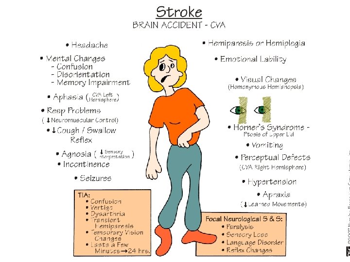
CVA? - Call 911 n Sudden numbness or weakness of face, arm, or leg, especially on one side of the body. n Sudden confusion or trouble speaking or understanding speech. n Sudden trouble seeing in one or both eyes. n n Sudden trouble walking, dizziness, or loss of balance or coordination Sudden severe headache with no known cause.

Cerebrovascular Accident Transient Ischemic Attack v v Temporary focal loss of neurologic function Caused by ischemia of one of the vascular territories of the brain v Microemboli with temporary blockage of blood flow v Lasts less than 24 hrs – often less than 15 mins v Most resolve within 3 hours v Warning sign of progressive cerebrovascular disease

Cerebrovascular Accident Transient Ischemic Attack v Diagnosis: v CT without contrast v v Cardiac Evaluation v v Confirm that TIA is not related to brain lesions Rule out cardiac mural thrombi Treatment: v Medications that prevent platelet aggregation v v ASA, Plavix Oral anticoagulants

Cerebrovascular Accident Classifications Based on underlying pathophysiologic findings

Cerebrovascular Accident Classifications v Ischemic Stroke Thrombotic v Embolic v v Hemorrhagic Stroke Intracerebral Hemorrhage v Subarachnoid Hemorrhage v v Aneurysm v Berry or Saccular

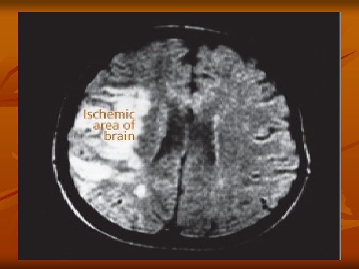
Cerebrovascular Accident Classifications v Ischemic Stroke—inadequate blood flow to the brain from partial or complete occlusions of an artery--85% of all strokes n n v Extent of a stroke depends on: n Rapidity of onset n Size of the lesion n Presence of collateral circulation Symptoms may progress in the first 72 hours as infarction & cerebral edema increase Types of Ischemic Stroke: Thrombotic Stroke Embolic Stroke

CVA Recognition

Cerebrovascular Accident Ischemic – Thrombotic Stroke Lumen of the blood vessels narrow – then becomes occluded – infarction v Associated with HTN and Diabetes Mellitus v >60% of strokes v 50% are preceded by TIA v Lacunar Stroke: development of cavity in place of infarcted brain tissue – results in considerable deficits – motor hemiplegia, contralateral loss of sensation or motor ability v

Cerebrovascular Accident Thrombotic Stroke

Cerebrovascular Accident Common Sites of Atherosclerosis

Cerebrovascular Accident Ischemic – Embolic Stroke Embolus lodges in and occludes a cerebral artery v Results in infarction & cerebral edema of the area supplied by the vessel v Second most common cause of stroke – 24% v Emboli originate in endocardial layer of the heart – atrial fibrillation, MI, infective endocarditis, rheumatic heart disease, valvular prostheses v Rapid occurrence with severe symptoms – body does not have time to develop collateral circulation v Any age group v Recurrence common if underlying cause not treated v

Cerebrovascular Accident Embolic Stroke

Cerebrovascular Accident Goals for Management n Immediate – assess & stabilize n n n n ABCs, VS Neurologic screening Oxygen if hypoxic IV access Check glucose Activate stroke team – CODE GREEN 12 -lead EKG Immediate Neuro Assessment n n Establish symptom onset Review hx Stroke Scale Facial droop; arm drift; abnormal speech

Cerebrovascular Accident Goals for Management n CT Scan – No hemorrhage: n Consider Fibrinolytic therapy n n Check for exclusions t. PA n n n No anticoagulants or antiplatelet therapy for 24 hours If not a candidate: Antiplatelet Therapy CT Scan – Hemorrhage: n n Neurosurgery? If no surgery: Stroke Unit n n Monitor BP and treat Hypertension Monitor Neuro status Monitor blood glucose and treat as needed Supportive therapy

Cerebrovascular Accident Goals for Management n Immediate – assess & stabilize n n n n n ABCs, VS Neurologic screening Oxygen if hypoxic IV access Check glucose Active stroke team Emergent CT scan of brain 12 -lead EKG Immediate Neuro Assessment n n Establish symptom onset Review hx Stroke Scale Facial droop; arm drift; abnormal speech

Cerebrovascular Accident Hemorrhagic Stroke v Hemorrhagic Stroke 15% of all strokes v Result from bleeding into the brain tissue itself v Intracerebral v. Subarachnoid v

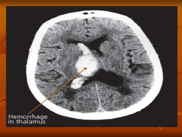
Cerebrovascular Accident Hemorrhage Stroke Intracerebral Hemorrhage Rupture of a vessel v Hypertension – most important cause v Others: vascular malformations, coagulation disorders, anticoagulation, trauma, brain tumor, ruptured aneurysms v Sudden onset of symptoms with progression v Neurological deficits, headache, nausea, vomiting, decreased LOC, and hypertension v Prognosis: poor – 50% die within weeks v 20% functionally independent at 6 months v

Cerebrovascular Accident Hemorrhage Stroke Intracerebral Hemorrhage

Cerebrovascular Accident Hemorrhagic-Subarachnoid v Hemorrhagic Stroke–Subarachnoid Hemorrhage v Intracranial bleeding into the cerebrospinal fluidfilled space between the arachnoid and pia mater membranes on the surface of the brain
")
Cerebrovascular Accident Hemorrhagic-Subarachnoid v Commonly caused by rupture of cerebral aneurysm (congenital or acquired) Saccular or berry – few to 20 -30 mm in size v Majority occur in the Circle of Willis v Other causes: Arteriovenous malformation (AVM), trauma, illicit drug abuse v Incidence: 6 -16/100, 000 v Increases with age and more common in women v

Cerebrovascular Accident Hemorrhagic-Subarachnoid Cerebral Aneurysm v v v Warning Symptoms: sudden onset of a severe headache – “worst headache of one’s life” Change of LOC, Neurological deficits, nausea, vomiting, seizures, stiff neck Despite improvements in surgical techniques, many patients die or left with significant cognitive difficulties
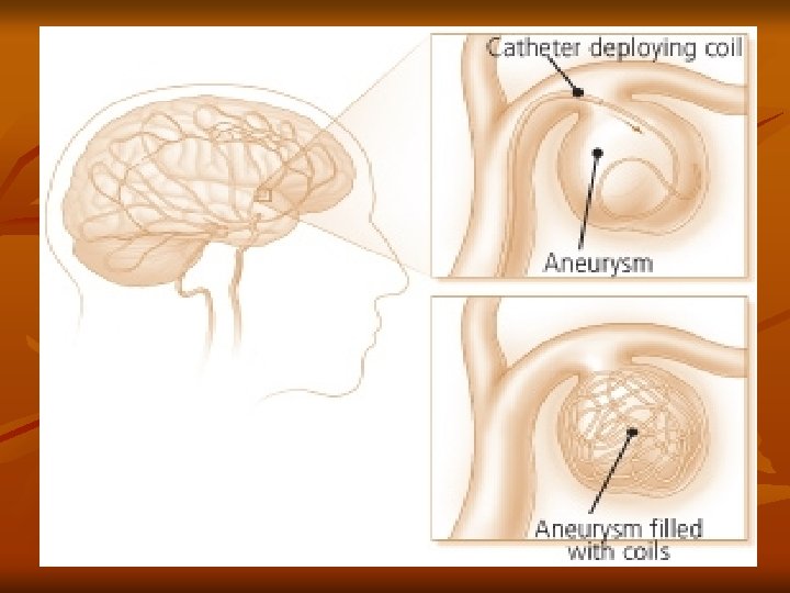

Hemorrhagic-Subarachnoid Cerebral Aneurysm v Surgical Treatment: Clipping the aneurysm – prevents rebleed v Coiling – platinum coil inserted into the lumen of the aneurysm to occlude the sac v v Postop: Vasospasm prevention – Calcium Channel Blockers

Hemorrhagic-Subarachnoid Cerebral Aneurysm – Surgical Tx

Hemorrhagic-Subarachnoid Cerebral Aneurysm – Coiling

Cerebrovascular Accident Classification

Cerebrovascular Accident Clinical Manifestations Middle Cerebral Artery Involvement v Contralateral weakness v Hemiparesis; hemiplegia Contralateral hemianesthesia v Loss of proprioception, fine touch and localization v Dominant hemisphere: aphasia v Nondominant hemisphere – neglect of opposite side; anosognosia – unaware or denial of neuro deficit v Homonymous hemianopsia – defective vision or blindness right or left halves of visual fields of both eyes v

Cerebrovascular Accident Clinical Manifestations Anterior Cerebral Artery Involvement v v v v Brain stem occlusion Contralateral v weakness of proximal upper extremity v sensory & motor deficits of lower extremities Urinary incontinence Sensory loss (discrimination, proprioception) Contralateral grasp & sucking reflexes may be present Apraxia – loss of ability to carry out familiar purposeful movements in the absence of sensory or motor impairment Personality change: flat affect, loss of spontaneity, loss of interest in surroundings Cognitive impairment

Cerebrovascular Accident Clinical Manifestations Posterior Cerebral Artery & Vertebrobasilar Involvement v v v v v Alert to comatose Unilateral or bilateral sensory loss Contralateral or bilateral weakness Dysarthria – impaired speech articulation Dysphagia – difficulty in swallowing Hoarseness Ataxia, Vertigo Unilateral hearing loss Visual disturbances (blindness, homonymous hemianopsia, nystagmus, diplopia)

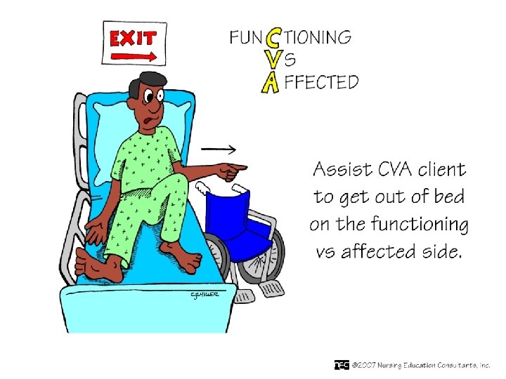
Cerebrovascular Accident Clinical Manifestations Motor Function Impairment v Caused by destruction of motor neurons in the pyramidal pathway (brain to spinal cord) v Mobility v Respiratory function v Swallowing and speech v Gag reflex v Self-care activities v

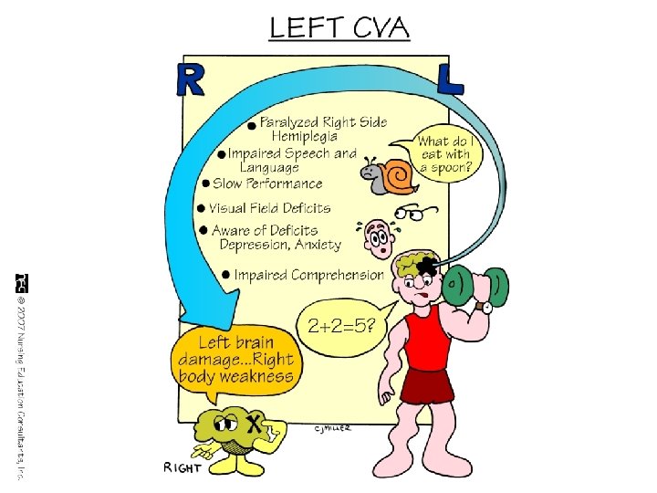
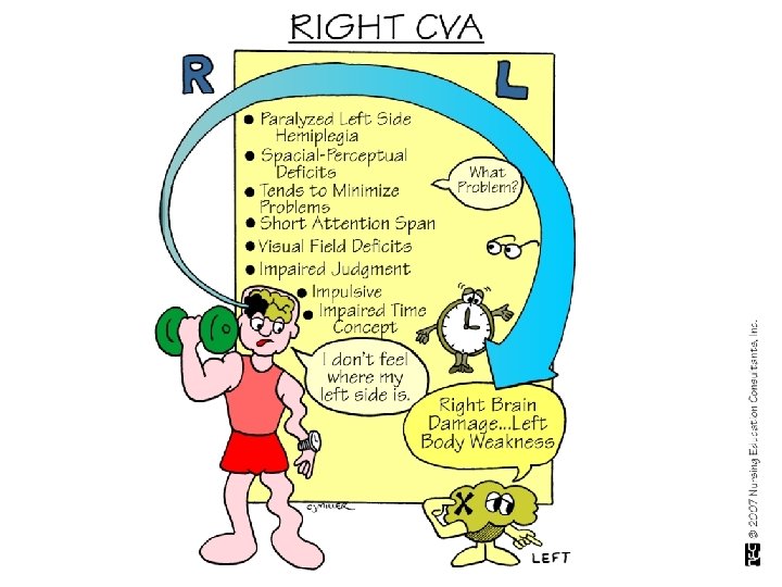
Cerebrovascular Accident Clinical Manifestations Right Brain – Left Brain Damage

Cerebrovascular Accident Clinical Manifestations v Affect Difficulty controlling emotions v Exaggerated or unpredictable emotional response v Depression / feelings regarding changed body image and loss of function v

Cerebrovascular Accident Clinical Manifestations v Intellectual Function v Memory and judgment Left-brain stroke: cautious in making judgments v Right-brain stroke: impulsive & moves quickly to decisions v v Difficulties in learning new skills

Cerebrovascular Accident Clinical Manifestations v v v Communication Left hemisphere dominant for language skills in the right-handed person & most left-handed persons -Aphasia/Dysphasia Involvement Expression & Comprehension v v v Receptive Aphasia (Wernicke’s area): sounds of speech nor its meaning can be understood – spoken & written Expressive Aphasia (Broca’s area): difficulty in speaking and writing Dysarthria: Affects the mechanics of speech due to muscle control disturbances – pronunciation, articulation, and phonation

Cerebrovascular Accident Clinical Manifestations v Spatial-Perceptual Alterations – 4 categories: 1. Incorrect perception of self & illness v 2. Erroneous perception of self in space – may neglect all input from the affected side (worsened by homonymous hemianopsia) v 3. Agnosia: Inability to recognize an object by sight, touch or hearing v 4. Apraxia: Inability to carry out learned sequential movements on command v

Homonymous Hemianopsia

Cerebrovascular Accident Clinical Manifestations v Elimination Most problems occur initially and are temporary v One hemisphere stroke: prognosis is excellent for normal bladder function v Bowel elimination: motor control not a problem – constipation associated with immobility, weak abdominal muscles, dehydration, diminished response to the defecation reflex v

Cerebrovascular Accident Treatment Goals v Prevention – Health Maintenance Focus: Healthy diet v Weight control v Regular exercise v No smoking v Limit alcohol consumption v Route health assessment v Control of risk factors v

Cerebrovascular Accident Treatment Goals Prevention v Drug Therapy v Surgical Therapy v Rehabilitation v

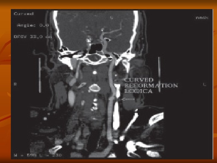
Cerebrovascular Accident Diagnostic Studies v Done to confirm CVA and identify cause PE: Neuro Assessment; Carotid bruit v Carotid doppler studies (ultrasound study) v CT – primary – identifies size, location, differentiates between ischemic and hemorrhagic v CTA – CT Angiography – visualizes vasculature v MRI – greater specificity than CT v v v May not be able to be used on all patients (metal, claustrophobia) Angiography: gold standard for imaging carotid arteries

Cerebrovascular Accident Treatment Goals v v Drug Therapy – Thrombotic CVA – to reestablish blood flow through a blocked artery Thrombolytic Drugs: t. PA (tissue plasminogen activator) v v v Administered within 3 hours of symptoms of ischemic CVA v v v produce localized fibrinolysis by binding to the fibrin in the thrombi Plasminogen is converted to plasmin (fibrinolysin) Enzymatic action digests fibrin & fibrinogen Results is clot lysis Confirmed DX with CT Patient anticoagulated ASA, Calcium Channel Blockers

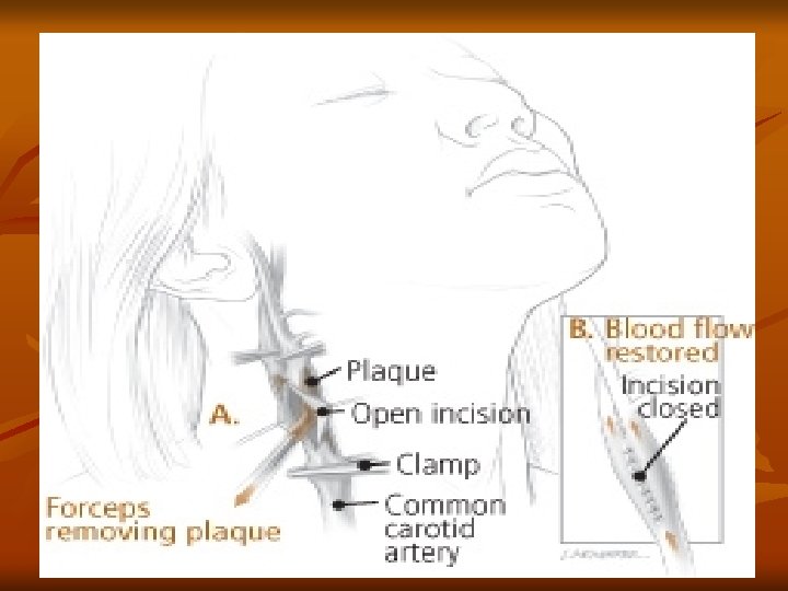
CVA - Treatment Goals v Surgical Treatment v Carotid endarterectomy – preventive – > 100, 000/year v v removal of atheromatous lesions Clipping, wrapping, coiling Aneurysm Evacuation of aneurysm-induced hematomas larger than 3 cm. Treatment of AV Malformations

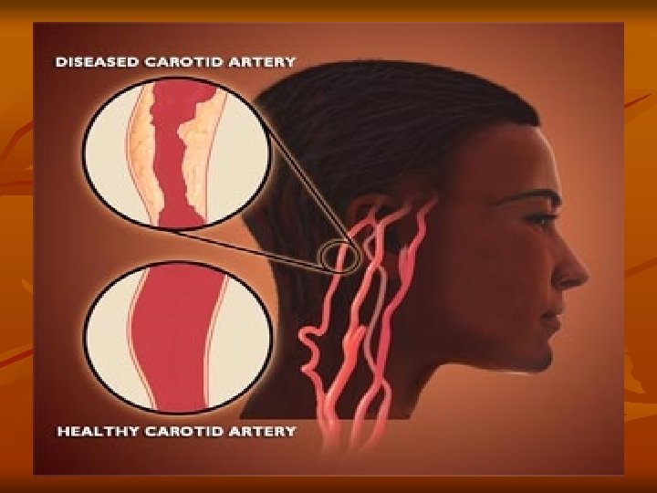
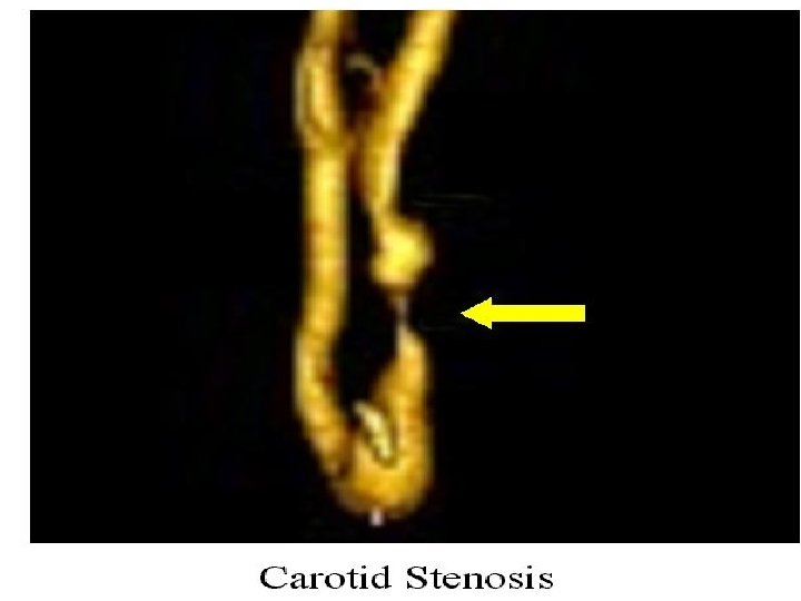
Carotid Artery Disease

Carotid Artery Disease n Carotid artery disease is the leading cause of strokes. n More than 50% of stroke victims present no warning signs. n n After age 55, the risk of stroke doubles every 10 years. 97% of the adult population cannot name a single warning sign of a stroke. 50% of nursing home admissions are stroke victims

Carotid Artery Stents

Carotid Endarterectomy

Cerebrovascular Accident Treatment Goals v Drug Therapy v Measures to prevent the development of a thrombus or embolus for “At Risk” patients: v Antiplatelet Agents v v Aspirin Plavix Combination Oral anticoagulation – Coumadin v Treatment of choice for individuals with atrial fibrillation who have had a TIA

Cerebrovascular Accident Nursing Diagnoses v v v v Ineffective tissue perfusion r/t decreased cerebrovascular blood flow Ineffective airway clearance Impaired physical mobility Impaired verbal communication Impaired swallowing Unilateral neglect r/t visual field cut & sensory loss Impaired urinary elimination Situational low self-esteem r/t actual or perceived loss of function

Cerebrovascular Accident Nursing Goals Maintain stable or improved LOC v Attain maximum physical functioning v Attain maximum self-care activities & skills v Maintain stable body functions v Maximize communication abilities v Maintain adequate nutrition v Avoid complications of stroke v Maintain effective personal & family coping v

Cerebrovascular Accident Warning Signs of Stroke Sudden weakness, paralysis, or numbness of the face, arm, or leg, especially on one side of the body v Sudden dimness or loss of vision in one or both eyes v Sudden loss of speech, confusion, or difficulty speaking or understanding speech v Unexplained sudden dizziness, unsteadiness, loss of balance, or coordination v Sudden severe headache v

Cerebrovascular Accident Acute Phase Assess: Frequently to assess CVA evolution Neuro — Glascow Coma Scale -- mental status, LOC, pupillary response, extremity movement, strength, sensation; ICP; Communication—speaking & understanding; sensory-perceptual alterations CV– cardiac monitoring; VS, PO, hemodynamic monitoring; Resp — airway/air exchange/aspiration; GI — swallowing—gag reflex; bowel sounds; bowel movement regularity GU — urinary continence Integumentary — skin integrity, hygiene Coping – individual and family v

Cerebrovascular Accident Acute Phase v Nsg Action: v Supportive Care Respiratory – spans from intubation to breathing on own v Musculoskeletal -- Positioning – side-to-side; HOB elevated; PROM exercise; splints; shoes/footboard v GI – enteral feedings initially v GU – foley catheter v Skin – preventive care v v Meds: anti platelet

Cerebrovascular Accident Acute Phase v Patient Education: Clear explanations for all care/treatments v Focus on improvements—regained abilities v Include family v

Cerebrovascular Accident Rehabilitation v v Assess: Swallowing; Communication; Complications; motor and sensory function Nsg Action: Coordinate resources: Speech Therapy—assess swallowing v Physical Therapy—ambulation/strengthening v Bowel/Bladder v Appropriate self-help resources v


Cerebrovascular Accident Rehabilitation v Comprehensive plan – v v v v v Physical Medicine & Rehabilitation / Inpatient Rehab Learn techniques to self-monitor & maintain physical wellness Demonstrate self-care skills Exhibit problem-solving skills with self-care Avoid complications of stroke Communication Maintain nutrition & hydration Use community resources Family cohesiveness

Cerebrovascular Accident Rehabilitation v Resources American Stroke Association v Association of Rehabilitation Nurses v National Institute of Neurological Disorders & Stroke v National Stroke Association v Society for Neuroscience v Stroke Clubs International v
- Slides: 79