CEREBROSPINAL FLUIDANATOMY PHYSIOLOGY AND DYNAMICS DR SUNITA DOLEY

CEREBROSPINAL FLUID-ANATOMY, PHYSIOLOGY AND DYNAMICS DR. SUNITA DOLEY MODERATED BY- DR. PRIYANKA

OUTLINE v. CSF SPACES v. CSF FORMATION – CIRCULATION – REABSORPTION v. METHODS OF DETERMINING Vf AND Ra v. EFFECTS OF DRUGS v. ALTERATION IN CSF DYNAMICS IN PATHOLOGY

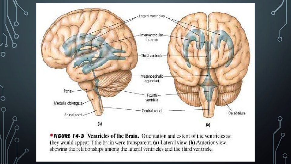
ANATOMY OF CSF SPACES CSF is clear, colourless liquid that is formed in brain and circulates through macroscopic & microscopic spaces that are in continuity. Macroscopic spaces (140 -150 ml): • Two lateral ventricles • Third ventricle • Aqueduct of sylvius • Fourth ventricle • Central canal of spinal cord Microscopic spaces: • Brain and spinal cord ECF space (300 -350 ml)

THREE DIMENSIONAL SHAPE OF THE VENTRICULAR SYSTEM

PROPERTIES OF CSF

COMPOSITION

• Na content peaks at 8: 00 am & 6: 00 pm • Relationship between Na concentration and migraine has been proposed as peaks correspond to migraine attacks . . Harrington MG, Salomon RM, et al. Cerebrospinal fluid sodium rhythms. Cerebrospinal fluid Res 2010

COMPOSITION • • Varies according to sampling site Altered during neuroendoscopy Na, Cl, Mg Glucose, Protiens, AA, K , HCO 3, Ca, Phosphate, Uric acid

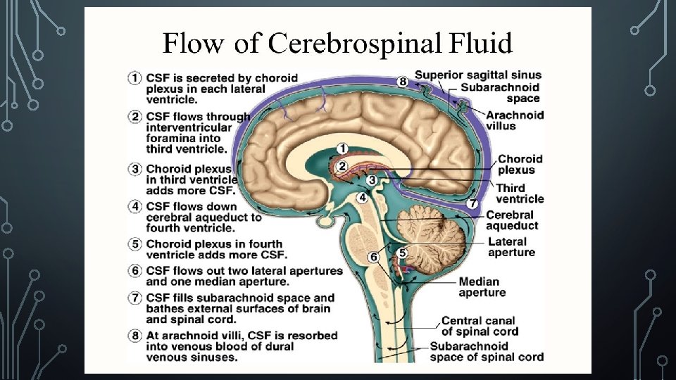
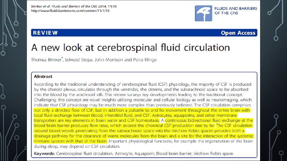
FORMATION OF CSF 3 Sites: • • • Choroid plexus ( 50 – 70 %) • Rate ( Vf ) 0. 35 -0. 40 ml/min Ependymal surfaces of ventricles Perivascular spaces 500 -600 ml/day • • Turnover time- 5 -7 hrs (4 times/day) 40%-70% enters macroscopic spaces via CP 30%-60% enters across ependyma and pia Recent studies – Bidirectional fluid exchange at BBB far exceeds CP csf formation - Brinker T. , et al. A new look at cerebrospinal fluid circulation. Fluids Barriers CNS. 2014; 11: 10 -15

Choroid plexus It is a cauliflower like growth of blood vessels covered by a thin layer of epithelial cells. It is made of 3 layers: • fenestrated capillary endothelium • extra cellular matrix • epithelial cells

Choroid plexus projects into: Ø Temporal horn of lateral ventricle Ø Post. Part of 3 rd ventricle Ø Roof of 4 th ventricle Blood supply: ant. & post choroidal artery (lateral & 3 rd ventricle) and supr cerebeller and PICA (temporal horn & 4 th ventricle) Nerve supply : branches of Vagus, Glossopharyngeal & Sympathetic N.

CSF FORMATION AT CHOROID PLEXUS Blood entering CP capillaries filtered form protein rich fluid similar to ISF in CP stroma Hydrostatic pressure & bulk flow enter cleft between epithelial cells stromal fluid transported across CP epithelium. Ultrafiltration & secretion ATP dependent membrane pump transport Na across luminal surface to macroscopic spaces in exchange for K & H. Water moves from stroma into CSF by conc gradient by ionic pump.

CSF FORMATION AT EXTRA CHOROIDAL SITES v. Derived from ECF & cerebral capillaries across BBB v. Oxidation of glucose (into H 2 O & CO 2) by brain [60%]. v. Ultrafiltration from cerebral capillaries[40%] TIGHT JUNCTIONS In blood-ECF interface Glucose /electrolyte/water/AA/lipid soluble material Large polar/protein Pass Stop Glucose rich and protein poor fluid diffuse through ECF space toward macroscopic spaces

• Water and other constituent of plasma crosses Blood brain Barrier into the brain ECF space by diffusion or transport. • Water and cellular metabolites added to the ECF from neurons and glial cells.

MOVEMENT OF GLUCOSE & PROTEIN • CSF glucose conc. is approx. 60% of that in blood • Ratio remains constant till 270 -360 mg/dl blood glucose • Glucose enters CSF -Facilitated transport & follows saturable kinetics (i. e rate depends on serum glucose conc. ) • Protein entry in CSF limited – conc. is 0. 5% or less of serum conc. • Protein in CSF transported with CSF & cleared from csf space to dural venous sinuses by • “Sink effect” – flowing CSF keeps CSF & brain protein conc. Low.

EFFECT OF INCREASED ICP ON CSF FORMATION Relation between Vf and ICP/CPP • ↑ICP • Vf • ↓CPP • Vf ØAs long as CPP remain > 70 mm of Hg, increase of ICP[upto 20 mm of Hg] has no major impact on Vf (rate of CSF formation). ØWhen CPP is significantly lowered <70 mm. Hg CBF↓ and CPBF ↓, Vf ↓

CIRCULATION OF CSF • Hydrostatic pressure of CSF formation 15 cm. H 20 produce CSF flow. • Cilia of ependymal cell generate current to propel CSF toward 4 th ventricle & its foramina into subarachnoid space. • Respiration variations • Vascular pulsation of cerebral arteries , CP • • • Additional CSF movement 15 cm H 20 CSF pressure of formation 9 cm H 20 superior sagittal sinus pressure 6 cm of pressure gradient across arachnoid villi Suction pump effect Reabsorption – arachnoid villi (SSS) & spinal dural sinusoids in dorsal nerve roots.

REABSORPTION • CSF pass from Subarachnoid spaces via Arachnoid villi & granulation into venous blood. • Arachnoid villi or granulations are protrusion of the arachnoid cells from subarachnoid space into & through wall of venous sinuses • Arachnoid villi are located: • Intracranial- Superior Sagittal sinus (85 -90% reabsorbed) • Spinal - dural sinusoids on dorsal nerve root (10 -15%)
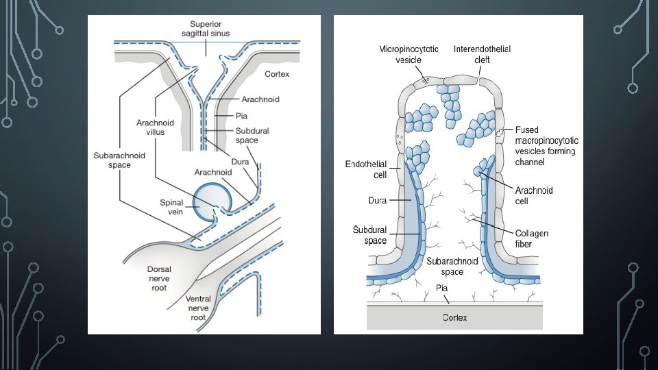

DETERMINANTS OF REABSORPTION • Normal intracranial pressure: • Endothelium covering the villus acts as a CSF- blood barrier • Rate of pass of CSF– 1. Trans villus hydrostatic pressure gradient (CSF pressure – venous sinus pressure) 2. Pressure sensitive resistance to CSF outflow at arachnoid villi CSF passes through endothelium via: 1. Pinocytotic vesicles 2. Transcellular openings
")
DETERMINANTS OF REABSORPTION • Increased intracranial pressure: • Rate of reabsorption of CSF (Va) ↑ if pressure gradient across villus↑ • Resistance to reabsorption of CSF(Ra) remains normal upto a CSF pressure of 30 cm of H 20; above this it is decreased.

FUNCTION OF CSF • Protection, Support, Nutrition • The low Specific gravity of CSF (1. 007) relative to that of the brain (1. 040) reduces the effective mass of a 1400 g brain to only 47 g. • Stable supply of nutrients, primarily glucose(active transport); also vitamins/ eicosanoids/monosaccharides/neutral & basic amino acids/monocarboxylic acid (specialized pump mechanism).

CONTROL OF CHEMICAL ENVIRONMENT • Exchange between neural tissue & CSF occurs readily by diffusion • (because distance b/w CSF and any brain area is max 15 mm & ISF spaces of brain and spinal cord is continuous with macroscopic CSF spaces. ) • Acid-base characteristics of CSF influence: • Respiration • CBF, CBF-AR • Cerebral metabolism

CONTROL OF CHEMICAL ENVIRONMENT HR Emotional BP CSF Ca/K/Mg/bicarb Vasomotor Muscle tone Respiration
 Secondary pumps (passive transport) • K/HCO")
CONTROL OF CHEMICAL ENVIRONMENT Primary pumps (active transport) Secondary pumps (passive transport) • K/HCO 3 • Ca/Mg • H+ • Cl-

EXCRETION • Removes metabolic products, unwanted drugs • BBB excludes out toxic, large , polar and lipid insoluble drug, humoral agents etc.

INTRACEREBRAL TRANSPORT MEDIAN EMINENCE CSF ECF Neuron Neurohormonal releasing factor formed in hypothalamus

METHODS OF DETERMINING CSF FORMATION RATE & RESISTANCE TO CSF ABSORPTION ØVentriculocisternal perfusion ØManometric infusion ØVolume injection or withdrawal

VENTRICULOCISTERNAL PERFUSION • EXPERIMENTAL ANIMALS: • 1 st described in 1960 by Heisey and Pappenheimer. • Cannula is placed in one/ both lateral ventricles & in cisterna magna. • Labelled mock CSF infused into the ventricle & mixed sample of labelled and native CSF collected from cisterna magna. • Conc. Of labelled CSF in outflow sample is measured & time of sample collection noted. • Vf, Va, Ra is measured using formulas.
 space and ventricular &")
IN HUMANS: • Outflow catheter is placed in lumbar subarachnoid(SA) space and ventricular & spinal CSF pressure closely monitored.

MANOMETRIC INFUSION • IN EXPERIMENTAL ANIMAL : • Described by Maffeo and Mann in 1970. • A manometric infusion device inserted into spinal or supracortical SA space. • Mock CSF infused into SA space, CSF pressure is measured at same site of infusion.

• IN HUMANS: • No. of infusion is reduced & infusion rate are limited to 0. 01 0. 1 ml/sec. • Infusion is restricted to 20 -60 secs. • Infusion discontinued at CSF pressure of 60 -70 cm H 2 O or rapid rise of CSF pressure.

VOLUME INFUSION OR WITHDRAWAL • IN EXPERIMENTAL ANIMALS • Described by Marmarou and Miller in mid 1970. • Ventricular/spinal SA catheter inserted to permit injection or withdrawal of CSF & measurement of CSF pressure change that accompanies injection or withdrawal.

IN HUMANS: • Previous two methods are less commonly used due to hazards associated with prolonged infusion of mock CSF. • Advantages: • In case of raised ICP- withdrawal of CSF is therapeutic • Calculate Vf, Ra, compliance (C) • Risk of infection is minimum(closed system) • Test can be use for repeated testing.
 and resistance")
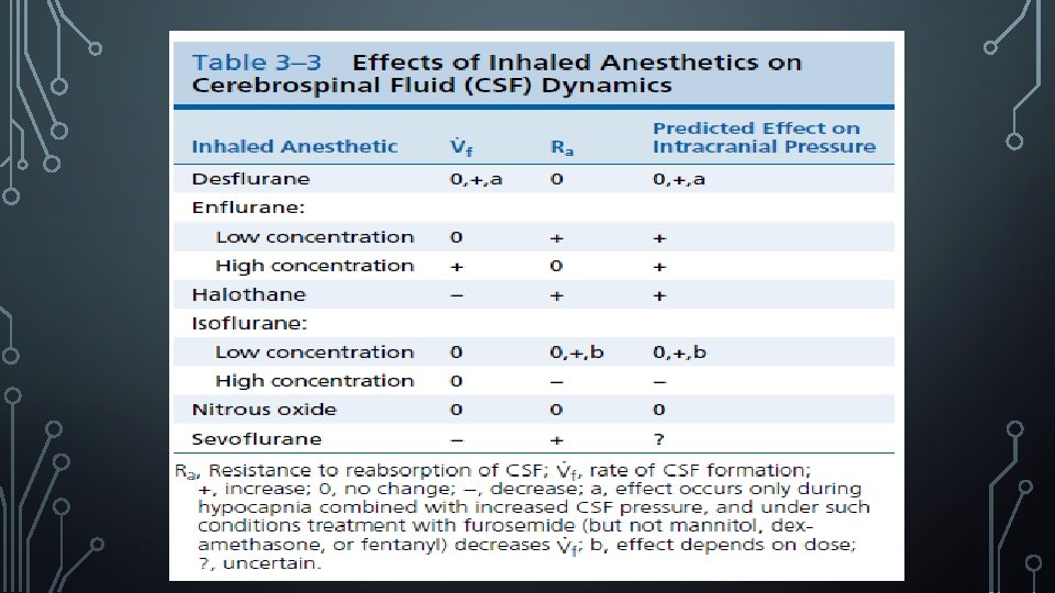
• Anaesthetic and drug induced changes in CSF formation rate (Vf) and resistance to CSF absorption (Ra)
![INHALED ANESTHETICS ENFLURANE Vf Ra ICP LOW[0. 9%-1. 8%] 0 + + HIGH[2. 65&](http://slidetodoc.com/presentation_image_h/bad64e901408489ae597c31e1ada799f/image-39.jpg "INHALED ANESTHETICS ENFLURANE Vf Ra ICP LOW[0. 9%-1. 8%] 0 + + HIGH[2. 65&")
INHALED ANESTHETICS ENFLURANE Vf Ra ICP LOW[0. 9%-1. 8%] 0 + + HIGH[2. 65& 3. 5 end expired] + (40%) 0 + ENFLURANE INCREASES METABOLISM
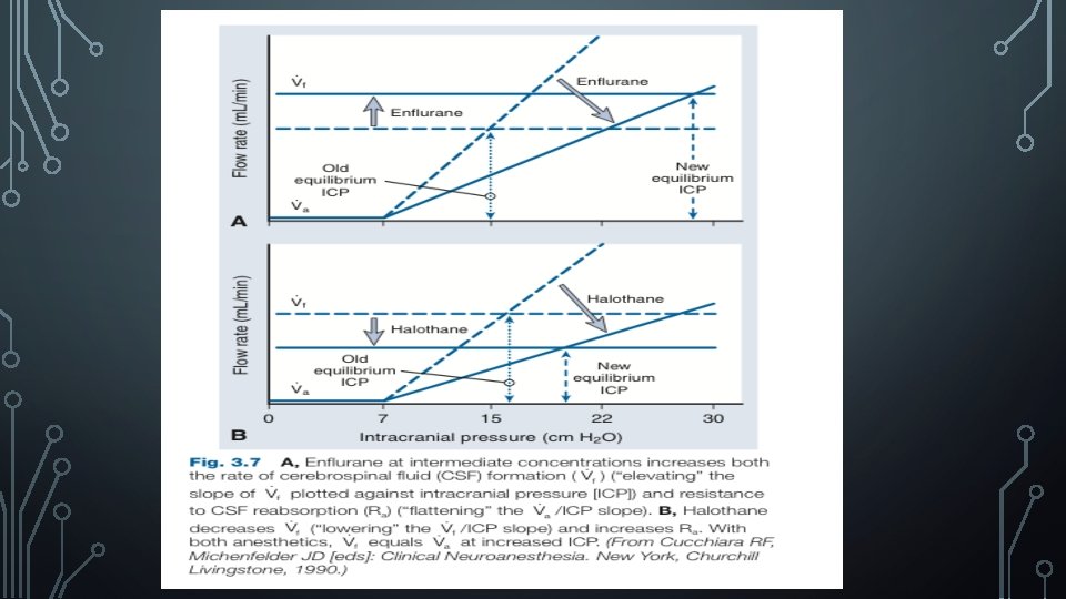

INHALED ANESTHETICS HALOTHANE Vf Ra ICP 1 MAC -- + + INCREASES GLUCOSE TRANSPORT INTO BRAIN INCREASES Na/Cl/H 2 O/ALBUMIN TRANSPORT INTO CSF HALOTHANE INDUCED STIMULATION OF VASOPRESSIN RECEPTORS DECREASE Vf
![INHALED ANESTHETICS ISOFLURANE Vf Ra ICP LOW[0. 6] 0 0 + + 0 --](http://slidetodoc.com/presentation_image_h/bad64e901408489ae597c31e1ada799f/image-42.jpg "INHALED ANESTHETICS ISOFLURANE Vf Ra ICP LOW[0. 6] 0 0 + + 0 --")
INHALED ANESTHETICS ISOFLURANE Vf Ra ICP LOW[0. 6] 0 0 + + 0 -- -- [1. 1%] HIGH[1. 7 -2. 2]

INHALED ANESTHETICS SEVOFLURANE Vf Ra ICP 1 MAC -- + ?

INHALED ANESTHETICS DESFLURANE Vf Ra ICP HYPOCAPNIA & ↑ CSF PRESSURE (0. 5 & 1 MAC) 0 + + OTHER SITUATI 0 NS 0 0 0

INHALED ANESTHETICS NITROUS OXIDE Vf Ra ICP 66% 0 0 0 DECREASES BRAIN GLUCOSE INFLUX AND EFFLUX

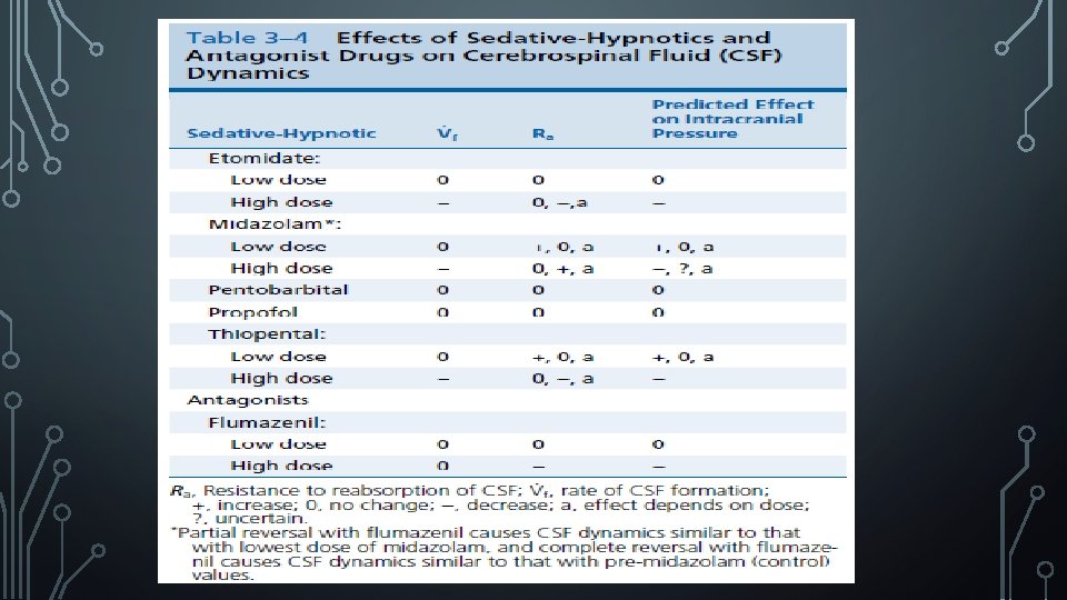
I. V. ANESTHETICS KETAMINE Vf Ra ICP 40 mg/kg/hr 0 + +

I. V. ANESTHETICS ETOMIDATE Vf Ra ICP Low dose. 86 mg/kg 0 0 0 High dose -- -- --

I. V. ANESTHETICS PROPOFOL Vf Ra ICP 6 mg/kg 12, 24, & 48 mg/kg/hr 0 0 0 PENTOBARBITA Vf L Ra ICP 40 mg/kg 0 0 0
")
I. V. ANESTHETICS THIOPENTAL Vf Ra ICP LOW (6 mg/kg F/B 6 -12 mg/kg/hr) 0 +/0 HIGH (1824 mg/kg/hr) -- -- --
 MIDAZOLAM Vf Ra ICP LOW (1. 6 mg/kg fb")
I. V. ANESTHETICS (SEDATIVES &HYPNOTICS) MIDAZOLAM Vf Ra ICP LOW (1. 6 mg/kg fb 0. 5 mg/kg/hr) 0 + + INTERMEDIATE (1 -1. 5 mg/kg/hr) 0 0 0 HIGH (2 mg/kg/hr) -- + --/? FLUMAZENIL Vf Ra ICP LOW (0. 0025 mg/kg) 0 0 0 HIGH (0. 16 mg/kg) 0 --
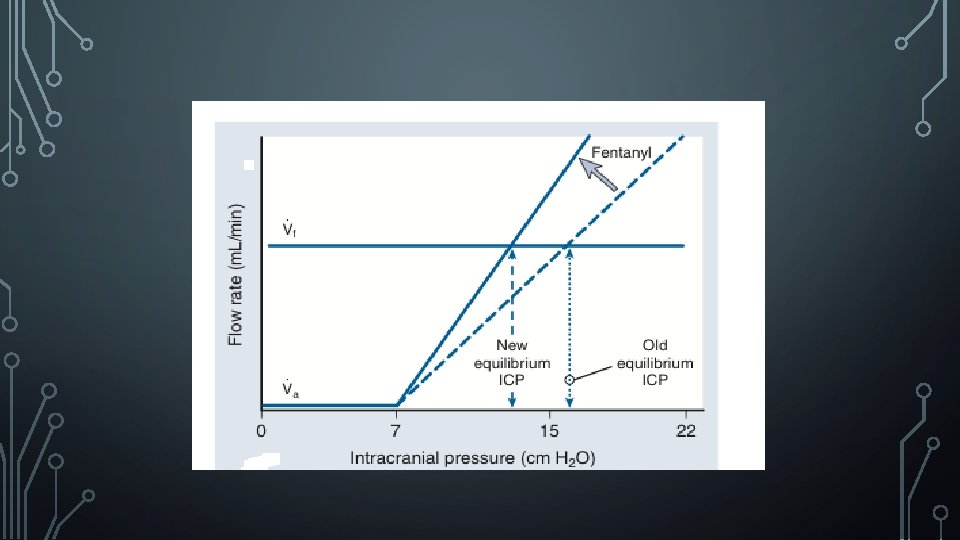
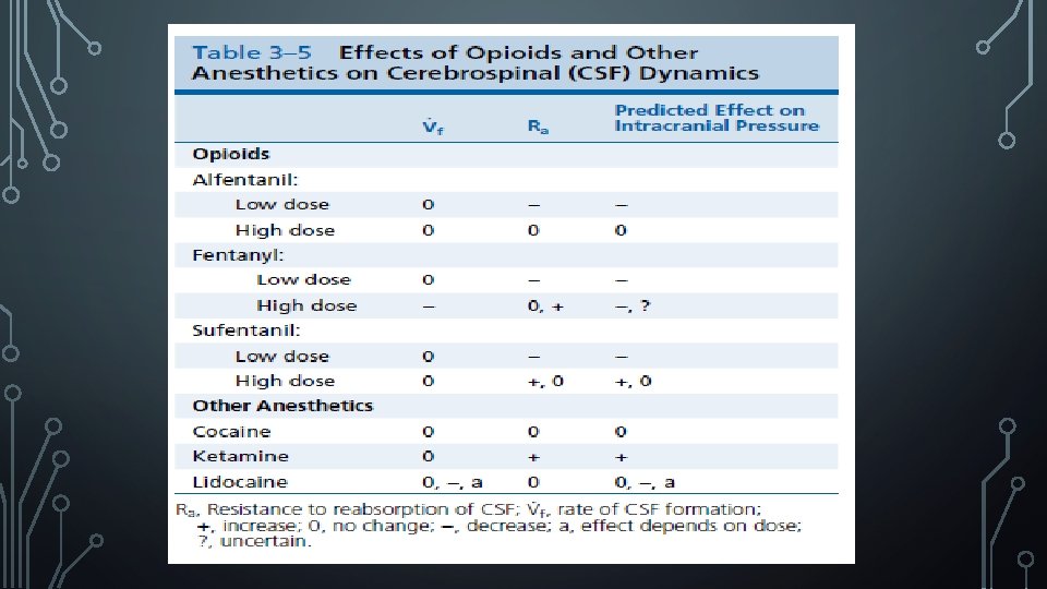
 FENTANYL Vf Ra ICP LOW DOSE 0 -- -- HIGH")
I. V. ANESTHETICS (OPIOIDS) FENTANYL Vf Ra ICP LOW DOSE 0 -- -- HIGH DOSE -- 0/+ --/? SUFENTANYL Vf Ra ICP LOW DOSE 0 -- -- HIGH DOSE 0 +/0 Al. FENTANYL Vf Ra ICP LOW DOSE 0 -- -- HIGH DOSE 0 0 0

I. V. ANESTHETICS LIDOCAINE Vf Ra ICP 0. 5 mg/kg 1µg/kg/min -- 0 0/+

CRUX OF VF AND RA • Vf increases • High dose Enflurane Both Vf & Ra • Ra increases • • • Halothane ( 1 MAC) DES( hypocapnia + increase csf pressure) Low dose enflurane Sevo ( 1 MAC ) Midazolam(low dose) Ketamine Fentanyl , Etomidate

I. V DRUGS • IV acetaminophen moves readily and attains peak conc. in an hour in CSF rapid central analgesia and antipyretic effect • Ibuprofen : peak at 30 -40 min

DIURETICS ACETAZOLAMIDE Vf MECHANISM -- BY 50% INHIBITION OF CA INDIRECT ACTION ON ION TRANSPORT(VIA HCO 3) CONSTRICTS CP ARTERIOLES & DECREASE CPBF METHAZOLAMIDE ACETAZOLAMIDE +OUABAIN ↓ Vf BY 95%= ADDITIVE

DIURETICS Vf MECHANISM FUROSEMIDE -- DECREASE Na+ OR Cl. TRANSPORT MANNITOL -- DECREASE CP OUTPUT AND ECF FLOW FROM BRAIN TO CSF COMPARTMENT

OTHERS DRUG Vf MECHANISM DIGOXIN, OUABAIN -- INHIBIT NA-K PUMP OF CP THEOPHYLLIN + PHOSPHODIESTERASE INHIBITOR ↑CAMP ST IMULATE CP NA-K PUMP VASSOPRESSIN -- CONSTRICTS CP BLOOD VESSELS 3% HYPER TONIC SALINE -- ↓OSMOLALITY GRADIENT FOR MOVEMENT OF FLUID PLASMA CP OR BRAIN TISSUE OR CSF DINITROPHENOL -- UNCOUPLE OXIDATIVE PHOSPHORYLATION ANP -- CGMP

MUSCLE RELAXANTV SCOLINE, VECURONIUM INFUSION Vf 0 Ra 0

STEROIDS • Decrese Ra • Methyl. Prednisolone/prednisolone/cortisone/dexamethasone • Probable mechanism : • Improved CSF flow in SA spaces/ A. villi • Reversal of metabolically induced changes in structure of villi, action at CP • Dexamethasone ↓ Vf by 50%(inhibition of Na-K ATPase)

ALTERATION IN VARIOUS PATHOLOGY • Intracranial volume changes • Volume of intracranial blood/gas/tissue ↑ CSF volume↓ • Mechanism: Translocation into spinal spaces • increased reabsorption • Volume of intracranial blood/gas/tissue ↓ CSF volume ↑ • Mechanism: Cephalad translocation • Decreased reabsorption

ACUTE SAH • Increases ICP • Intrathecal injection: Whole blood, plasma, diasylate of plasma, serum & saline--- Va & Ra values measured by Manometric method • Whole blood and plasma raised ICP and caused a 3 to 10 fold rise in Ra respectively • Fibrin deposits within villi

CHRONIC CHANGES AFTER SAH • Extensive fibrosis of villi leptomeningeal scarring functional narrowing or blockage of CSF outflow tracts [ Ra is increased] hydrocephalus

BACTERIAL MENINGITIS • Animal study with S. pnemoniae, E coli • ICP & Ra increased in both • Even with antibiotic Ra remained high for 2 weeks post Rx • Methyl prednisolone ↓ed Ra to a value that was intermediate between control and infected.

PSEUDOTUMOR CEREBRI • Increased ICP Increased 1. Ra, 2. Vf, 3. greater water movement into brain 4. CBF & CBV, 5 glial or cellular edema. • Impaired reabsorption is the principal cause • Prednisone decreases Ra

HEAD INJURY Ra increased and Vf within normal limits in 75% patients. 20% of the raised ICP derived from changes in Ra /Vf.

IN SUMMARY • CSF plays a key role in brain well being • Vf changes : changes ICP • Ra changes: Changes ICP, alters pressure buffering capacity of brain • In raised ICP, Anesthetics induced changes in Vf & Ra significantly alter the effectiveness of treatments employed to reduce ICP.

- Slides: 72