Cerebrospinal Fluid CSF Cerebrospinal Fluid CSF Is a
")
Cerebrospinal Fluid (CSF)
. . - Is a clear, colorless body fluid. . - bathes")
Cerebrospinal Fluid (CSF). . - Is a clear, colorless body fluid. . - bathes the. . B rain Spinal cord

- Formed and Secreted by the choroid plexus, special tissue that has many blood vessels and lines the small cavities (ventricles) in the brain. .

- It is continually produced, circulated, and then absorbed into the blood system. . - About 500 m. L is produced each day. This rate of production means that all of the CSF is replaced every few hours. .

The primary function of CSF is to. . Buoyancy : The actual mass of the human brain is about 1400 grams; however , the net weight of the brain suspended in the CSF is equivalent to a mass of 25 grams. which allows the brain to maintain its density without being impaired by its own weight

Protection: CSF protects the brain tissue from injury. Chemical stability: CSF maintain the distribution of necessary substance and waste product between CNS and Blood stream Prevention of brain ischemia : made by decreasing the amount of CSF in the limited space inside the skull. This decreases total and pressure

A protective barrier separates the brain from circulating blood and regulates the distribution of substances between the blood and the CSF.

If this protective barrier disrupts may result in a change in the normal level or type of constituents of CSF. Because CSF surrounds the brain and spinal cord, testing a sample of CSF can be very valuable in diagnosing a variety of conditions affecting the central nervous system (CNS). .

Doctors order CSF analysis when some combination of the following signs and symptoms appear. . - Changes in mental status and consciousness. . - Confusion, - Muscle weakness or lethargy, fatigue. . - Nausea. . - Flu-like symptoms that intensify over a few hours to a few days. . - Fever or rash. . - Sudden, severe or persistent headache or a stiff neck. .

- Sensitivity to light. . - Numbness - Dizziness. . - Difficulties with speech. . - Difficulty walking, lack of coordination. . - Mood swings, depression. . - Infants may be irritable, cry when they are held, have body stiffness, refuse food.
 is collected only by the doctor from the")
A sample of cerebrospinal fluid (CSF) is collected only by the doctor from the lower back between third, fourth, fifth lumbar vertebrae using a procedure called a lumbar puncture or spinal tap. It requires certain precautions and careful technique to prevent the infection or the damage of neural tissue

CSF usually collected in three sterile tubes Label 1 / Tube 1 – used for chemical and serologic • test ( tubes are frozen) Label 2 / Tube 2 – used for microbiology lab • ( room temp. ) Label 3 / Tube 3 – used for hematology (cell count) • ( refrigerated)

CSF analysis usually involves an initial basic set of tests performed when CSF analysis is requested: - CSF color, clarity and pressure during collection - CSF protein - CSF glucose - CSF cell count - CSF differential If infection is suspected, CSF gram stain and culture. .

Each of these tests can be grouped according to the type of exam that is performed: Infectious Physical characteristics Microscopic disease test examination the appearance of the CSF. Chemical tests

Chemical tests. . detect or measure the chemical substances found in spinal fluid. CSF is basically an ultafiltrate of the blood, so it can also be affected by what is going on in the blood. Normally, certain constituents of CSF such as protein and glucose are a percentage of blood levels, so CSF levels are often evaluated in relation to blood levels. .
—any cells that may be present are counted")
Microscopic examination. . (Cell count and differential)—any cells that may be present are counted and identified by cell type under a microscope. Infectious disease test detect and identify microorganisms if an infection is suspected
 Color")
Examination Of Cerebrospinal Fluid To estimate sugar, protein and chloride in CSF. 1) Color - Normally CSF is colorless 2) Appearance - the appearance of CSF i. e. turbidity, suspension etc
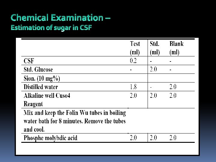

Dilute up to 25 ml with distilled water. Mix well and take the reading on colorimeter blue filter Calculations:

Estimation of Protein in CSF

Calculations

Estimation of chlorides in CSF Take 1 ml of CSF in a test tube. Add 5 drops of 5% potassium chromate. Titrate against std. Ag. No 3 (0. 29%) End point Lemon Yellow to Bolt Red colored precipitate Reading in ml. x 100 = mg% chloride. CSF chloride estimation can also be done directly as tests using standard units by calorimetric other estimation.

Normal Results Normal Values: Normal values typically range as follows: Pressure: 70 - 180 mm H 20 Appearance: clear, colorless CSF total protein: 15 - 60 mg/100 m. L Gamma globulin: 3 - 12% of the total protein CSF glucose: 50 - 80 mg/100 m. L (or greater than 2/3 of blood sugar level) CSF cell count: 0 - 5 white blood cells (all mononuclear), and no red blood cells Chloride: 110 - 125 m. Eq/L
 In severe")
Clinical Interpretation Color: In pathological conditions it tums to various colours i) In severe obstructive jaundice – Yellow coloration ii) Due to obstruction and hemorrhage in subarachnoid space –Yellow coloration due to conversion of Hb to bilirubin Yellow coloration of CSF is called as Xanthochromia. iii) In pneumococcal meningitis (Bacterial infection)– greenish coloration. v) Reddish coloration of CSF due to injury of subarachnoid space. iv) Obstruction and spinal tumor – called erythrochromia cause a reddish color)

In various of meningitis, due to increase in cells, CSF turns cloudy, turbid is possibility of presence of fibrinogen which turns into fibrin clot on standing. All these estimations can be done by using kits
.")
-Increased CSF pressure may be due to increased intracranial pressure (pressure inside the skull). -Decreased CSF pressure may be due to spinal cord tumor, shock, fainting, or diabetic coma. -Increased CSF protein may be due to blood in the CSF, diabetes, polyneuritis, tumor, injury, or any inflammatory or infectious condition. -Decreased protein is a sign of rapid CSF production. -Increased CSF glucose is a sign of high blood sugar. -Decreased CSF glucose may be due to hypoglycemia (low blood sugar), bacterial or fungal infection (such as meningitis), tuberculosis, or certain other types of meningitis. -Increase in nephritis -Desrease in meningitis particularly tuberculous meningitis

-Increased white blood cells in the CSF may be a sign of meningitis, acute infection, beginning of a chronic illness, tumor, abscess, stroke, or multiple sclerosis Red blood cells in the CSF sample may be a sign of bleeding into the spinal fluid or the result of a traumatic lumbar puncture. Increased CSF gamma globulin levels may be due to diseases such as multiple sclerosis, neurosyphilis, or Guillain-Barre syndrome.

References 1. http: //labtestsonline. org/understanding/analytes/csf/tab/test 2 - www. yxjc. sdu. edu. cn/jiaoxuekejian/CSF. ppt 3 -Techniques In Biochemistry Lab 4 - http: //www. umm. edu/ency/article/003428 res. htm

Done by: Wesam oteeda 0876071 Warda yousef 0874592
- Slides: 29