CEREBRALCEREBELLAR ANATOMY SCALP Dural Sinuses SULCUS GYRUS FRONTAL

CEREBRAL-CEREBELLAR ANATOMY

SCALP

Dural Sinuses

SULCUS & GYRUS
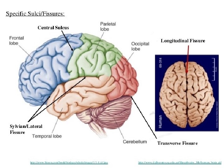

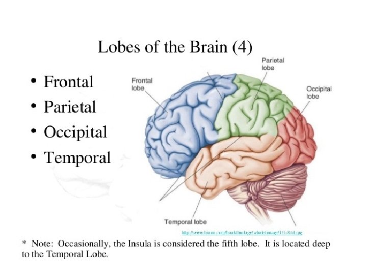

FRONTAL LOBE • Frontal lobe lies anterior to the central sulcus and superior to the sylvian fissure.
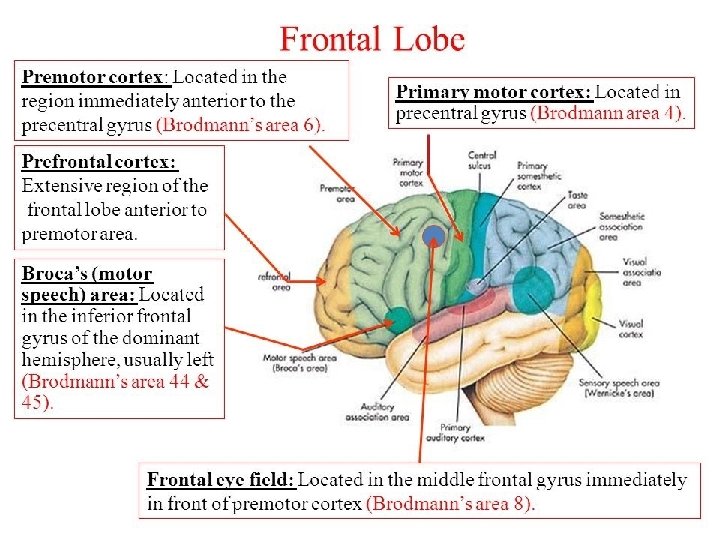

FRONTAL LOBE

TEMPORAL LOBE • The temporal lobe lies beneath the lateral fissure.

TEMPORAL LOBE


TEMPORAL LOBE

TEMPORAL LOBE

TEMPORAL LOBE

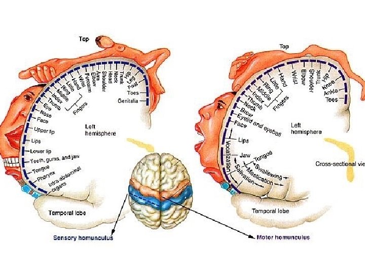
PARIETAL LOBE • Behind the central sulcus, and above the lateral fissure, lies the parietal lobe. • Its most anterior part is the postcentral gyrus, which is the site of the primary somatosensory cortex. (Broadmann’s 3, 1, 2) • Somesthetic association area occupies superior parietal lobule(Broadmann’s 5, 7) • Somatotopic representation
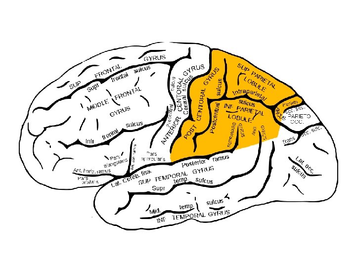

PARIETAL LOBE

PARIETAL LOBE

PARIETAL LOBE

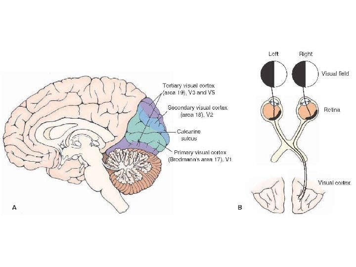
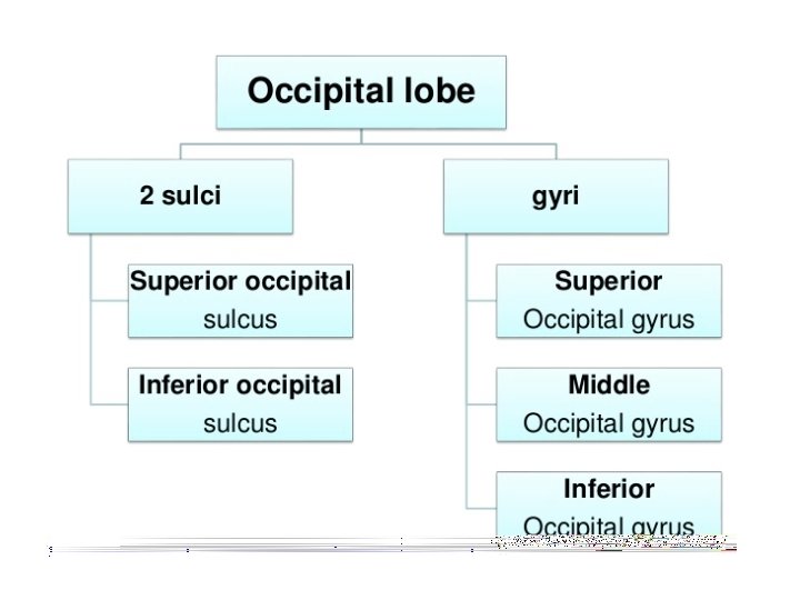
OCCIPITAL LOBE • The occipital lobe is the smallest lobe • It is separated from the parietal and temporal lobes on the medial surface by the parietooccipital sulcus and on the lateral side by the lateral parietotemporal line • The tentorium cerebelli forms its inferior border and the occipital bone its posterior margin.
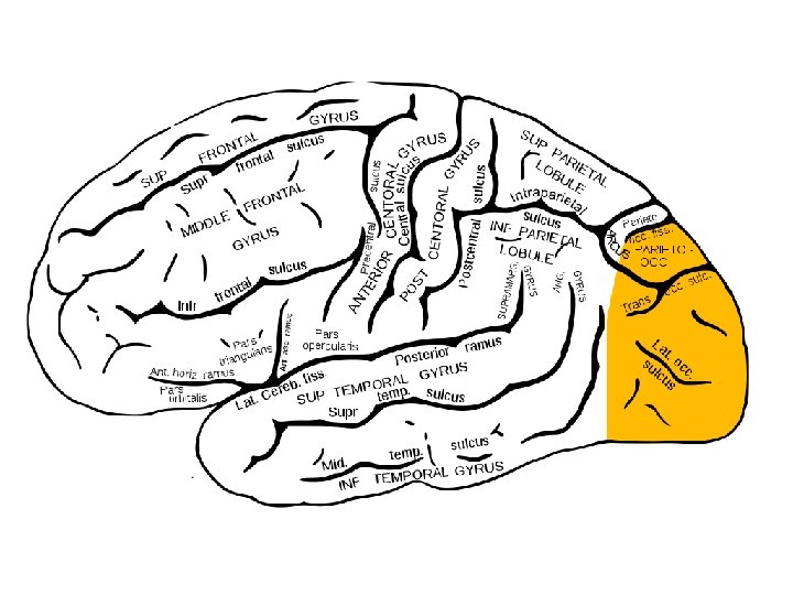

OCCIPITAL LOBE • 1. Primary visual area Broadmann’s area 17 Walls of the posterior part of the calcarine sulcus Receives fibers from lateral geniculate body • 2. Secondary visual area Broadmann’s areas 18 and 19 Surrounds the primary visual area Interpret and relate the visual information received by primary visual area

CEREBELLUM • Latin for ‘little brain’ • Largest part of hindbrain • Occupies most of posterior cranial fossa • Lies behind pons & medulla – forming roof of 4 th ventricle • Separated from posterior part of cerebrum – by tentorium cerebelli

Diagram depicting the anatomical position of cerebellum
 – Connects with midbrain")
Connection with the brainstem • Superior Cerebellar Peduncle (brachium conjunctivum) – Connects with midbrain • Middle Cerebellar Peduncle (brachium pontis) – Connects with pons • Inferior Cerebellar Peduncle (restiform body) – Connects with medulla

External features • Two cerebellar hemispheres. • Median vermis. • Two surfaces ----superior and inferior • 3 fissures: – fissura prima, – horizontal fissure and – posterolateral fissure • 3 lobes in each hemisphere – anterior , – posterior and – flocculonodular.

Flattened view of the cerebellar cortex showing the main cerebellar lobes, lobules, and fissures. B: Relationship between the diagram in (A) and the cerebellum. (courtesy- snell’s clinical neuroanatomy)

TEŞEKKÜRLER EGEMEN SAV
- Slides: 35