Cerebral Cortex Total surface area 2200 cm 2
 about 1/3")
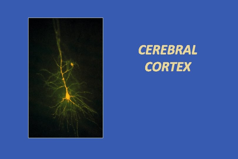
Cerebral Cortex Total surface area: 2200 cm 2 (2. 5 ft 2) about 1/3 ------ surface area about 2/3 ------ hidden in the sulci Thickness: 1. 5 mm (V I) - 4. 5 mm (M I) thicker over the convolution, thinner in the depth of sulci Weight: 600 gm (40 % of total brain weight) 180 gm ----- neurons 420 gm ----- glial cells Number of neuronal cells in cerebral cortex neurons ------ 10 -15 billion glial cells ----- 50 billion

What is mind? No matter. What is matter? Never mind.

- The most complex 1500 grams in the universe; - A particle of a sand-sized brain contains 100, 000 neurons and 1 million synapses between them; 250, 000 neurons per minute during the fetal period; 100 billion neurons at birth and about 1 quadrillion (1 million billion) connections between them, with each synapse activated ~ 10 x per second; -100 million photoreceptor cells in the retina - 10 events x second = -1 billion pieces of information x second.

Human brain Although it is only 2% of total body weight, the human brain consumes: • 87% of the body's energy in newborns; • 50% of energy in five-year-olds; • 25% of energy in adults. "The brain is the most powerful computer we know, and understanding it is one of the major challenges in science. It is the one that makes people special. " Luis Bettencourt, National laboratory Los Alamos, professor in Santa Fe Institue.

Human brain and memory One neuron - one memory = space shortage (i. Pod or USB flash drive). The neurons are combined, each helping memory at a time = increasing brain memory capacity to about 2. 5 petabytes (one million gigabytes). If the brain works as a digital video recorder, 2. 5 petabytes of memory will be able to support 3 million hours of television broadcasts.

What we know is a negligible part of what it is… -the brain is not a digital machine, and the synapse data transmission is probably of the analog type, with a huge number of input stimuli; -the connection is selective; -only a small fraction of neurons function at a given time, which limits "energy consumption"

The human cerebral cortex operates at about one petaflop - one quadrillion operations per second. • Tianhe-1 A – (Milky Way, Sky River) (Китай) -2, 57 petaflops maximal productivity; • Blue Waters (Илинойс) – up to 10 petaflops.

10 septillion operations per second - the absolute maximum; D-Wave, British Columbia - 64, 000 operations simultaneously in parallel "universes"; Processor with only 16 qubits (quantum bit - the smallest unit of information in quantum calculations); Qubit contains an exponentially larger amount of information than traditional bits; Systems with hundreds of qubits - will process more input than atoms in the universe.

CLARITY project Karl Deisseroth and team, Stanford, California. A powerful push in the field of "connectomics" – mapping of brain contacts. Creates optically transparent brains or blocks of brain tissue. Allows to observe the smallest details without losing the overall image of the structure. Observing large networks of neurons with unprecedented ease and accuracy. New brain research opportunities from patients and healthy donors. “We can transform mouse brain into a completely transparent form within four to five days. " Karl Deisseroth

CLARITY project CLARITY enables molecular analysis of the brain. Each color represents a different molecular marking. Image of a hippocampus showing networks of neurons expressing different molecules (green, red) and astrocytes (blue).

Human connectome project

BRAIN Initiative
 Palaeocortex (Paleopallium) Isocortex Neocortex (Neopallium) cf. mesocortex,")
Subdivision of Cerebral Cortex Allocortex Archicortex (Archipallium) Palaeocortex (Paleopallium) Isocortex Neocortex (Neopallium) cf. mesocortex, juxtallocortex, mesallocortex

Isocortex – typical 6 layered cortex I. Molecular Layer II. External Granular Layer III. External Pyramidal Layer IV. Internal Granular Layer V. Internal Pyramidal Layer VI. Polymorphic Layer

Cerebral Cortex Histological Organization Cellular Elements 1. Pyramidal Cell - output neuron giant pyramidal cell of Betz 2. Fusiform Cell --- modified pyramidal cell 3. Granular (Stellate) Cell basket cell, double bouquet cell, bipolar cell, chandlier cell, neurogliform cell 4. Horizontal Cell of Cajal (Retzius-Cajal cell) 5. Cells of Martinotti

I. Molecular Layer II. External Granular Layer III. External Pyramidal Layer Line of Kaes-Bechterew IV. Internal Granular Layer Outer band of Baillarger - Line of Gennari in area 17 V. Internal Pyramidal Layer Giant pyramidal cell of Betz Inner Band of Baillarger VI. Polymorphic Layer Golgi Nissl Weigert

Cortical Afferent Fibers 1. corticocortical fiber association fiber commissural fiber 2. thalamocortical fiber - specific and non-specific 3. extrathalamic subcortical fiber cholinergic fiber - acetylcholine - basal nucleus of Meynert mesolimbic dopaminergic fiber - dopamine - ventral tegmental area serotonergic fiber – serotonine - raphe nuclei norepinephrinergic fiber - norepinephrine - nucleus locus ceruleus

Cortical Efferent Fibers 1. Corticofugal Fiber - Projection Fiber corticostriate fiber corticothalamic fiber corticorubral fiber corticotectal fiber corticopontine fiber cortico-olivary fiber corticobulbar fiber corticospinal fiber 2. Corticocortical Fiber Association fiber Commissural fiber

Columnar Cortical Unit and Cortical Circuitary A. pyramidal neuron B. excitatory granular cell C. inhibitory granular cell 1. afferent fiber 2. efferent fiber 3. corticothalamic fiber

Regional Variation of Cortical Lamination A. Homotypical isocortex ------- association cortex B. Heterotypical isocortex 1. granular cortex --- primary sensory cortex V I (17), S I (3), A I (41) 2. agranular cortex --- motor cortex M I (4), PM (6)

Von Economo’s classification of cortical types 1. agranular, 2. frontal, 3. parietal, 4. polar, 5. granular
 Jacksonian epilepsy (1864) Experimental")
Functional Localization of Cerebral Cortex Clinical evidences Broca’s area (1861) Jacksonian epilepsy (1864) Experimental evidences Fritsch and Hitzig (1870) --- motor cortex von Gudden (1870) ---- visual cortex Ferrier (1873) ---- auditory cortex
 scan")
PET (positron emission tomography) scan
 ------ most popular")
Morphological Classification of Cortical Areas based on cytoarchitectonic studies Brodmann (1909) ------ most popular 47 areas
")
Brodmann’s cytoarchitectorial map (Lateral & Medial surface)

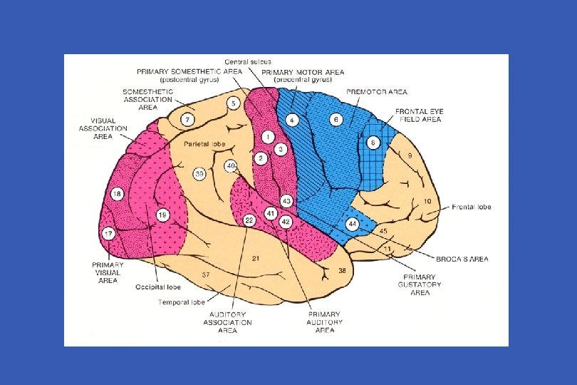
Functional Localization of Cerebral Cortex Sensory area • primary sensory area • secondary sensory area Motor area • primary motor area • secondary motor area • supplementary motor area Association area • parietal, occipital and temporal cortex - conceptual elaboration of sensory data • prefrontal (frontal) cortex - judgement, foresight
 S I, S II Visual Area (vision) V I,")
Sensory Areas Somesthetic Area (Somesthesia) S I, S II Visual Area (vision) V I, V II Auditory Area (Hearing) A I, A II Vestibular Area (Equilibrium) Gustatory Area (Taste) Olfactory Area (Smell)
 afferernts: ventrobasal complex (VPLc,")
Somesthetic Area S I ----- 3, 1, 2 (postcentral gyrus) afferernts: ventrobasal complex (VPLc, VPM) discrimination of position and intensity of sensation S II ---- superior bank of lateral fissure no clinical disorders Somesthetic Association Cortex ------- 5, 7 (parietal lobule, precuneus) afferents: S I, LP of thalamus integration of geneal sensation with past experience tactile agnosia, astereognosis

Sensory Homunculus
 greatly thickened")
Visual Cortex V I ----- 17 (striate cortex - line of Gennari) greatly thickened outer band of Baillarger heterotypical isocortex afferent: LGd of thalamus visual field defect: homonymous quadranopsia and macular sparing V II ---- 18, 19 (visual association area) afferents: V I, pulvinar of thalamus integration of vision with past experience visual agnosia cf. occipital eye field

Visual fields
 Face recognition Perceive Facial Expression")
Visual association fields V 4 (color) Face recognition Perceive Facial Expression
 heterotypical isocortex")
Auditory Cortex A I ----- 41, 42 (trannsverse temporal gyrus of Heschl) heterotypical isocortex afferents: MGv of thalamus - core projection slight diminution in auditory acuity A II ---- 22 (Wernike's area of original connotation) not well-defined afferents: non-laminar part (MGm, MGd) – belt projection AI auditory agnosia - sensory aphasia

Auditory fields A I ----- 41, 42 A II ---- 22

Other Primary Sensory Areas Vestibular Area 3 a and 2 v of S I afferents: VPLo [superior temporal gyrus anterior to A I] Gustatory Area 43 (inferior end of postcentral gyrus) afferents: VPMpc Olfactory Area Piriform Lobe - Limbic System
 Premotor Area (PM) Supplementary Motor Area (SMA)")
Motor Areas primary Motor Area (M I) Premotor Area (PM) Supplementary Motor Area (SMA) Frontal Eye Field

Motor Homunculus

Primary Motor Area M I ------- 4 precentral gyrus of lateral surface anterior part of paracentral lobule heterotypical agranular cortex giant pyramidal cell of Betz afferents: premotor area, SMA, S I VLc, VPLo of thalamus Motor Homunculus Upper Motor Neuron (UMN) syndrome
 ------ lateral surface of 6 afferents: VLc, VPLo")
Other Motor Areas Premotor Area (PM) ------ lateral surface of 6 afferents: VLc, VPLo of thalamus from cerebellum Supplementary Motor Area (SMA) ------------- medial surface of 6 afferents: VLo, Vapc of thalamus from basal ganglia Frontal Eye Field ----- 8 voluntary tracking movement

Brodman’s Map of Motor and Sensory Areas

Association Areas Language Areas ----- 22, 39, 40, 44, 45 Posterior Parietal Association Area ------ 5, 7 (39, 40) body image Temporal Association Area ------ 20, 21, 37, 38 (22) multisensory integration, conceptual ideation Prefrontal Association Area ----- 9, 10, 11, 12, 46, 47 (44, 45) judgement, foresight, personality

1 3 3 2 1 2 3 Order of Cortical Maturation 1

Disorders of Association Cortex Agnosia Tactile agnosia Visual agnosia Alexia Auditory agnosia Apraxia Aphasia Wernicke’s (receptive) aphasia Broca’s (Motor) aphasia conduction aphasia global aphasia

Apraxia The inability to execute a voluntary motor movement despite being able to demonstrate normal muscle function.
 ---- 22, 39, 40 Receptive Aphasia -")
Language Areas Sensory Language Area (Wernike's area) ---- 22, 39, 40 Receptive Aphasia - area 22 defect in comprehension, good spontaneous speech Anomic Aphasia - word finding difficulty Jargon aphasia - fluent, but unintelligiable jargon 39 (supramarginal gyrus), 40 (angular gyrus) Superior Longitudinal Fasciculus Conduction Aphasia good comprehension, good spontaneous speech poor repetition, poor response Motor Language Area (Broca’s area) --- 44, 45 Motor Apahsia good comprehension, no speech
")
Language Areas (Geschwind Model)
 Dominant Hemisphere Language – speech, writing Calculation Non-dominant Hemisphere Spatial")
Cerebral Dominance (Lateralization, Asymmetry) Dominant Hemisphere Language – speech, writing Calculation Non-dominant Hemisphere Spatial Perception (3 D subject) Singing Playing musical instrument

Language 3 D perception Speech Singing Writing Playing Musical Calculation instrument
 Two minds in one brain? Roger Sperry (1913")
Split Brain Commissuratomy (split corpus callosum) Two minds in one brain? Roger Sperry (1913 -1994) 1981 Nobel Laureate

Prefrontal Association Areas § Frontal Granular Cortex Lateral Prefrontal Association Area ------ 9, 10, 46 judgement, foresight, problem solving Orbitofrontal Cortex ------ 11, 12, 47 emotion, olfaction, personality Case of Phineas Gage Prefrontal Leucotomy of Moniz and Freeman
")
Phineas Gage (1823 -1861, accident in 1848)
")
Phineas Gage’s lesion reconstructed (H. Damasio and R. Frank, 1992)
 Antonio Egas Moniz")
Prefrontal Leucotomy (Frontal Lobotomy) Antonio Egas Moniz
- Slides: 55