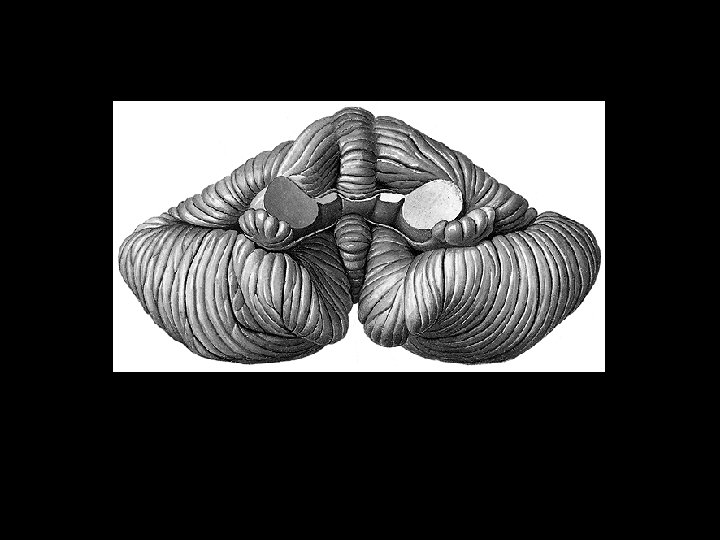
Cerebellum Coordination of movements Margo ant incisura cerebelli

Cerebellum Coordination of movements

Margo ant. , incisura cerebelli ant. Margo post. , incisura cerebelli post.

Vermis Hemispheres Pars flocculonodularis Folia, lobuli, lobi

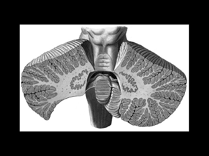
Fissura prima Lobus ant. Lobus post. flocculonod. ra at. u s rol s i F ste po

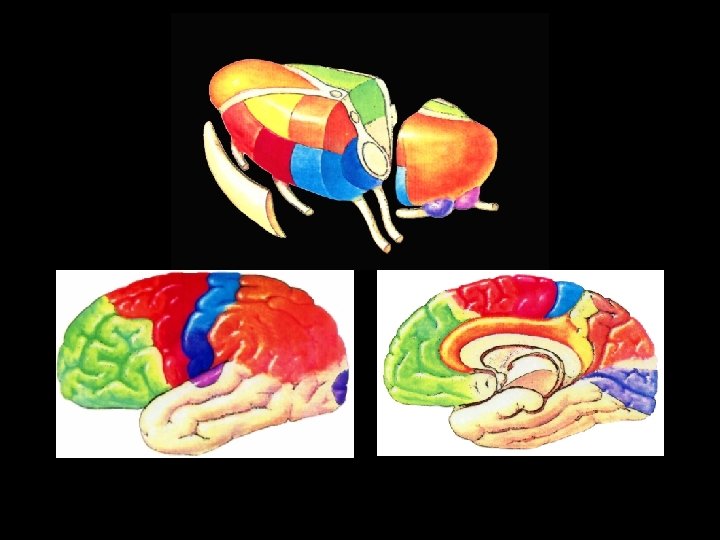
Developmental anatomy Afferents from vestib. labyrinth fish, amphibians Archi cerebellum VESTIBULO CEREBELLUM Afferents from spinal cord and brainstem reptiles, birds, mammals Paleo SPINO cerebellum CEREBELLUM Afferents from cortex telencephali Neo cerebellum PONTO CEREBELLUM

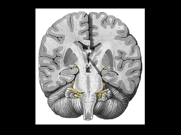
Structure of the cerebellum Grey matter Cortex cerebelli str. moleculare str. ganglionare str. granulare Nuclei cerebellares White matter Subst. medullaris laminae albae (arbor vitae) Pedunculi cerebellares

4 3 2 1 1 ncl. dentatus 2 ncl. emboliformis 3 ncll. globosi 4 ncl. fastigii Nuclei cerebelli

Spinocerebellum 3 2 1 Ponto cerebellum Vestibulocerebellum 1 median zone 2 paramedian zone 3 lateral zone L. flocculonodularis

Spinocerebellum Ponto cerebellum Vestibulocereb. ncll. vestibulares Spinocereb. ncll. fastigii, emboliformes, globosi Neocereb. ncl. dentatus
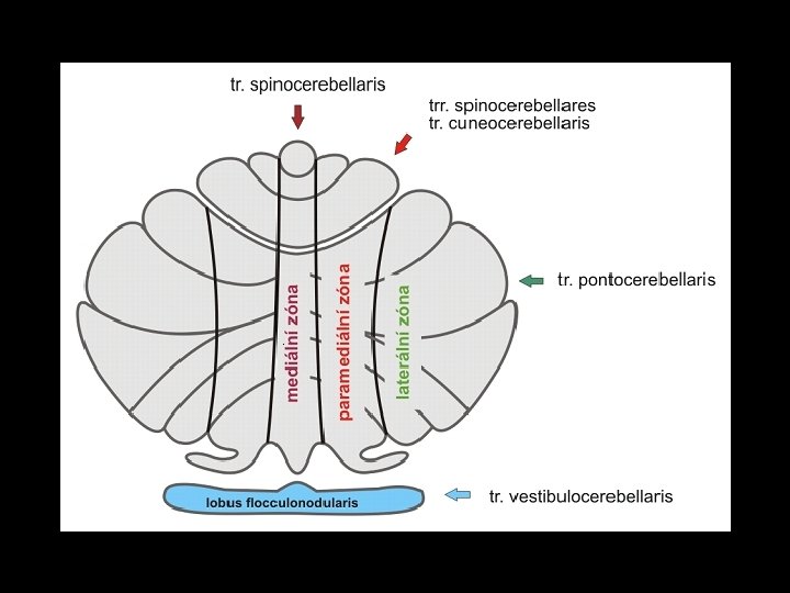

Pedunculi cerebel. inf. tr. sp-ce post. , cuneo-ce, bulbo-ce, ve-ce, re-ce, olivo -ce from lobus flocculonodul. do ncll. vestibulares (tr. ceve) to RF of the brainstem (tr. ce-re) Pedunculi cerebel. medii tr. ponto-ce Pedunculi cerebel. sup. tr. sp-ce ant. , ru-ce a afferents from ncl. mesenceph. CN V from ncll. emboliformes, globosi and dentatus Afferents : efferents = 40: 1

Pathways of the cerebellum Afferents to the cortex cerebelli from vestib. labyrinth from spinal cord and brainstem from cortex of the brain Efferents from the nuclei to brainstem, thalamus

Function of the cerebellum ■ archicerebellum > posture and eye movements ■ paleocerebellum > progressive movements (walking, swimming etc. ) ■ neocerebellum > manipulative movements and speech

CEREBELLAR DISORDERS Ataxia inability to stand upright without support Dysmetria „overshooting“ - the hand may travel past the target Dyssynergia incoordination Adiadochokinesia inability to perform rapid alternating movements
")
DIENCEPHALON ■ thalamus ■ epithalamus ■ subthalamus ■ hypothalamus (metathalamus)
")
Thalamus ■ tuberculum ant. ■ pulvinar ■ stria medullaris (tela choroidea ventr. III. ) fissura transversa cerebri ■ taenia choroidea (tela choroidea ventr. lat. ) ■ lamina affixa thalami ■ stria terminalis

Epithalamus ■ stria medullaris thalami ■ trigona habenularum ■ commissura habenularum et post. ■ corpus pineale (epiphysis cerebri)

Subthalamus Grey matter ■ zona incerta ■ ncl. subthalamicus ■ part of subst. nigra ■ part of globus pallidus White matter ■ Fasc. thalamicus ■ Fasc. lenticularis ■ Ansa lenticularis ■ Fasc. subthalamicus Involved in motor circuits

su lcu sh yp oth al.

Hypothalamus Corp. mamillaria Infundibulum Tuber cinereum Hypophysis cerebri

Metathalamus 2 1 1 corp. geniculatum med. brachium colliculi inf. – colliculus inf. 2 corp. geniculatum lat. brachium colliculi sup. – colliculus sup.

THALAMUS relay station of ascending pathways involved in motor circuits reciprocal connections to the association areas of the cerebral cortex – functions related to memory, cognition, judgement, mood

Anterior group A ncll. ant. A LD Lateral group dorsal row LD ncl. lat. dors. LP ncl. lat. post. VA VL R ventral row VA ncl. ventr. ant. VL ncl. ventr. lat. VP ncl. ventr. post. : VPL ncl. ventr. post-lat VPM ncl. ventr. post-med CGL ncl. corporis gen. lat. CGM ncl. corporis gen. med. DM LP CM VP: VPM VPL P CGL CGM Medial group DM ncl. dorsomed. Posterior group P ncll. pulvinari, post. Intralaminar group CM ncl. centromed. R ncll. reticulares

Functional groups of nuclei ■ specific nuclei somatosenzory motor ■ non-specific nuclei ■ association nuclei

Specific nuclei VA VL VPM GP cereb ellum BG SS: VPL, VPM CGL visual O Rpathway S: CGM, CGL M: VA, VL tr. sp-th LM auditory pathway tr. trig-th tr. so-th (taste)

Non-specific nuclei ncll. intralaminares ncl. medianus R from FR of the brainstem and other thalamic nuclei to BG, thalamus, cortex (ARAS)

Projection to the cortex through specific and non-specific thalamic nuclei CORTEX THA LAM US specific pathway non-specific pathway

Association nuclei A DM LD LP P ■ integration of GSA a SA inputs to cortex ■ reciprocal connections with the association cortex

Function of association nuclei cortex Ncl. ant. thalami Interconnection of association areas of the cortex

Hypothalamus control of: - ANS - endocrine system Function of the hypothalamus is related to: ■ control of vital functions that maintain homeostasis ■ control of emotions

Hypothalamic nuclei at the frontal section Periventricular row Lat. row Med. row Fornix III. Med. Lat. row III. ventricle Fornix

ant. medial post. Hypothalamic nuclei - sagittal section Anterior nuclei Periventricul row: ncl. suprachiasmat. Medial row: ncl. preopticus, ncl. supraopticus, ncl. ant. , ncl. paraventr.

Medial nuclei Periventricular row: ncl. arcuatus Medial row: ncl. ventromed. et ncl. dorsomed.

Posterior nuclei Periventricular + med. rows: ncl. post. et ncl. mamillaris

HYPOTHALAMIC NUCLEI 1. ncl. preopticus 2. ncl. paraventricularis 3. ant. hypoth. area 4. ncl. supraopticus 5. lat. hypoth. area 6. dorsal hypoth. 7. ncl. dorsomedialis 8. ncl. ventromed. 9. post. hypoth. area 10. corpus mammilaris 11. chiasma opticum 12. lamina terminalis 13. commissura ant. 14. sulcus hypothal. 15. adhesio interthal. 16. fornix 17. septum pellucidum 18. fossa interped. 19. thalamus 20. tuber cinereum 21. n. opticus 22. infundibulum 23. lobus ant. 24. lobus post.

White matter of the diencephalon Fornix Stria medullaris Stria terminalis FLD

Hypophysis cerebri Lobus ant. adenohypophysis Pars intermedia Lobus post. neurohypophysis (eminentia mediana infundibular stalk lobus post. )

Adenohypophysis Secretion of hormones: Thyreotropin Gonadotropic Growth Adrenocorticotropic ■ cells of adenohypophysis are stimulated or inhibited by „releasing“ and „inhibiting“ hormones producing in some hypothalamic nuclei (neurosecretion) parvocellular neurons reach the median eminence from the infundibulum are transported to the adenohypophysis by the portal vessels

ncl. paraventricularis ncl. preopticus ncl. arcuatus ncll. tuberales eminentia med. a. hypophysea sup. primary vascular plexus „releasing hormones“ sinus cavernosus hypophyseal portal vessel „inhibiting hormones“ secondary vascular plexus

aa. hypoph. sup. primary plx. long port. vessel short port. vessels lobus ant. sec. plx. drainage into the sinus cavernosus

Neurohypophysis ■ receives axons of magnocellular neuroendocrine cells of the supraoptic and paraventricular hypoth. nuclei ■ developmentally – part of diencephalon ■ oxytocin and ADH ■ neuroendocrine cells reach the posterior lobe of the hypophysis through tr. hypothalamo-hypophysialis
 neurohypophysis a. hypophysea")
Tr. hypoth. -hypophysialis Ncl. paraventricularis Oxytocin Ncl. supraopticus Antidiuretic h. (Vasopresin) neurohypophysis a. hypophysea inf. sinus cavernosus

Telencephalon

Structure of telencephalon Gray matter Basal ganglia Cortex White matter pathways Projection Commissural Association

Basal ganglia 1 2 3 4 ■ 1 5 6 3 2 ncl. caudatus globus pallidus putamen claustrum corp. amygdaloideum 4 Functionally 5 ncl. subthalamicus 6 substantia nigra globus pallidus + putamen = ncl. lentif. ncl. caudatus + putamen = corpus striatum
 = globus pallidus lat. + med. segment – dorsal")
Development of BG Palleostriatum (pallidum) = globus pallidus lat. + med. segment – dorsal pallidum ventral pallidum Neostriatum (striatum) ncl. caudatus, putamen – dorsal striatum ncl. accumbens – ventral striatum Archistriatum corpus amygdaloideum

C Ncl caudatus + putamen = dorsal striatum Pu GP VP VS Globus pallidus = dorsal pallidum VS = ventr. striatum (ncl. accumbens septi) VP = ventral pallidum (ncl. basalis Meynerti)

Corpus amygdaloideum

Functional connections of BG CORTEX STRIATUM THALAMUS PALLIDUM Function of BG inhibition of cortical and subcortical motor functions
 palleocortex (rhinencephalon) archicortex")
Cerebral cortex ne ALLOCORTEX oc ort ex 3 -4 layers a) palleocortex (rhinencephalon) archicortex palleocortex b) archicortex NEOCORTEX 6 layers
 ■ 11 regiones ■ 52 areae")
Brodman’s map (cytoarchitectonic map of cortex) ■ 11 regiones ■ 52 areae
, primary somatic sensory c. (a")
Functional regions of cortex Primary motor c. (a 4), primary somatic sensory c. (a 3, 1, 2), primary visual c. (a 17), primary auditory c. (a 41, 42) Secondary and association areas

Representation of contralateral body parts rty zuby, dásně, čelist stí a ě p ruk zá t kolen gyrus precentralis o ke l če ra pře lokpeaže men zá o d t rukapěstíloktí ec bér 2 -5 prst , o hn palec stenohay t prsitál oko gen nos tvář o en el rup č m t ky ra lo k t týl rk rup ky gyrus postcentralis kotníky prsty ty prs c pale krk obočí o , ok víčko tvář rty hltan y útrob „sensory homunculus“ žv sl ýká in ěn ní í jazyk „motor homunculus“

cent rum semi oval e White matter of the telencephalon - corpus medullare Fibers commissural projection association

Commissural fibers 1 corpus callosum neocortex 1 3 2 2 commissura ant. pars ant. - paleocortex pars post. - neocortex 3 commissura fornicis archicortex

Corpus callosum - 300 million fibers forceps minor connection of frontal lobes forceps major connection of occipital lobes tapetum roof of the posterior horn

Commissura fornicis et anterior

Projection fibers short ■ connections between cortex and BG ■ reciprocal connections between cortex and thalamus long tr. co-sp tr. co-ncl tr. co-ret tr. co-tec tr. co-ru tr. co-bulb tr. co-po capsula interna

Caps ula i nt.

crus ant. fr- po genu co-ncl crus post. , to , p po radi atio opti ca ra ac diat us io tic a p, s co e ru, r
, long fibrae arcuatae cingulum f. longit. sup. f. uncinatus")
Association fibers: short (fibrae arcuatae), long fibrae arcuatae cingulum f. longit. sup. f. uncinatus f. longit. inf.

Illustrations were copied from: Atlas der Anatomie des Menschen/ Sobotta. Putz, R. , und Pabst, R. 20. Auflage. München: Urban & Schwarzenberg, 1993 Netter: Interactive Atlas of Human Anatomy. Windows Version 2. 0
- Slides: 66