
Central nervous system Guanghui Yu Radiology college Central
- Slides: 64
Central nervous system Guanghui Yu Radiology college
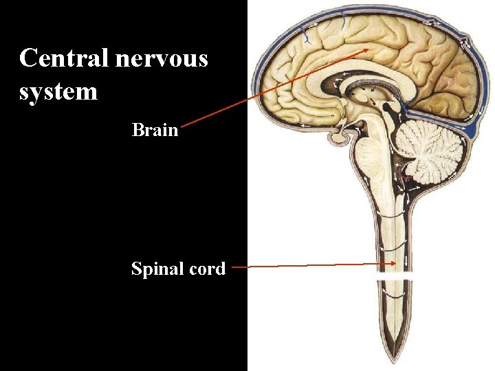
Central nervous system Brain Spinal cord
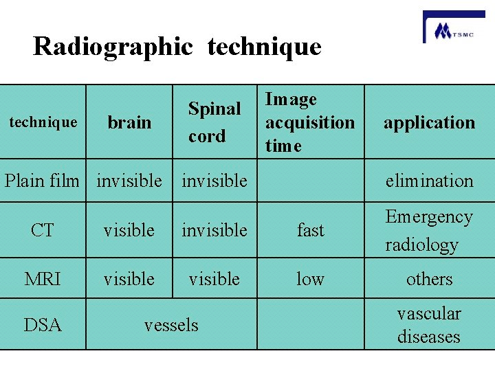
Radiographic technique brain Spinal cord Image acquisition time Plain film invisible application elimination CT visible invisible fast Emergency radiology MRI visible low others DSA vessels vascular diseases
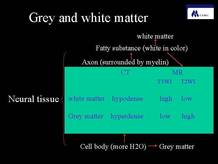
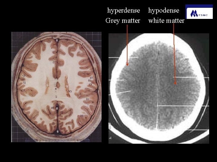
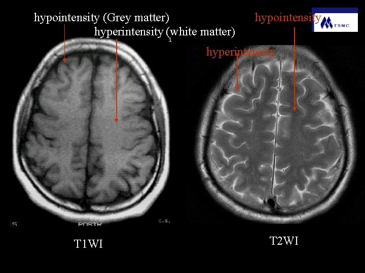
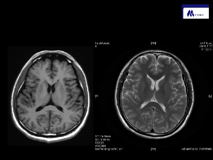
Grey and white matter Fatty substance (white in color) Axon (surrounded by myelin) CT MR Neural tissue T 1 WI T 2 WI white matter hypodense high low Grey matter hyperdense low high Cell body (more H 2 O) Grey matter
hyperdense Grey matter hypodense white matter
hypointensity (Grey matter) hyperintensity (white matter) hypointensity hyperintensity T 1 WI T 2 WI
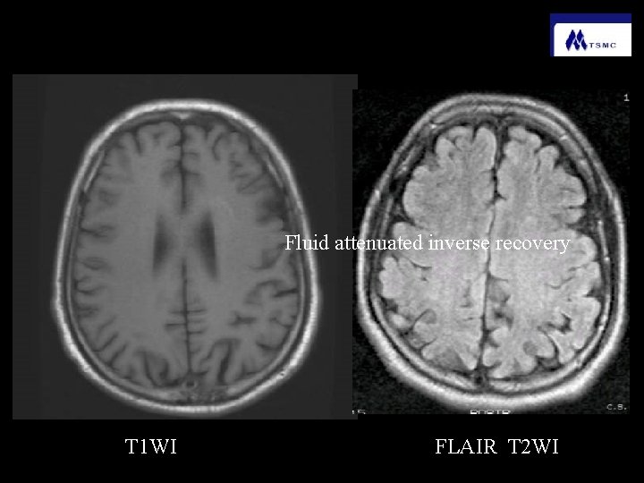
Fluid attenuated inverse recovery T 1 WI FLAIR T 2 WI
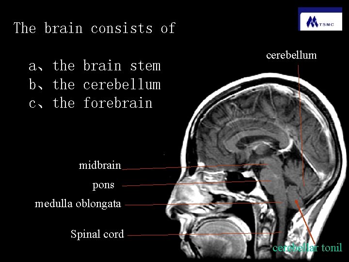
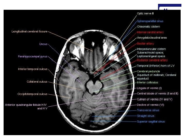
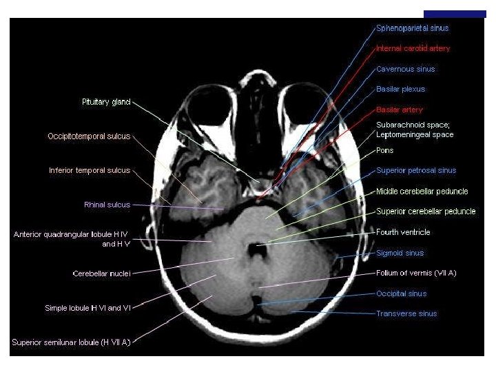
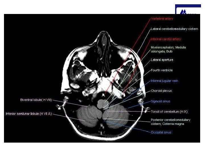
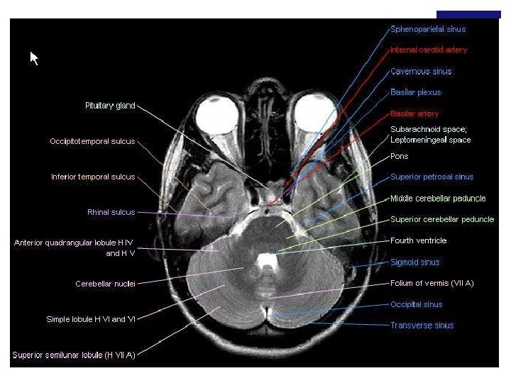
The brain consists of a、the brain stem b、the cerebellum c、the forebrain cerebellum midbrain pons medulla oblongata Spinal cord cerebellar tonil
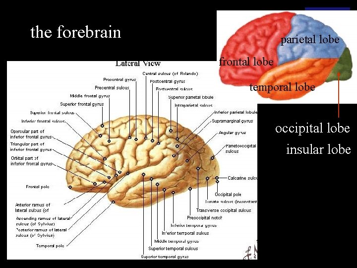
the forebrain parietal lobe frontal lobe temporal lobe occipital lobe insular lobe
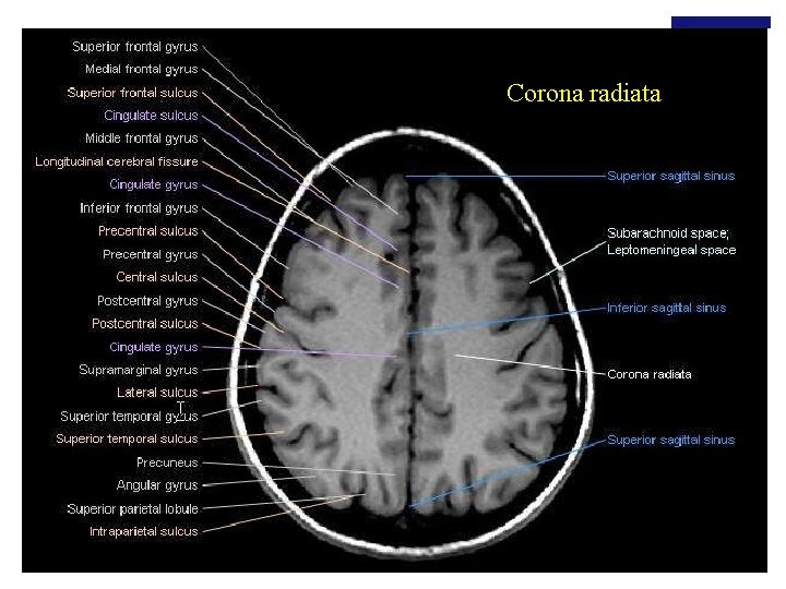
Corona radiata
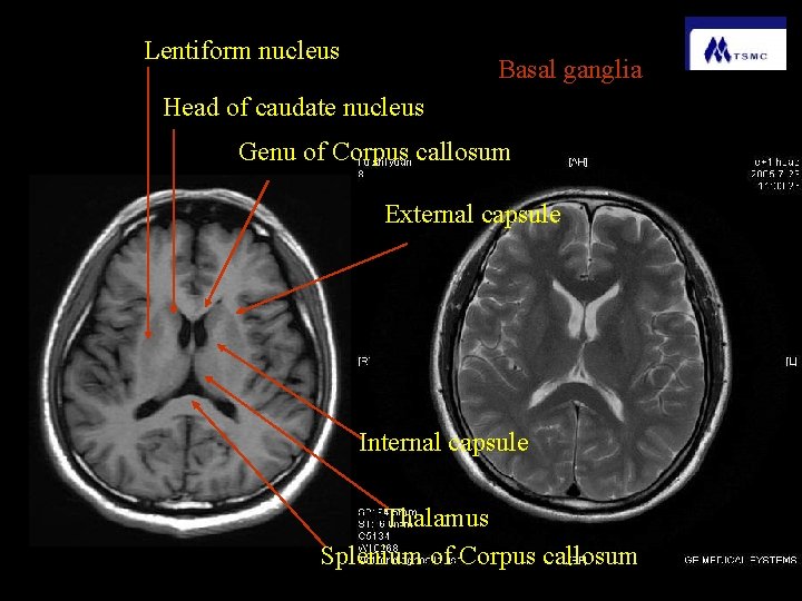
Lentiform nucleus Basal ganglia Head of caudate nucleus Genu of Corpus callosum External capsule Internal capsule Thalamus Splenium of Corpus callosum
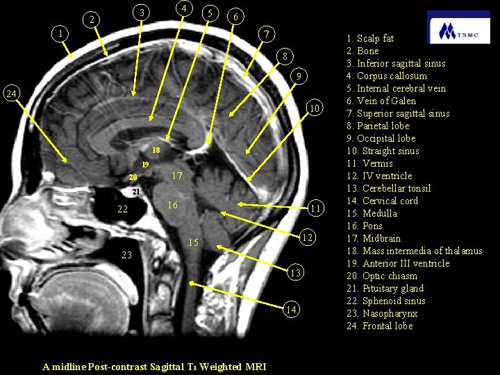
4 3 2 5 1 6 7 8 9 24 10 18 19 20 17 21 22 16 11 12 15 23 13 14 A midline Post-contrast Sagittal T 1 Weighted MRI 1. Scalp fat 2. Bone 3. Inferior sagittal sinus 4. Corpus callosum 5. Internal cerebral vein 6. Vein of Galen 7. Superior sagittal sinus 8. Parietal lobe 9. Occipital lobe 10. Straight sinus 11. Vermis 12. IV ventricle 13. Cerebellar tonsil 14. Cervical cord 15. Medulla 16. Pons 17. Midbrain 18. Mass intermedia of thalamus 19. Anterior III ventricle 20. Optic chiasm 21. Pituitary gland 22. Sphenoid sinus 23. Nasopharynx 24. Frontal lobe
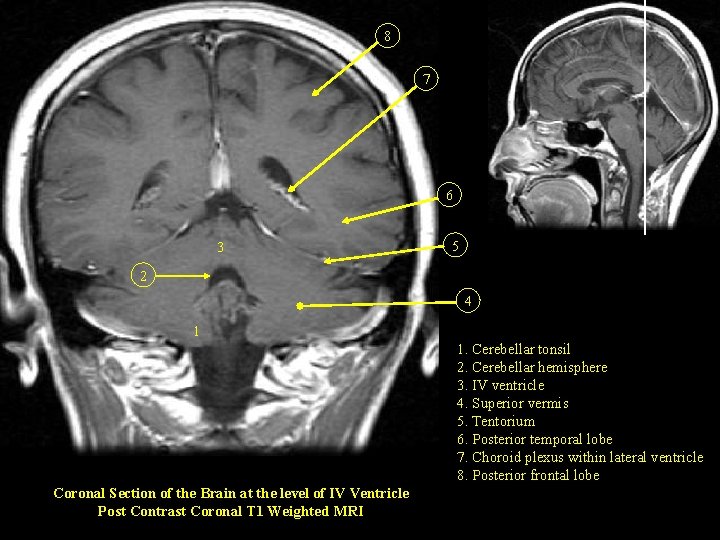
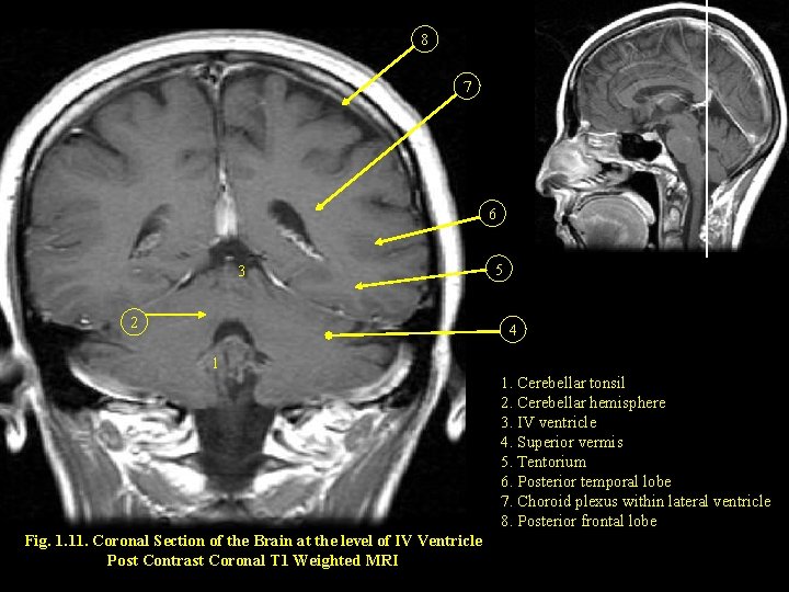
8 7 6 3 5 2 4 1 1. Cerebellar tonsil 2. Cerebellar hemisphere 3. IV ventricle 4. Superior vermis 5. Tentorium 6. Posterior temporal lobe 7. Choroid plexus within lateral ventricle 8. Posterior frontal lobe Coronal Section of the Brain at the level of IV Ventricle Post Contrast Coronal T 1 Weighted MRI
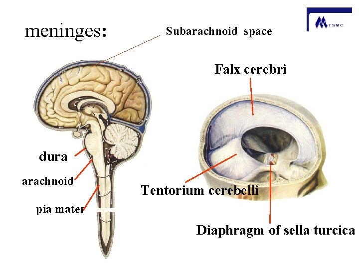
Meninges dura mater the outer layer arachnoid mater the middle layer pia mater the inner layer inseparable from the brain The cranial dura is formly adherent to the periosteum of skull bones The arachnoid mater is loosely adherent to the dura mater Between the arachnoid and the pia is the subarachnoid space
A double layer of dura projects into the cranial cavity. These dural folds form: Falx cerebri Tentorium cerebelli Diaphragm of sella turcica
meninges: Subarachnoid space Falx cerebri dura arachnoid Tentorium cerebelli pia mater Diaphragm of sella turcica
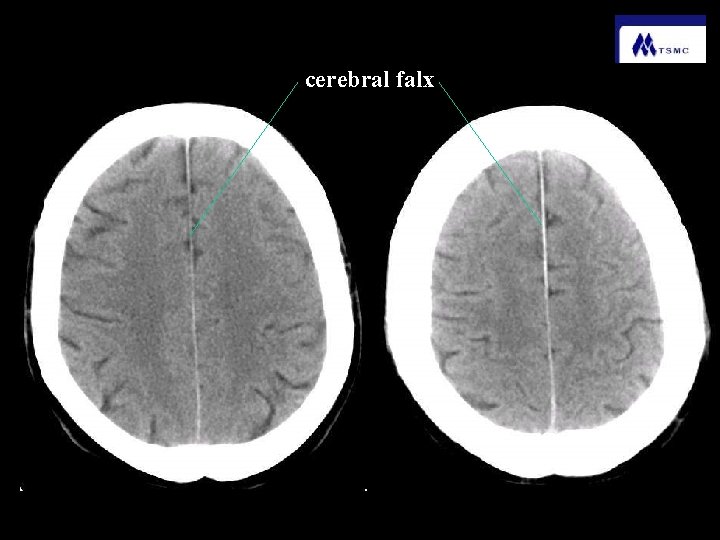
cerebral falx
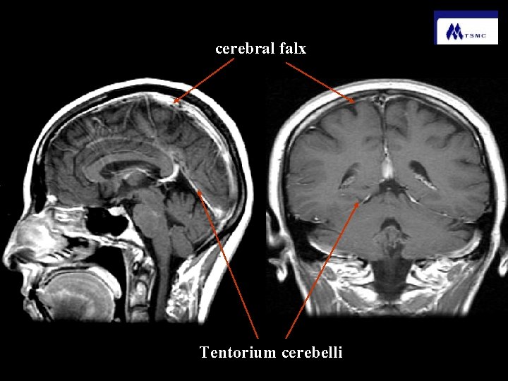
cerebral falx Tentorium cerebelli
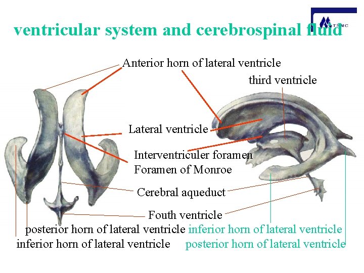
ventricular system and cerebrospinal fluid Anterior horn of lateral ventricle third ventricle Lateral ventricle Interventriculer foramen Foramen of Monroe Cerebral aqueduct Fouth ventricle posterior horn of lateral ventricle inferior horn of lateral ventricle posterior horn of lateral ventricle
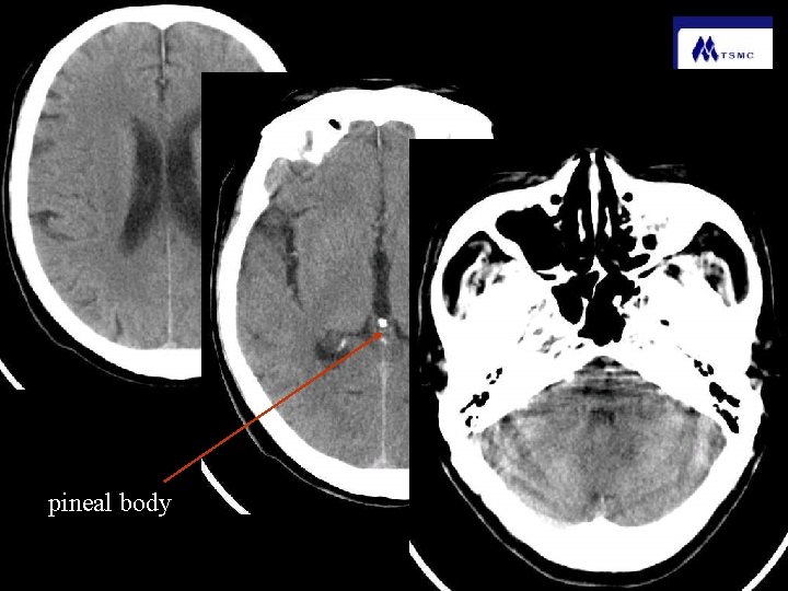
pineal body
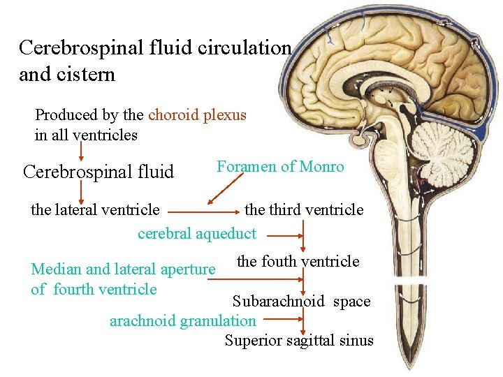
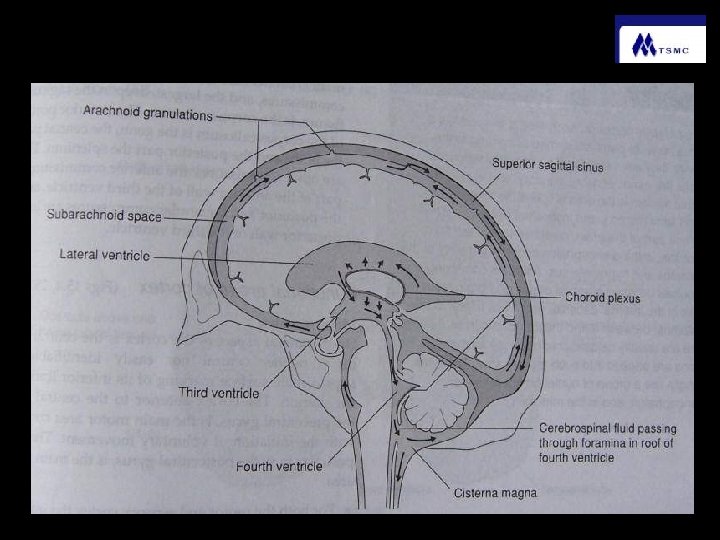
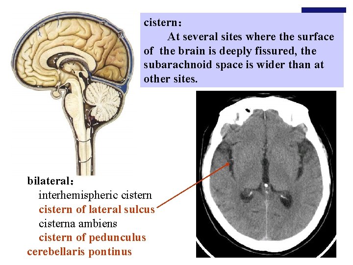
Cerebrospinal fluid circulation and cistern Produced by the choroid plexus in all ventricles Cerebrospinal fluid Foramen of Monro the lateral ventricle third ventricle cerebral aqueduct Median and lateral aperture of fourth ventricle the fouth ventricle Subarachnoid space arachnoid granulation Superior sagittal sinus
cistern: At several sites where the surface of the brain is deeply fissured, the subarachnoid space is wider than at other sites. bilateral: interhemispheric cistern of lateral sulcus cisterna ambiens cistern of pedunculus cerebellaris pontinus
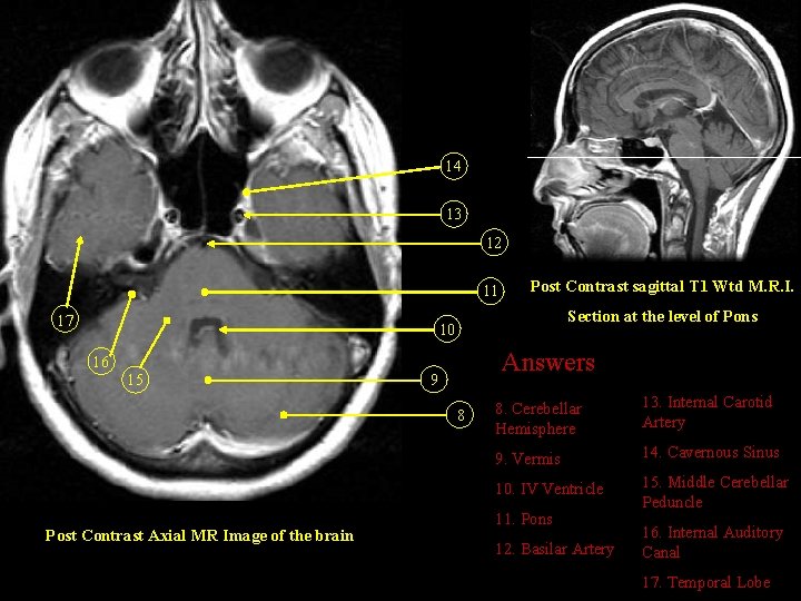
14 13 12 11 17 Post Contrast sagittal T 1 Wtd M. R. I. Section at the level of Pons 10 16 15 Answers 9 8 Post Contrast Axial MR Image of the brain 8. Cerebellar Hemisphere 13. Internal Carotid Artery 9. Vermis 14. Cavernous Sinus 10. IV Ventricle 15. Middle Cerebellar Peduncle 11. Pons 12. Basilar Artery 16. Internal Auditory Canal 17. Temporal Lobe
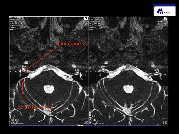
facial nerve acoustic nerve
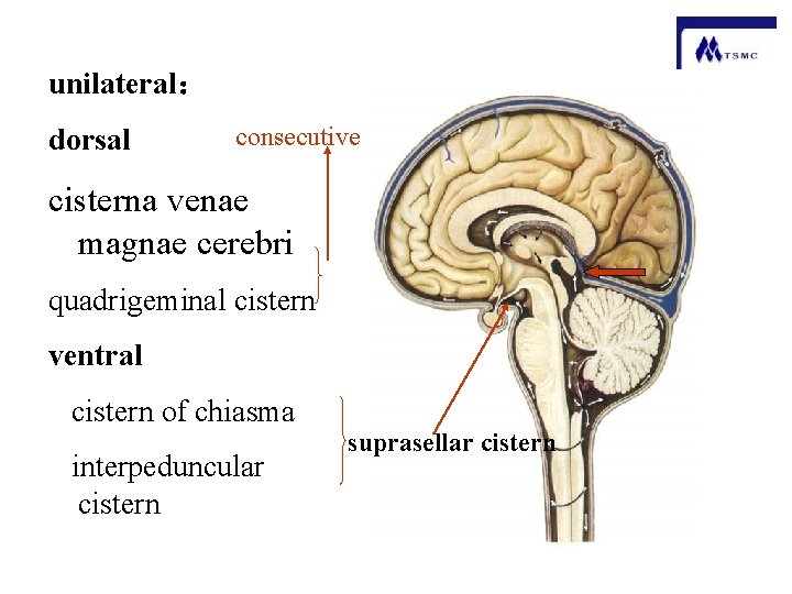
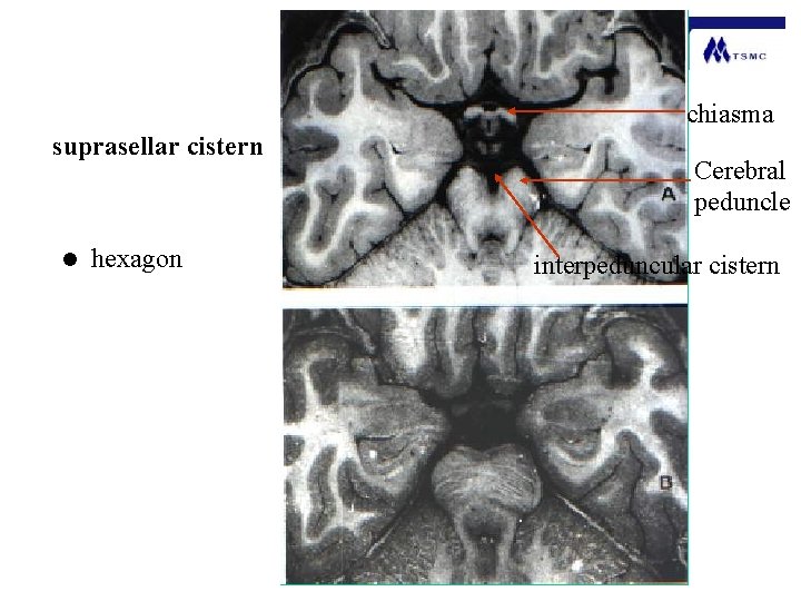
unilateral: dorsal consecutive cisterna venae magnae cerebri quadrigeminal cistern ventral cistern of chiasma interpeduncular cistern suprasellar cistern
chiasma suprasellar cistern l hexagon Cerebral peduncle interpeduncular cistern
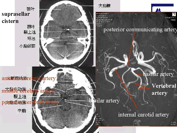
suprasellar cistern posterior communicating artery basilar artery anterior cerebral artery Vertebral artery middle cerebral artery posterior cerebral artery basilar artery internal carotid artery
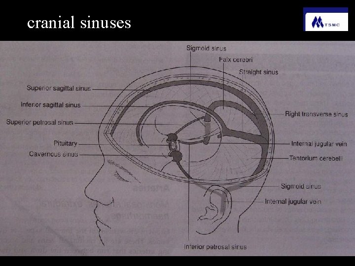
cranial sinuses
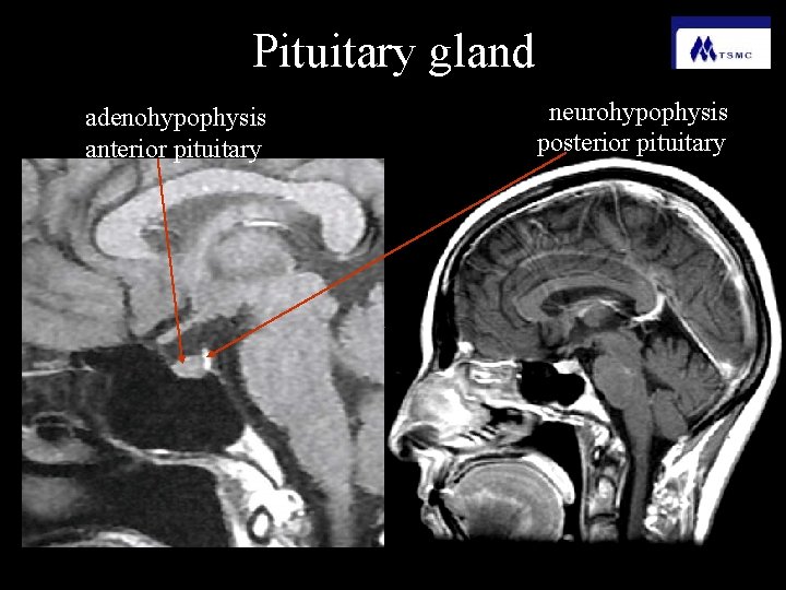
Pituitary gland adenohypophysis anterior pituitary neurohypophysis posterior pituitary
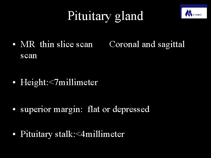
Pituitary gland • MR thin slice scan Coronal and sagittal • Height: <7 millimeter • superior margin: flat or depressed • Pituitary stalk: <4 millimeter
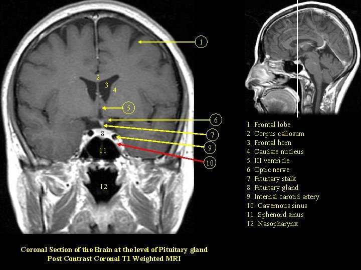
1 2 3 4 5 6 8 7 9 11 10 12 Coronal Section of the Brain at the level of Pituitary gland Post Contrast Coronal T 1 Weighted MRI 1. Frontal lobe 2. Corpus callosum 3. Frontal horn 4. Caudate nucleus 5. III ventricle 6. Optic nerve 7. Pituitary stalk 8. Pituitary gland 9. Internal carotid artery 10. Cavernous sinus 11. Sphenoid sinus 12. Nasopharynx
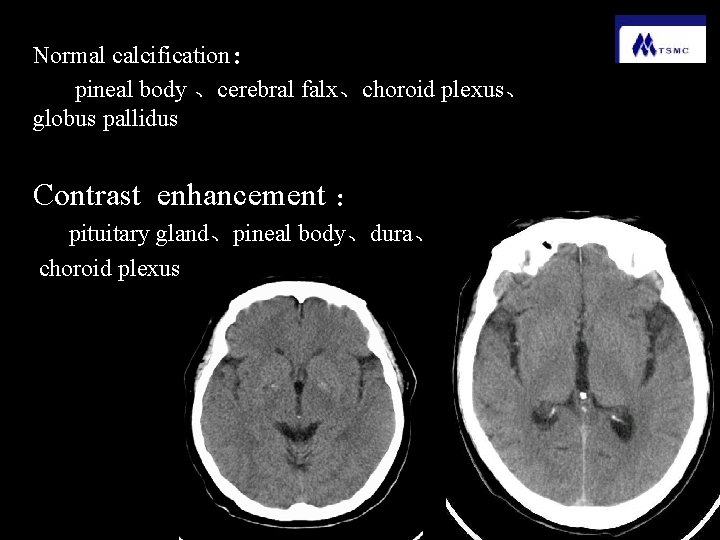
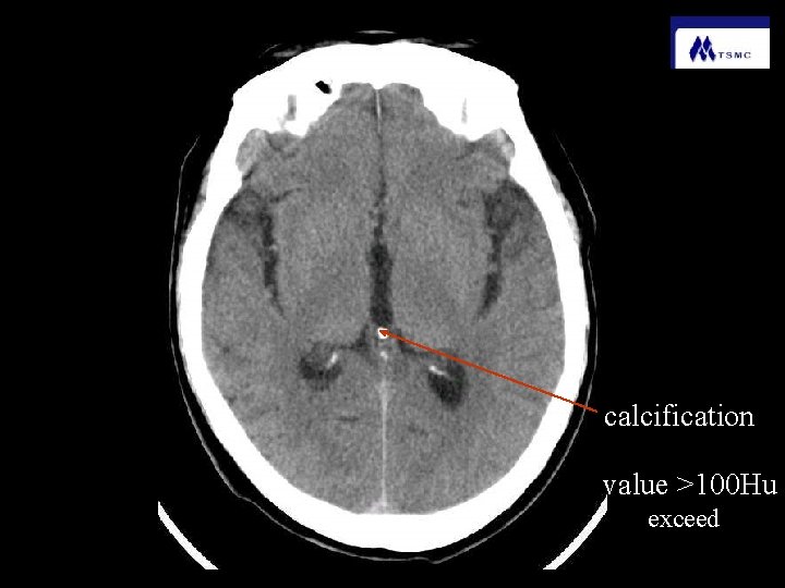
Normal calcification: pineal body 、cerebral falx、choroid plexus、 globus pallidus Contrast enhancement : pituitary gland、pineal body、dura、 choroid plexus
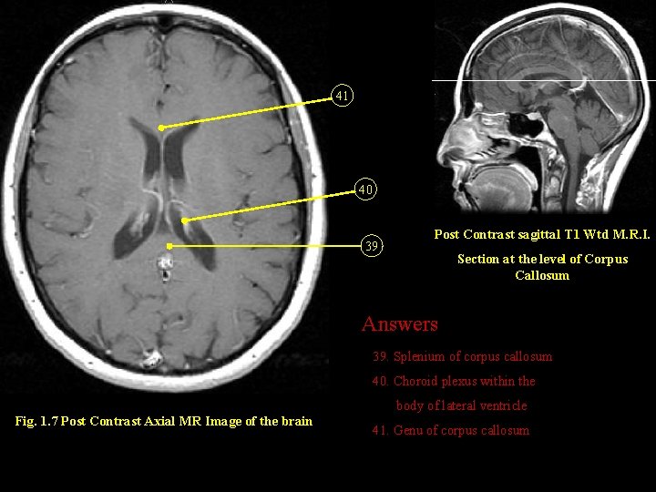
41 40 39 Post Contrast sagittal T 1 Wtd M. R. I. Section at the level of Corpus Callosum Answers 39. Splenium of corpus callosum 40. Choroid plexus within the body of lateral ventricle Fig. 1. 7 Post Contrast Axial MR Image of the brain 41. Genu of corpus callosum
8 7 6 3 2 5 4 1 1. Cerebellar tonsil 2. Cerebellar hemisphere 3. IV ventricle 4. Superior vermis 5. Tentorium 6. Posterior temporal lobe 7. Choroid plexus within lateral ventricle 8. Posterior frontal lobe Fig. 1. 11. Coronal Section of the Brain at the level of IV Ventricle Post Contrast Coronal T 1 Weighted MRI
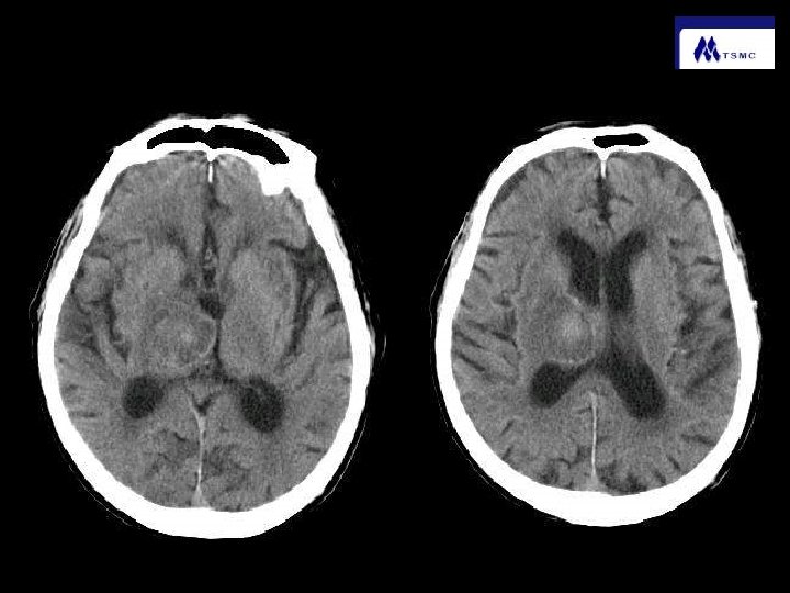
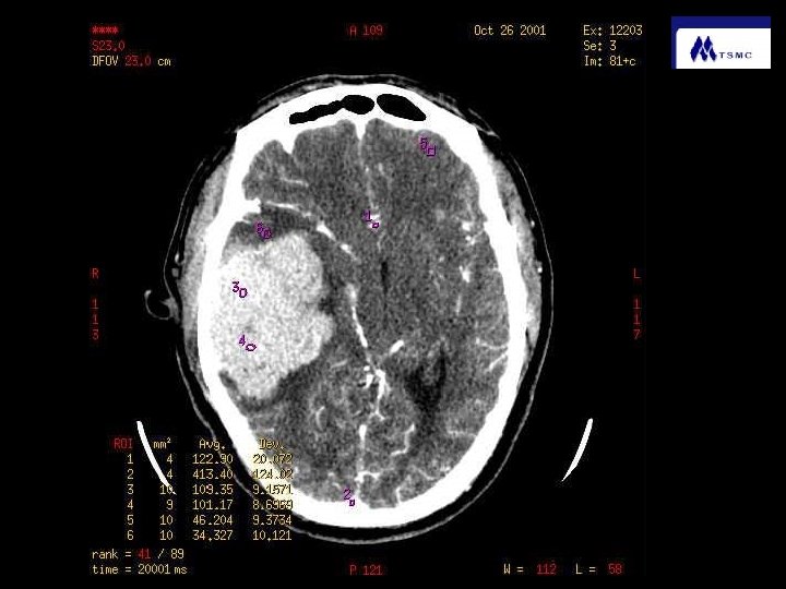
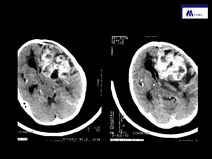
Abnormal changes of CT scan a、hyperdense:calcification、hemorrhage、 tumor b、 hypodense :edema、 tumor 、cystis、 liquefactive necrosis、lipoma、 fluid c、 isodense :chronic hamorrhage、 tumor d、heterogeneous density :
calcification value >100 Hu exceed
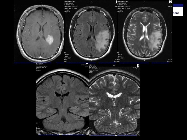
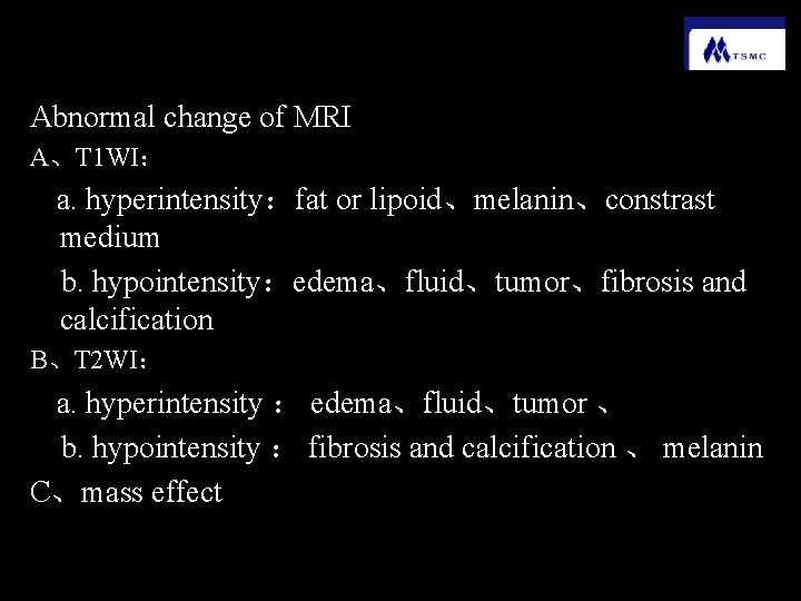
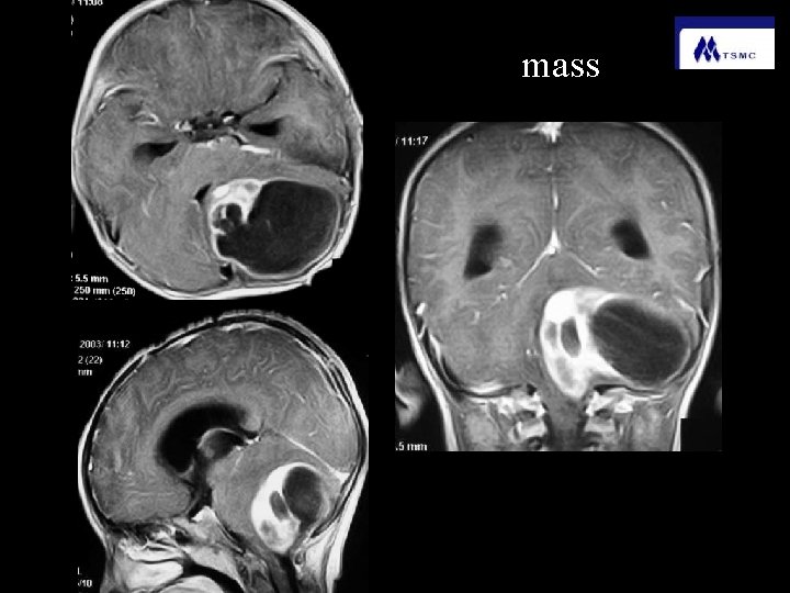
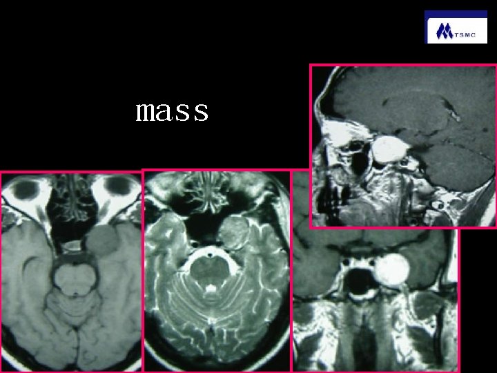
Abnormal change of MRI A、T 1 WI: a. hyperintensity:fat or lipoid、melanin、constrast medium b. hypointensity:edema、fluid、tumor、fibrosis and calcification B、T 2 WI: a. hyperintensity : edema、fluid、tumor 、 b. hypointensity : fibrosis and calcification 、 melanin C、mass effect
mass
mass
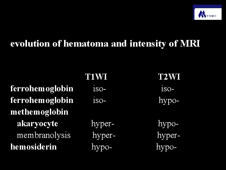
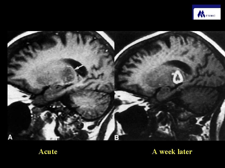
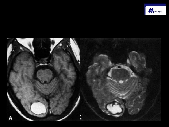
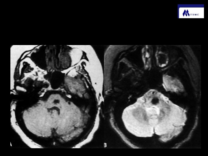
evolution of hematoma and intensity of MRI T 1 WI T 2 WI ferrohemoglobin iso- isoferrohemoglobin iso- hypomethemoglobin akaryocyte hyper- hypomembranolysis hyper- hyperhemosiderin hypo-
Acute A week later
methemoglobin
hemosiderin
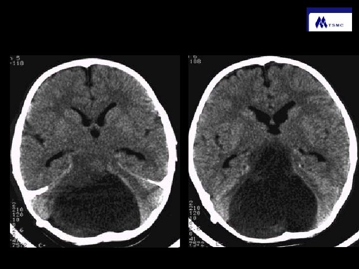
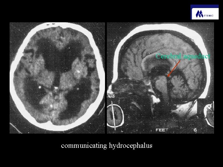
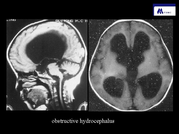
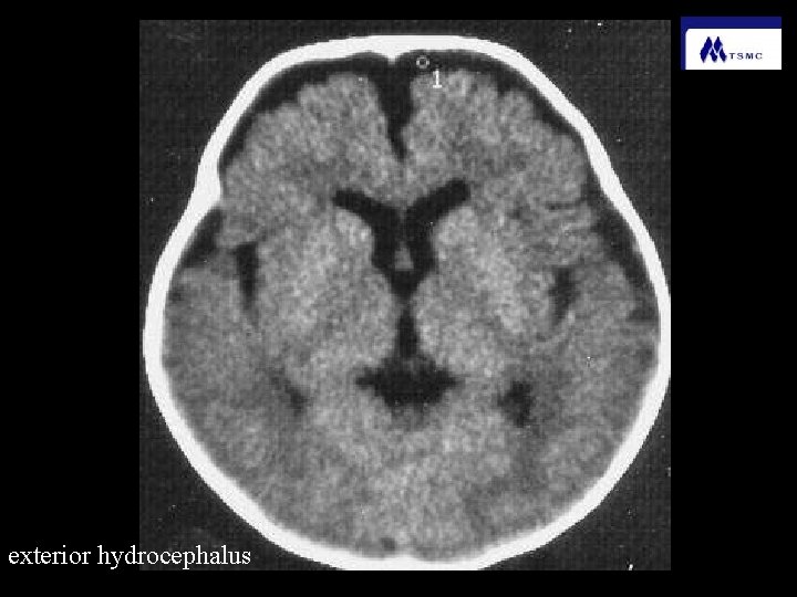
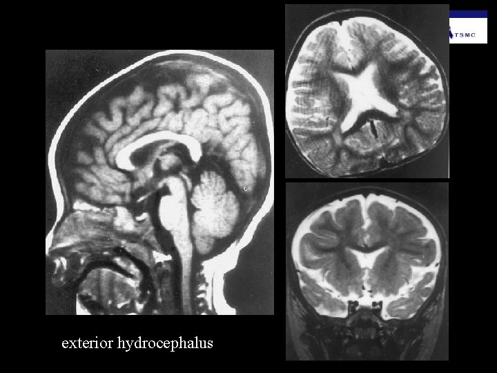
Hydrocephalus (increased volume of cerebrospinal fluid) 1、obstructive hydrocephalus 2、communicating hydrocephalus 3、exterior hydrocephalus
Cerebral aqueduct communicating hydrocephalus
obstructive hydrocephalus
exterior hydrocephalus
exterior hydrocephalus
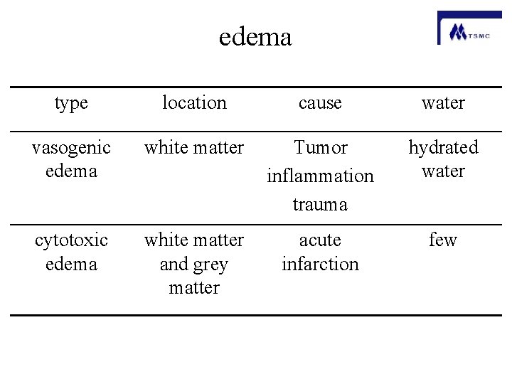
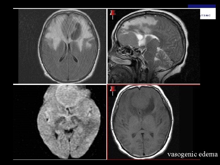
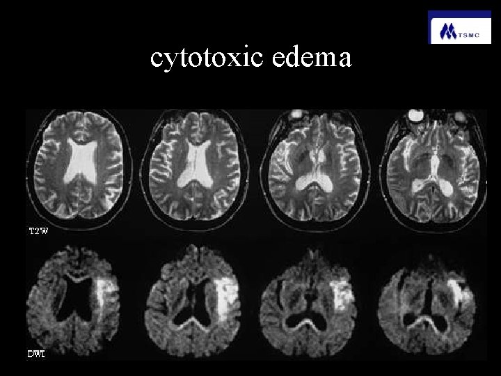
edema type location cause water vasogenic edema white matter Tumor inflammation trauma hydrated water cytotoxic edema white matter and grey matter acute infarction few
vasogenic edema
cytotoxic edema