Central Nervous System CNS Brain and Spinal Cord
: Brain and Spinal Cord Comparative Anatomy Tony Serino, Ph. D.")
Central Nervous System (CNS): Brain and Spinal Cord Comparative Anatomy Tony Serino, Ph. D. Biology Dept. Misericordia Univ.
 • Gray vs. White matter • Protection of CNS –")
Central Nervous System (CNS) • Gray vs. White matter • Protection of CNS – Meninges – CSF flow • Brain – Development – Selected structures • Spinal cord – Selected structures
 • Brian and spinal cord • Displays gray and white")
CNS (Central Nervous System) • Brian and spinal cord • Displays gray and white matter – Gray matter areas of CNS with many cell bodies of neurons present (little myelinated nerve fibers) – White matter area of CNS with few cell bodies but many myelinated nerve fibers • Protected by bone and membranes

Meninges • Dura Mater –outermost; tough, fibrous CT – In brain, divided into two layers (periosteal and meningeal) – In spine, only one layer with fat filled space above the layer called the epidural space • Arachnoid –middle; web-like appearance – Potential space between Dura and arachnoid is the subdural space • Pia Mater –innermost, delicate membrane fused with CNS surface – Space between Pia and Arachnoid is the subarachnoid space

Brain Meninges

Brain Ventricles

Choroid Plexus
 Flow")
CSF (cerebral-spinal fluid) Flow

Hydrocephalus • Blockage of CSF flow can lead to severe brain and/or head enlargement. • In an adult, such swelling would be fatal.

Brain • Development • Structures • Functional Areas

Brain Vesicles
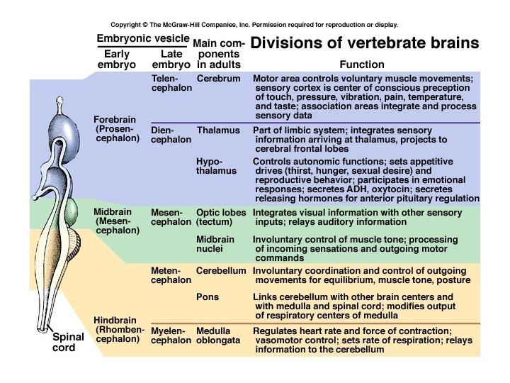

Major Divisions of Brain Stem = midbrain + pons + medulla
")
Brain Anatomy (req’d)

Projections vs. Commissures

Functional Anatomy of Brain

Functional Areas of Cerebrum

Primary Motor and Somatosensory Gyri

Basal Nuclei: cerebral nuclei Putamen and Globus Pallidus Subthalamic nuclei and the Substantia nigra are usually included

Limbic System: functional system; responsible for emotion and memory Cingulate Gyrus Fornix Mammillary body

Circle of Willis

Spinal Cord • Receives and generates signals to body through the spinal nerves
")
Spinal Cord (X. S. )

Cord in Spinal Canal Posterior Median Sulcus Posterior Root Denticulate Ligament Dorsal Root Ganglion Anterior Root Spinal Nerve

Spinal Cord Segments • 4 segments: Cervical, Thoracic, Lumbar, and Sacral (only 1 coccygeal nerve) • 31 pairs of spinal nerves

Cauda Equina

Lumbar Puncture

Functional Arrangement of SC

Neurotransmission Scenario

ANS Divsions
")
Parasympathetic (Craniosacral)
")
Sympathetic (Thoracolumbar)

Sympathetic Ganglia
- Slides: 34