Central diabetes insipidus Maryamkabootari MD Research Institute for

Central diabetes insipidus Maryam-kabootari MD Research Institute for Endocrine Sciences Shahid Beheshti University of Medical Sciences mordad 95
 belongs to the spectrum of polyuric and polydipsic diseases, a")
Ø Diabetes insipidus (DI) belongs to the spectrum of polyuric and polydipsic diseases, a group of hereditary or acquired disorders mainly associated with an inadequate arginine vasopressin (AVP) secretion or renal response to AVP, which clinically results in hypotonic polyuria and a compensatory or underlying polydipsia. Current State and Future Perspectives in the Diagnosis of Diabetes Insipidus: A Clinical Review. J Clin Endocrinol Metab, October 2012, 97(10): 3426– 3437

Differential Diagnosis clinical presentation: Ø Patients with hypothalamic DI often have a sudden onset of symptoms and persistent thirst throughout the day and night associated with a desire for cold liquids. " Ø Patients with DI usually have serum Na in the high range of normal, whereas patients with primary polydipsia have serum Na in the low range of normal.

Ø The blood urea nitrogen concentration is often low in both hypothalamic DI and primary polydipsia because of the high renal clearance. Ø Serum uric acid is elevated in hypothalamic DI because of the modest volume contraction and the absence of the normal action of vasopressin on V 1 receptors in the kidney to increase urate clearance. Ø A serum uric acid value greater than 5 µ/d. L was reported to separate hypothalamic Dr from primary polydipsia.

Ø Urine volume greater than 18 L is highly suggestive of primary polydipsia. Ø Most patients with hypothalamic Dl have modest dehydration, decreased GFR, and urine volumes in the range of 6 to 12 L/day.
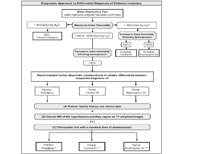

some times lower than expected urinary response to injected vasopressin in CDI : Ø chronic polyuria itself can decrease the urinary concentration capacity, probably through a washout mechanism of the renal medullary concentration gradient Ø by down-regulation of kidney AQP 2 water channels Current State and Future Perspectives in the Diagnosis of Diabetes Insipidus: A Clinical Review. J Clin Endocrinol Metab, October 2012, 97(10): 3426– 3437

Causes of multiple ring-enhancing lesions of the brain Multiple ring-enhancing lesions of the brain. JPGM. 2015; 56(4): 307 -316
 is affected by")
CNS involvement in multiple myeloma Ø The central nervous system (CNS) is affected by multiple myeloma in approximately 1% of all cases, and such involvement is a dire prognostic sign Ø Intracranial myeloma constitutes less than 1% of intracranial tumors and can occur as a solitary (primary) plasmacytoma in addition to being a manifestation of systemic multiplemyeloma. Central nervous system myeloma takes many forms, including: v localized intraparenchymal v dural-based lesions and CNS myelomatosis involving leptomeninges v cranial nerves invovement v a combination of different sites of lesions such as intraparenchymal and dural based Imaging of extraosseous intracranial and intraspinal multiple myeloma, including central nervous system involvement. Clinical Imaging 39 (2015) 213– 219

Ø The central nervous system may be involved at all stages of multiple myeloma by direct invasion from contiguous bone lesions or by hematogenous spread with parenchymal infiltration. Ø Autopsy studies in patients with leptomeningeal myeloma demonstrated that circulating myeloma cells can infiltrate arachnoid veins diffusely, spilling over into the cerebrospinal fluid (CSF). Imaging of extraosseous intracranial and intraspinal multiple myeloma, including central nervous system involvement. Clinical Imaging 39 (2015) 213– 219

Clinical manifestations are nonspecific and may include: Ø Headache Ø nausea and vomiting Ø Paraparesis Ø cranial nerve palsies Ø mental status changes Ø rarely, seizures v. ROLE OUT: hypercalcemia, hyperviscosity, drug neurotoxicity, or spinal cord compression Imaging of extraosseous intracranial and intraspinal multiple myeloma, including central nervous system involvement. Clinical Imaging 39 (2015) 213– 219

Intracranial lesions may have perilesional edema, focal calcifications, or intralesional hemorrhage. Differential diagnosis includes: Ø Meningioma Ø Sarcoma Ø Lymphoma Ø metastatic carcinoma Ø plasma cell granuloma Ø infectious meningitis Ø leptomeningeal carcinomatosis Imaging of extraosseous intracranial and intraspinal multiple myeloma, including central nervous system involvement. Clinical Imaging 39 (2015) 213– 219

Magnetic resonance imaging findings are often nonspecific, and differentiation of leptomeningeal myelomatosis from infectious meningitis requires clinical and laboratory correlation. Final diagnosis is made by direct biopsy of localized masses and/or by detection of monoclonal plasma cells in the CSF Imaging of extraosseous intracranial and intraspinal multiple myeloma, including central nervous system involvement. Clinical Imaging 39 (2015) 213– 219

Ø The median interval from diagnosis of multiple myeloma to diagnosis of CNS myeloma is 6– 18 months, although, rarely, CNS involvement may be the initial presentation of multiple myeloma. Ø While CNS myeloma is usually associated with a higher tumor burden and circulating plasma cells, a significant minority of patients with CNS myeloma appear to be in clinical remission at the time of lesion detection. Imaging of extraosseous intracranial and intraspinal multiple myeloma, including central nervous system involvement. Clinical Imaging 39 (2015) 213– 219

Ø Despite the available therapies, patients with CNS myeloma have a poor prognosis, with a median survival ranging from 1 month for those who show no initial response to therapy to up to 5 months for those who have at least an initial response to therapy. Imaging of extraosseous intracranial and intraspinal multiple myeloma, including central nervous system involvement. Clinical Imaging 39 (2015) 213– 219

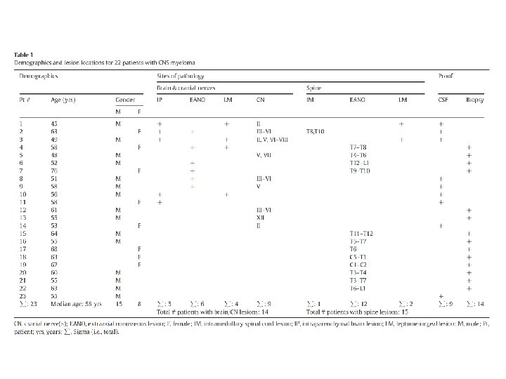
Purpose : to investigate the imaging manifestations of extraosseous intracranial and intraspinal multiple myeloma, including involvement of the central nervous system. Method: 23 patients included 15 males (65%) and 8 females (35%) ranging in age from 45 to 76 years (median age, 58 years) who had CSF positive for multiple myeloma and had neuroimaging studies available for our review were included in this study. Patients without positive CSF were included only if they had biopsy-proven intracranial or intraspinal myelomatous lesions that were not primarily osseous
 Sagittal T 1 -weighted (T")
Fig. 1. Patient 2. A 63 -year-old woman. (A) Sagittal T 1 -weighted (T 1 W) postcontrast MRI demonstrates an enhancing mass within the body of the corpus callosum. (B) Coronal T 1 W postcontrast MRI shows an enhancing nodule in the left perimesencephalic cistern (arrow) in addition to the callosal mass.

Ø In A a 65 -year-old man was admitted to our department because of anorexia, easy fatigue and shortness of breath. Ø A laboratory investigation showed hemoglobin 8. 7 g/dl, white blood cells 7800/ml with normal differential, platelet counts 234, 000/ml, and ESR 134 mm/h. Blood urea was 68 mg/dl and serum creatinine 1. 5 mg/dl, while serum calcium and serum CRP were within normal range. Ø A skeletal X-ray survey showed a single osteolytic lesion in the left humerus. Total serum proteins were 8. 20 g/dl with a monoclonal spike in the b-region of the electrophorogram representing an Ig. A k paraprotein. Ø Bone marrow aspirates and trephine biopsy showed 50% infiltration by plasma cells without plasmablastic morphology.

With the diagnosis of a stage IIA disease, the patient was started on the VMCP protocol (vincristine, cyclophosphamide, melphalan, prednisone) receiving the regimen every 4 – 5 weeks. This protocol was retracted after the second cycle because of protracted hematological toxicity, and treatment was continued with the classical orally administered MP protocol (melphalan, prednisone). 2 days after the fifth course, the patient experienced frontal headache, fever, and vomiting. A neurological examination showed a right sixth nerve palsy with concomitant papilledema.

A solid mass in the sellar region is shown. The mass extends upwards to the suprasellular fossa, comes into contact with the optic chiasm and infiltrates the sphenoid sinus. Meningeal uptake is also noted, especially in the anterior cranial fossa. (A) Post-contrast coronal T 1 -W magnetic resonance image, (B) transverse T 2 -W magnetic resonance image.

Ø The CSF analysis showed protein 228 mg/dl with a small Ig. Ak fraction on immunofixation, glucose 82 mg/dl, and 137/ml nucleated cells, mainly plasma cells. Ø The diagnosis of intracranial plasmacytoma associated with myelomatous meningitis was considered and the patient was subjected to cranial radiotherapy. Ø However, 5 weeks later, the sellar mass remained unchanged in size. Ø Hormonal evaluation demonstrated hypopituitarism. Ø Moreover, the patient developed polydipsia and hypotonic poluyria, suggestive of central diabetes insipidus.

Ø Appropriate hormone replacement therapy was administered, i. e. thyroxine 100 mg/day, desmopressin 0. 1 mg/day and hydrocortisone 35 mg/day. Ø The patient died 2 years later, due to disease progression, but with no signs of deterioration in the CNS.

Ø A 64 -year-old man presented with diagnosis of advanced Ig. A lambda multiple myeloma (MM) Ø The patient commenced chemotherapy with cyclophosphamide, dexamethasone and bortezomib. Ø On the day of his third weekly dose of the first cycle of chemotherapy, he required hospital admission with profound dehydration and hypotension. Ø He then underwent a formal 7 -hour water deprivation test, as per the local protocol.

A diagnosis of central DI was made and nasal desmopressin prescribed. The patient reported resolution of his nocturia and return to a normal fluid intake. There was no evidence of anterior pituitary dysfunction. He had a normal plasma glucose and none of his medications were known to cause central DI.
 : anomalous cerebral")
Figure 1: MRI pituitary demonstrating persistent primitive trigeminal artery (solid lines) : anomalous cerebral vasculature with a primitive trigeminal artery that caused distortion of the pituitary gland mottled changes of the clivus (broken line)
 The bone lesions")
Figure 2: CT head demonstrating lytic lesions of the clivus (arrow) The bone lesions were consistent with myeloma.

Ø Chemotherapy was continued with good effect associated with a rapid fall in serum free light chains after three cycles of four doses each. Ø With this improvement the polydipsia and polyuria also decreased and the patient stopped his desmopressin nasal spray after 6 weeks without a recurrence of symptoms.

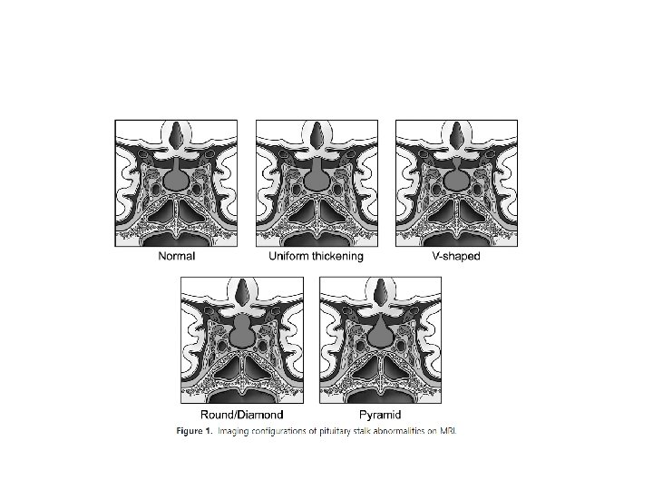
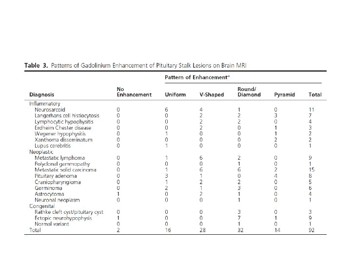
Design: A retrospective review of patients with pituitary stalk lesions seen at Mayo Clinic Rochester between 1987 and 2006 was conducted. Demographic, clinical presentation, imaging, laboratory, operative, and pathology data were reviewed and are reported using descriptive statistics. Results: Of the 152 pituitary stalk lesions included, 49 (32%) were neoplastic, 30 (20%) were inflammatory, 13 (9%) were congenital anomalies, and 60 (39%) were of unclear etiology. Diabetes insipidus was diagnosed in 43(28%)of the 152 patients, and 49(32%) patients had at least one anterior pituitary hormone deficit. Secondary hypogonadism was the most common endocrine deficiency.


Thank you
- Slides: 36