Central Abdominal Pain and masses Supervised by DR

Central Abdominal Pain and masses Supervised by : DR. HAMED AL QAHTANI

Objectives � Approach a patient with central abdominal pain and mass � Differential diagnosis of central abdominal pain and mass ç Appendicitis ç Small Bowel Obstruction ç Mesenteric Ischemia

Abdominal pain is frequently a benign complaint, but it can also indicate serious acute pathology. It is very commonly due to Irritable bowel syndrome, however, other possible pathologies should be taken in consideration.

q The history is the most important clue to the source of abdominal pain. Starting from the outer surface to the inner surface of the abdomen, the pain could be : cutaneous, musculoskeletal, vascular, neurological or organic.

Central Abdominal Pain �Referred to midgut structures , which begin from second part of duodenum to splenic flexure

Generally, abdominal pain can be categorized by its underlying mechanism: -Visceral -Parietal -Referred - Radiating

Visceral pain is usually dull and aching in character, although it can be colicky, poorly localized. • It arises from distention or spasm of a hollow organ such as the discomfort experienced early in intestinal obstruction or cholecystitis. • • • Parietal pain is sharp and very well localized. It arises from peritoneal irritation such as the pain of acute appendicitis with spread of inflammation to the parietal peritoneum.

�Referred pain is aching and perceived to be near the surface of the body. Radiating pain: is at site of pathology and other site

What are the possible DDx of central abdominal pain?

History • Age, gender. Pain analysis: location, q radiation, nature of the pain, q duration, onset, mode, aggrevating and relieving factors, associated symptoms. • q Associated syptoms: q nausea, vomitting, q dyspepsia, constipation, diarrhea, change in stool color, change in urine color, q abdomenal distention, fever, loss of weight, loss of appetite. •

Cont. History �Past history: - Medical: Diabetes, hypertention, hyperlipidemia, history of previous similar complaint, co-existing medical diseases. - Surgical: abdomenal procedures. - Drugs: eg. steriods, PPI’s, paracetamol. - Allergies.

Cont. History �Social history: Alcohol, diet and socioeconomical status, pain in relation to psychological factros and stress. �Family history. �Systemic review.

Examination �General: - Appearance: jaundice, pallor, body mass, hydration, bruises, respiratory or cardiac distress, patient looking in pain discomfort, IV fluids. - Vital signs.

Cont. Examination • Abdomen: - Inspection: abdominal distention, symmetry, visible pulsations, hernia, scars. - Palpation: superficial (rigidity, rebound tenderness, masses). Deep (Murphy’s sign, masses, organomegaly) - Percussion: tenderness, dullness/ tympany. - Auscultation: bruit, bowels sounds. - PR examination.

Investigation �Labs: - CBC - Serum U&E - LFT - Amylase - Lipase - Blood glucose level
. - X-ray with")
Cont. Investigation �Imaging: - Abdominal Xray (air-fluid levels, distended bowel, stones). - X-ray with contrast (follow-through). - CT with contrast.

�Interventional investigations: - Endoscopy. - Laproscopy

Causes of Central abdominal pain: �Gastroenteritis. �Peptic Ulcer Disease. �Pancreatitis. �Appendicitis. �Abdomenal Aortic Aneurism. �Mesenteric Ischemia. �Small Bowel Obstruction. �Intussusception.

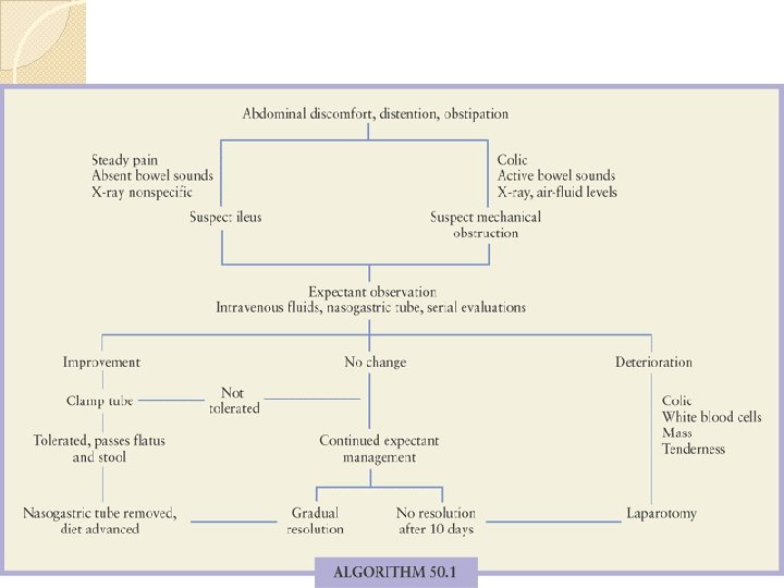
Small Bowel Obstruction

Definition Interruption of the passage of intestinal contents.

Small Bowel Obstruction Clinical features Colicky central abdominal pain Vomiting - early in high obstruction Abdominal distension - extent depends on level of obstruction � Absolute constipation - late feature of small bowel obstruction � Dehydration associated with tachycardia, hypotension and oliguria � Features of peritonitis indicate strangulation or perforation � � �

Small Bowel Obstruction Investigation Supine abdominal X-ray shows dilated small bowel • May be normal • Valvulae coniventes differentiate small from large intestine • Erect abdominal film is very important to show the presence of air fluid level to differentiate if there is true obstruction or adynamic ileus • • Contrast studies(water soluble gastograffin not barium) & CT. are very helpful

Small Bowel Obstruction Pathophysiology �Hypercontractility--hypocontractility �Massive third space losses ◦ oliguria, hypotension, hemoconcentration �Electrolyte depletion �bowel distension--increased intraluminal pressure--impedement in venous return--arterial insufficiency
, in bowel habits(LBO) �Clinical picture")
Site? Small Bowel vs. Large Bowel �Scenario �prior operations(SBO), in bowel habits(LBO) �Clinical picture �scars, masses/ hernias(SBO), amount of distension(more distension more distal the obstruction usually )/ vomiting(more w/ SBO) �Radiological studies �gas in colon(LBO), mass(according to its site) �(Almost) always operate on LBO, often treat SBO non-operatively

Etiology? �Outside the wall �Inside the lumen
(usually postoperative) �Hernia (2 nd")
Lesions Extrinsic to Intestinal Wall �Adhesions (most common cause )(usually postoperative) �Hernia (2 nd most common) �External (e. g. , inguinal, femoral, umbilical, or ventral hernias) �Internal (e. g. , congenital defects such as paraduodenal, and diaphragmatic hernias or postoperative secondary to mesenteric defects) �Neoplastic �Carcinomatosis, extraintestinal neoplasm �Intra-abdominal abscess/ diverticulitis �Volvulus (small bowel )

Lesions Intrinsic to Intestinal Wall �Congenital ◦ Malrotation ◦ Duplications/cysts �Traumatic ◦ Hematoma ◦ Ischemic stricture �Infections ◦ Tuberculosis ◦ Actinomycosis ◦ Diverticulitis �Neoplastic ◦ Primary neoplasms ◦ Metastatic neoplasms �Inflammatory ◦ Crohn's disease �Miscellaneous ◦ Intussusception ◦ Endometriosis ◦ Radiation enteropathy/stricture

Intraluminal Lesions �Gallstone �Enterolith �Foreign body

Is there strangulation? 4 Cardinal Signs: � fever � tachycardia � localized abdominal tenderness � leukocytosis
 �Admission �NPO �Fluid resuscitation �Electrolyte, acid-base correction �Close monitoring �Foley")
Management of SBO (Principles) �Admission �NPO �Fluid resuscitation �Electrolyte, acid-base correction �Close monitoring �Foley +/- central line �NGT decompression �? Surgery

Resuscitation �Massive third space losses as fluid and electrolytes accumulate in bowel wall and lumen �Depend on site and duration �proximal- vomiting early, with dehydration, hypochloremia, alkalosis �distal- more distension, vomiting late, dehydration profound, fewer electrolyte abnormalities �Requirements ongoing loses = deficit + maintenance +

TO OPERATE OR NOT TO OPERATE �The rule in SBO is to manage the pt conservatively w/ observation & give the pt time up to 48 hrs then reevaluate if still obstructed.

Indications for surgery Peritoneal findings. Rapidly progressing abdominal pain or distension. Visceral perforation. . (evident by increase amylase level) Irreducible hernia Development of: - Fever. - Diminished urine output. - Metabolic acidosis.

Paralytic ileus • • Functional obstruction most commonly seen after abdominal surgery, or w/ hypokalemia & sepsis Small bowel is distended throughout its length Absorption of fluid, electrolytes and nutrients is impaired. . Abdominal distension is often apparent Pain is often not a prominent feature Auscultation will reveal absence of bowel sounds Water soluble contrast study may be helpful to differentiate if in doubt is it mechanical or functional obstruction

Management : � for ilius conservative (it resolve 2 -3 days after surgery � mechanical : 1 -adhesive conservative wait for 48 h 2 - non-adhesive CT scan & imm surgery

Acute Mesenteric Ischemia

Acute Mesenteric Ischemia �Definition: It is defined as an occlusive or nonocclusive mechanism leading to hypoperfusion of one or more mesenteric vessels.

Acute Mesenteric Ischemia � Incidence: relatively rare. More in older population � Survival & Mortality: Survival is v. bad, although there has been a reduction in mortality but it remained around 60 -70% since then. . � Mortality is high because usually the diagnosis is made after infarction, damage proceeds even after revascularization, and concomitant medical problems affect long-term outcomes � There is significant morbidity associated with acute mesenteric ischemia and up to 30% of patients become TPN dependent. � Recurrence of disease is common

Mesenteric Ischemia �CAUSES: 1. Arterial embolic disease 2. Arterial thrombotic disease 3. Low flow status. non-occlusive disease. 4. Venous thrombotic disease 5. Atherosclerosis. (chronic)
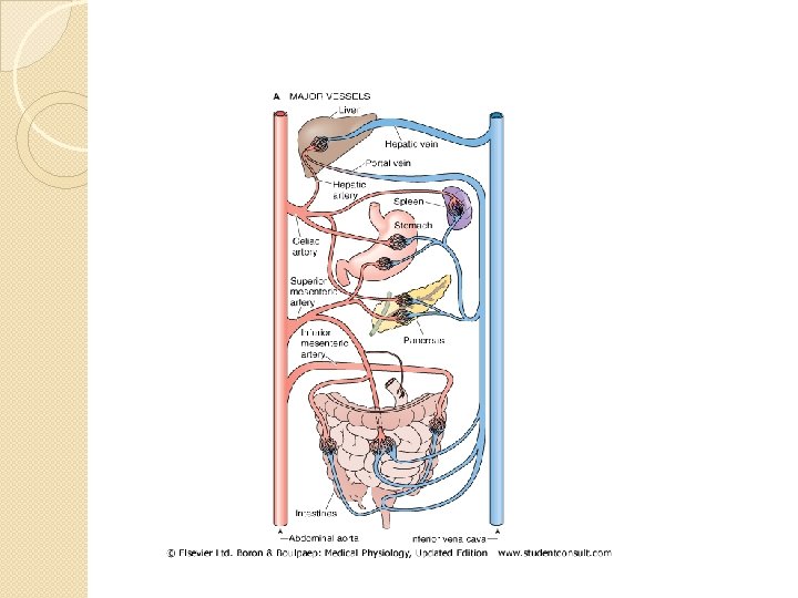

Mesenteric arterial embolism • • • The classic presentation is severe abdominal pain that is out of proportion to minimal or absent physical signs Most common cause of acute mesenteric ischemia Embolic sources: 80% cardiac. Others. . in SMA: Jejunal & ileal branches of SMA are affected more cuz they r end arteries (no anastomosis ) History: 1. Sudden and severe epigastric or mid-abdominal pain 2. Vomiting and explosive diarrhea 3. 25% of patients have had previous embolic events

Mesenteric arterial embolism Examination findings: �Cardiac �The abdominal examination: - may be normal initially with signs of acute abdomen later - Slight to moderate abdominal distension is common - Bowel sounds are highly variable - Peritoneal signs or blood in the stools are late ominous signs implying infarction

Mesenteric arterial embolism Investigations: • • • The diagnosis usually depends on clinical suspicion Initially the standard hematological and biochemical studies are unrewarding. . Plain AXR CT Scan(It is the most imp & the Ix of choice here) Occasionally US Angiography: Embolic lodging in thr SMA is often just past the inferior pancreaticoduodenal and middle colic arteries thus isolting the small bowel from its major collateral circulation

Plain AXR � The purpose of doing it is mainly to exclude other pathologies that could present in the same way. � Shown here is the thumb print sign which is a late sign that indicates infarction of the bowel

CT Scan SMA embolus wall thickening Bowel

Angiography
 3. Resect non-viable tissue")
Principles of Treatment 1. Diagnose 2. Restore Flow (surgical embolectomy) 3. Resect non-viable tissue 4. Supportive Care 5. Reevaluation( second look operation)

Acute Arterial Mesenteric Thrombosis �A less common cause �Follows thrombosis of an underlying diseased SMA (Found at ostium of SMA) Cause: Thrombosis on top of an ruptured atheromatous plaque w/ exposed intima

Mesenteric venous thrombosis Clinically: �The presentation is of an acute abdominal catastrophe less abrupt than seen with the SMA embolus with eventual development of severe mid-abdominal pain �These symptoms may occur de novo or be superimposed on a background of chronic intestinal ischemia

Mesenteric venous thrombosis Investigations The venous phase of selective angiography may reveal the thrombus. CT Scan often demonstrates a thrombus within the portal vein and the superios mesenteric vein

�Treatment: �Surgery: resection of non viable bowel, thrombectomy and anticoagulants. �Correction of hypercoagulable states (heparinization)

Low-flow nonocclusive mesenteric ischemia � 20 -30% of acute intestinal ischemia � Response to systemic hypoperfusion Sympathetic adrenergic system mediated visceral vasoconstriction/shunting for cerebral protection � Causes: any severe systemic illness: Diminished cardiad output Shock Hypovolemia Dehydration Use of vaso-active medications �Mucosal sloughing and bleeding may be present �The diagnosis may be established with angiography

Low-flow nonocclusive mesenteric ischemia Treatment �Optimize hemodynamics and volume status � Correct contributing medical conditions � Eliminate adverse pharmacologic agents �Pharmacologic support of the circulation with the relief of the vasoconstriction � Selective intra-arterial perfusion of vasodilators as papaverine and glucagon

Iatrogenic acute splanchnic ischemia Results from catheter related procedures as: � 1. Diagnostic or theraputic angiography may cause ischemia due to dissection or embolization � 2. Aortic aneurysm resection �These patients often present with diarrhea and the stools are usually grossly bloody �If the ischemia is profound and infarction occurs resection is required

Chronic arteriosclerotic splanchnic ischemia �Due to atherosclerosis affecting the origin of: Celiac, SMA, IMA � There is food fear and intestinal angina �Profound weight loss. �Investigations: Duplex scan, CT Scanning support the diagnosis Aortogram Treatment: Elective intestinal revascularization

Thank You
- Slides: 57