Cellular Division Mitosis and Meiosis Cellular Communication Types


 Cell-cell recognition. Two cells in an animal may communicate by")


 Transduction Inactive G protein Active")


 followed")
 receives and identical set of genetic material")
 Haploid (n) Diploid (2 n) Ovum (n) Sperm")


 C M yto ito ki si ne s si s")
 G 1 - Gap 1")

 Five phases - actually a continuum (one continuous change)")
 Prophase: l chromatin condenses forming chromosomes l nuclear envelope breaks down l nucleolus")
 Prometaphase: (before metaphase) - microtubules attach to the centromeres on a structure called")
 Metaphase: \"meta\" means \"middle\" - chromosomes are lined up in the middle of")

 Chromatin (duplicated) Nucleolus Nuclear Plasma envelope")
 Anaphase: l separation of sister chromatids l move toward the opposite poles via")

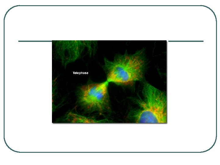
 Telophase: l daughter nuclei begin to reform l each cell has identical sets")


 If")


 Normal mammalian cells. The availability of nutrients, growth factors, and a substratum for")
 Cancer cells usually")


 Sister chromatids")


 Parent cell (before chromosome replication) MEIOSIS I")


- Slides: 75
Cellular Division Mitosis and Meiosis
Cellular Communication Types of Communication 1. Direct Contact: exchange of materials between cells a. Plasmodesmata b. Gap junctions c. Surface to Surface contact: interaction of peripheral proteins on the cellular membrane (immune response)
Plasma membranes Gap junctions between animal cells Plasmodesmata between plant cells Figure 11. 3 (a) Cell junctions. Both animals and plants have cell junctions that allow molecules to pass readily between adjacent cells without crossing plasma membranes.
Figure 11. 3 (b) Cell-cell recognition. Two cells in an animal may communicate by interaction between molecules protruding from their surfaces.
2. Distant Contact a. Local: a cell secretes a chemical signal to a nearby cell - Neurotransmitters b. Hormonal: cell secretes a chemical signal that travels through the organism by the circulatory system and causes a response in a cell that is farther away
Local signaling Target cell Electrical signal along nerve cell triggers release of neurotransmitter Neurotransmitter diffuses across synapse Secretory vesicle Local regulator diffuses through extracellular fluid Figure 11. 4 A B (a) Paracrine signaling. A secreting cell acts on nearby target cells by discharging molecules of a local regulator (a growth factor, for example) into the extracellular fluid. Target cell is stimulated (b) Synaptic signaling. A nerve cell releases neurotransmitter molecules into a synapse, stimulating the target cell.
Long-distance signaling Endocrine cell Blood vessel Hormone travels in bloodstream to target cells Target cell Figure 11. 4 (c) Hormonal signaling. Specialized endocrine cells secrete hormones into body fluids, often the blood. Hormones may reach virtually all C body cells.
General Signal-Transduction Pathway 1. Reception – cell receives a signal from an outside source – signal binds to a surface protein (LIGAND) 2. Transduction – changes inside of the cell – usually a conformational change of an enzyme or protein (often due to phosphorylation) • • change may be one or more steps – in cases where there are more than one step the intermediate carries are called relay molecules these may act as allosteric inhibitors or activators
EXTRACELLULAR FLUID Reception CYTOPLASM Plasma membrane Transduction Response Receptor Activation of cellular response Relay molecules in a signal transduction pathway Signal molecule Figure 11. 5
3. Cellular Response – action that the cell takes due to the transduction • • Secretion/absorption of a material Building of a protein or enzyme – activation of a Transcription Factor – begins the process of protein synthesis Change in the structure of the cell Growth or Division Signal Transduction
Complex Signal Transductions: l G-Protein-Linked Receptors – page 206 Signal-binding site Segment that interacts with G proteins G-protein-linked Plasma Membrane Receptor GDP CYTOPLASM G-protein (inactive) Enzyme Activated Signal molecule Receptor GDP GTP Activated enzyme GTP Figure 11. 7 Cellular response GDP Pi Inctivate enzyme
l Receptor Tyrosine Kinase – page 207 Signal-binding sitea molecule Helix in the Membrane Tyr Tyrosines CYTOPLASM Tyr Tyr Figure 11. 7 Tyr Tyr 6 Signal molecule Tyr Tyr Tyr Tyr Receptor tyrosine kinase proteins (inactive monomers) ATP P Tyr 6 ADP Activated tyrosinekinase regions (unphosphorylated dimer) Tyr P Fully activated receptor tyrosine-kinase (phosphorylated dimer) Dimer P Tyr P Tyr P Activated relay proteins Cellular response 1 Cellular response 2 Inactive relay proteins
l Phosphorylation Cascade – page 209 Signal molecule Activated relay molecule Receptor Inactive protein kinase 1 de ca Pi ATP s ca Inactive protein kinase 3 P ion PP Active protein kinase 2 lat Pi ADP ry ho ATP p os Inactive protein kinase 2 Ph Active protein kinase 1 Active P protein kinase 3 ADP PP Inactive protein ATP Pi PP ADP Active protein P Cellular response
Reception Binding of epinephrine to G-protein-linked receptor (1 molecule) Transduction Inactive G protein Active G protein (102 molecules) Inactive adenylyl cyclase Active adenylyl cyclase (102) ATP Cyclic AMP (104) Inactive protein kinase A Active protein kinase A (104) Inactive phosphorylase kinase Active phosphorylase kinase (105) Inactive glycogen phosphorylase Active glycogen phosphorylase (106) Response Glycogen Glucose-1 -phosphate (108 molecules)
Signal molecule Receptor Relay molecules Cell A. Pathway leads to a single response Response 1 Response 2 3 Cell B. Pathway branches, leading to two responses Activation or inhibition Cell C. Cross-talk occurs between two pathways Response 4 Response 5 Cell D. Different receptor leads to a different response
Cell Division: basis for the continuity of life Functions: 1. Reproduction - making of new individuals (asexual) - generation of gametes (sexual) 2. Growth - more cells - bigger organism 3. Repair - new cells to fix old or damaged
l l Focus of Cellular Division is KARYOKINESIS (division of the nuclear material) followed by CYTOKINESIS (division of the cytoplasm - cytosol and organelles)
Mitosis: - each new cell (daughter cell) receives and identical set of genetic material from the old cell (parent cell) -forms SOMATIC CELLS (body cells) Meiosis: - daughter cells receive half the amount of genetic material from the parent cell - forms GAMETES (sex cells)
Key Haploid gametes (n = 23) Haploid (n) Diploid (2 n) Ovum (n) Sperm Cell (n) FERTILIZATION MEIOSIS Ovary Testis Mitosis and development Multicellular diploid adults (2 n = 46) Diploid zygote (2 n = 46)
Division of the nuclear material involves the separation of condensed DNA chromatin l CHROMOSOMES: “colored bodies” – condensed chromatin l - each species has a particular number l SOMATIC Cells: 2 sets of each chromosome (2 N) – diploid GAMETES: 1 set of each chromosome (N) haploid l
Structure of Chromosomes l Chromosome contains two sets of the same genetic material - each set of sister chromatids - linked by centromere - narrow waste helps identify different chromosomes Karyotype
A eukaryotic cell has multiple chromosomes, one of which is represented here. Before duplication, each chromosome has a single DNA molecule. Once duplicated, a chromosome consists of two sister chromatids connected at the centromere. Each chromatid contains a copy of the DNA molecule. Mechanical processes separate the sister chromatids into two chromosomes and distribute them to two daughter cells. 0. 5 µm Chromosome duplication (including DNA synthesis) Centromere Separation of sister chromatids Centromeres Sister chromatids
Karyotype Pair of homologous chromosomes Centromere Sister chromatids Figure 13. 3 5 µm
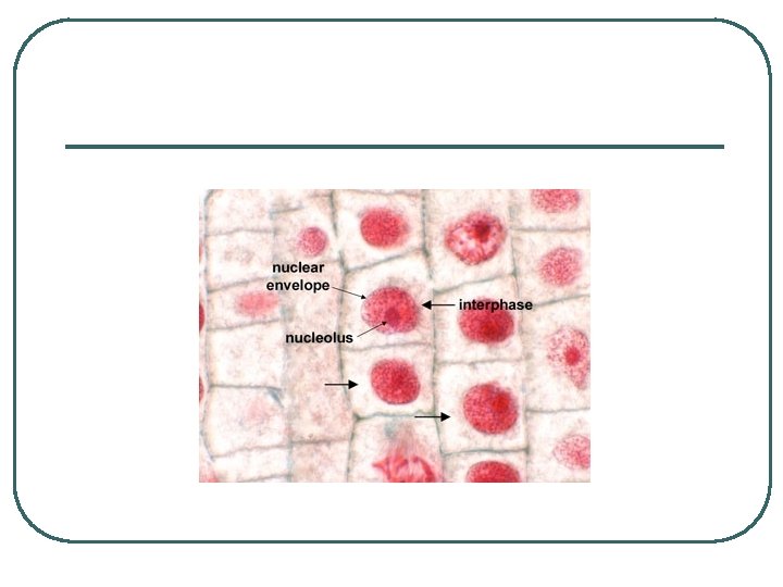
MITOTIC CELL CYCLE l l separtion of chromosomes into identical daughter cells Two phases: Interphase: 90% of cell cycle Mitotic Phase: division of nuclear material and cytokinesis
INTERPHASE S (DNA synthesis) C M yto ito ki si ne s si s G 1 MI (M TOT ) P IC HA SE G 2
INTERPHASE: - made up of three distinct sub-phases 1) G 1 - Gap 1 2) S - Synthesis 3) G 2 - Gap 2
l l l Gap 1: growth of cell after cytokinesis: protein synthesis and generation of new organelles Synthesis: DNA is copied (DNA replication) forming sister chromatid Gap 2: growth and development: preparation for mitosis
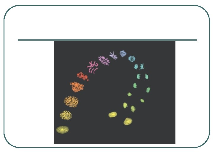
Mitosis (Karyokinesis) Five phases - actually a continuum (one continuous change)
1) Prophase: l chromatin condenses forming chromosomes l nuclear envelope breaks down l nucleolus dissolves l centrioles (in animals) begin to migrate toward the ends of cells l microtubules begin to form
2) Prometaphase: (before metaphase) - microtubules attach to the centromeres on a structure called the KINETOCHORE - those microtubules are called kinetochore microtubules and those not attached to the chromosomes are called non-kinetochore micortubules - chromosomes continue to condense - nuclear envelope almost all gone
3) Metaphase: "meta" means "middle" - chromosomes are lined up in the middle of the cell equidistant from the centroile pairs (centrosome) - metaphase plate - move due to interactions of the microtubules
Aster Sister chromatids Centrosome Metaphase Plate Kinetochores Overlapping nonkinetochore microtubules Kinetochores microtubules Microtubules 0. 5 µm Figure 12. 7 Centrosome 1 µm Chromosomes
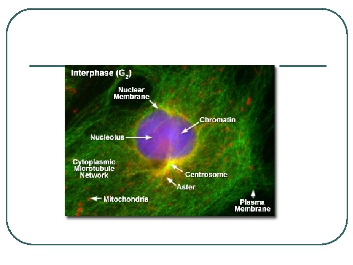
G 2 OF INTERPHASE Centrosomes (with centriole pairs) Chromatin (duplicated) Nucleolus Nuclear Plasma envelope membrane PROPHASE Early mitotic spindle Aster Centromere Chromosome, consisting of two sister chromatids PROMETAPHASE Fragments Kinetochore of nuclear envelope Nonkinetochore microtubules Kinetochore microtubule
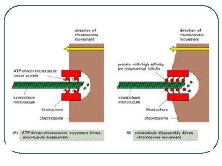
4) Anaphase: l separation of sister chromatids l move toward the opposite poles via the shortening of kinetochore microtubules - motor molecules in kinetochore move down the microtubule - as they do they break it down l each chromatid now considered a separate chromosome (each has the same genetic infomation as the other chromatid) • l Cells are genetically identical non-kinetochore microtubules lengthen and push on one another lengthening the cell's poles
EXPERIMENT 1 The microtubules of a cell in early anaphase were labeled with a fluorescent dye that glows in the microscope (yellow). Kinetochore Spindle pole Figure 12. 8
5) Telophase: l daughter nuclei begin to reform l each cell has identical sets of genetic information l nuclear envelope begins to reform l chromosomes unwind l nucleoli reform l beginning of cytokinesis
METAPHASE ANAPHASE Metaphase plate Spindle Centrosome at Daughter one spindle pole chromosomes TELOPHASE AND CYTOKINESIS Cleavage furrow Nuclear envelope forming Nucleolus forming
MAKING SENSE OF CHROMOSOME NUMBERS Ex: 10 Chromosomes l just divided daughter cell - 10 chromosomes l synthesis of DNA • 10 Chromosomes of duplicated DNA in sister chromatids l separate into 2 sets of 10
CYTOKINESIS l l division of the cytoplasm Animal cells: use of cleavage furrow • contractile ring of actin filaments and myosin • contract and pull the cell membrane in - like a • draw string Ex: clay and string
Cleavage furrow Contractile ring of microfilaments Figure 12. 9 A 100 µm Daughter cells (a) Cleavage of an animal cell (SEM)
l Plant Cells: use of cell plate • vesicles from the golgi fuse at the metaphase plate • build new cell wall • Ex: dividing a room with a wall
Vesicles forming cell plate 1 µm Wall of patent cell Cell plate New cell wall Daughter cells Figure 12. 9 B (b) Cell plate formation in a plant cell (SEM)
Chromatine Nucleus Nucleolus condensing Chromosome Metaphase. The 2 Prometaphase. 3 1 Prophase. spindle is complete, 4 The chromatin We now see discrete and the chromosomes, is condensing. chromosomes; each attached to microtubules The nucleolus is consists of two at their kinetochores, beginning to identical sister are all at the metaphase disappear. chromatids. Later plate. Although not in prometaphase, the yet visible nuclear envelop will in the micrograph, fragment. the mitotic spindle is staring to from. Figure 12. 10 Anaphase. The 5 chromatids of each chromosome have separated, and the daughter chromosomes are moving to the ends of cell as their kinetochore microtubles shorten. Telophase. Daughter nuclei are forming. Meanwhile, cytokinesis has started: The cell plate, which will divided the cytoplasm in two, is growing toward the perimeter of the parent cell.
l l l Mitosis Video
REGULATION OF THE CELL CYCLE l Cell-Cycle Control System: dependent upon the concentration of various chemicals in the cytoplasm Evidence: • combine cells in various stages resulting in activity of the cell in the later stage EX: • G 1 + Metaphase = G 1 nucleus began condensation of chromosomes • G 1 cell skipped S and G 2 phases
EXPERIMENTS In each experiment, cultured mammalian cells at two different phases of the cell cycle were induced to fuse. Experiment 1 Experiment 2 S G 1 M S M G 1 RESULTS S When a cell in the S phase was fused with a cell in G 1, the G 1 cell immediately entered the S phase—DNA was synthesized. M When a cell in the M phase was fused with a cell in G 1, the G 1 cell immediately began mitosis— a spindle formed and chromatin condensed, even though the chromosome had not been duplicated. CONCLUSION The results of fusing cells at two different phases of the cell cycle suggest that molecules present in the Figure 12. 13 A, B cytoplasm of cells in the S or M phase control the progression of phases.
l l Signals build up and initiate the next phase. Once the next phase has begun, it does not stop.
G 1 checkpoint Control system G 1 M G 2 M checkpoint Figure 12. 14 G 2 checkpoint S
G 0 G 1 checkpoint G 1 Figure 12. 15 A, B (a) If a cell receives a go-ahead signal at the G 1 checkpoint, the cell continues on in the cell cycle. G 1 (b) If a cell does not receive a go-ahead signal at the G 1 checkpoint, the cell exits the cell cycle and goes into G 0, a nondividing state.
CONTROL SIGNALS Internal: Kinetochore signals l Anaphase cannot start until all microtubules l l are attached to kinetochore Kinetochores that are not attached to a microtubule send out signals that prevent anaphase from occuring - when all are attached, signals cease and separation begins by the activation of an Anaphase promoting complex
l l l External: Chemical: bind to cell membranes hormones from other tissues that trigger mitotic growth Ex: platelete derived growth factor - stimulates healing of wounds - made by platelets which release the factor at a wound gibberellins - stem elongation Physical: not enough room in the kitchen Density Dependent Inhibition: Growth is inhibited when cells become crowded Anchorage Dependency: cells must be attached to something - can’t just grow in a solution EXCEPTION: Cancer does not follow density-dependent inhibition or anchorage dependency divide indefinitely - spread - malignant – metastasis
(a) Normal mammalian cells. The availability of nutrients, growth factors, and a substratum for attachment limits cell density to a single layer. Cells anchor to dish surface and divide (anchorage dependence). When cells have formed a complete single layer, they stop dividing (density-dependent inhibition). If some cells are scraped away, the remaining cells divide to fill the gap and then stop (density-dependent inhibition). Figure 12. 18 A 25 µm
Cancer cells do not exhibit anchorage dependence or density-dependent inhibition. (b) Cancer cells usually continue to divide well beyond a single layer, forming a clump of overlapping cells. Figure 12. 18 B 25 µm
MEIOSIS Meiotic Cell Cycle: reduction of chromosomes Major Processes l Replication of Chromosomes l Pairing of Homologous Chromosomes (Synapsis) l Crossing Over at Chiasmata l Separation of Homologous Chromosomes l Separation of Sister Chromatids
l l 2 Phases of Meiosis: Meiosis I and Meiosis II Meiosis I: Separation of Homologous Chromosomes l each chromosome has one sister chromosome • • Chomosomes that carry the same basic genetic content Humans: 46 chromosomes: 22 homologous pairs and the Sex Chromosomes – X and Y Interphase I: Like mitosis: Replication of DNA forms sister chromatids
l l l Prophase I: Like mitosis except 1. Longer: 90% of meiosis 2. Synapsis: Pairing of homologous chromosomes (each made of two sister chromatids - makes tetrad - four chromatids) non-sister chromatids of homologous pairs cross one another chiasma leads to crossing over - chromatids break and exchange genes - happens in the same place - synaptonemal complex lines up chromosomes gene by gene
Metaphase I: l Like mitosis except homologous pairs line up along metaphase plate and microtubules only attach to one side of kinetocore Anaphase I: l Separation of homologous pairs: sister chromatids remain attached Telophase I and Cytokinesis: l may or may not reform nucleus and nucleolus l cell divides and forms haploid cells (only a half set)
MEIOSIS I: Separates homologous chromosomes INTERPHASE PROPHASE I Centrosomes (with centriole pairs) Sister chromatids Nuclear envelope METAPHASE I Chiasmata ANAPHASE I Sister chromatids remain attached Centromere (with kinetochore) Spindle Metaphase plate Homologous Microtubule chromosomes attached to Chromatin separate kinetochore Pairs of homologous Chromosomes duplicate Tertads line up chromosomes split up Homologous chromosomes (red and blue) pair and exchange segments; 2 n = 6 in this example Tetrad
Prophase II: l spindle reforms Metaphase II: l line up along metaphase plate Anaphase II: l separation of sister chromatids Telophase II/Cytokinesis: l nuclei form and cells divide
MEIOSIS II: Separates sister chromatids TELOPHASE I AND CYTOKINESIS PROPHASE II Cleavage furrow Two haploid cells form; chromosomes are still double METAPHASE II ANAPHASE II Sister chromatids separate TELOPHASE II AND CYTOKINESIS Haploid daughter cells forming During another round of cell division, the sister chromatids finally separate; four haploid daughter cells result, containing single chromosomes
Interphase Homologous pair of chromosomes in diploid parent cell Chromosomes replicate Homologous pair of replicated chromosomes Sister chromatids Diploid cell with replicated chromosomes Meiosis I 1 Homologous chromosomes separate Haploid cells with replicated chromosomes Meiosis II 2 Sister chromatids separate Haploid cells with unreplicated chromosomes
l Meiosis Video
COMPARISON OF MITOSIS AND MEIOSIS Mitosis: one phase: 2 daughter cells: Diploid Meiosis: two phases: 4 daughter cells: Haploid
MITOSIS MEIOSIS Chiasma (site of crossing over) Parent cell (before chromosome replication) MEIOSIS I Prophase Chromosome replication Duplicated chromosome (two sister chromatids) Chromosome replication Tetrad formed by synapsis of homologous chromosomes 2 n = 6 Metaphase Chromosomes positioned at the metaphase plate Anaphase Telophase Sister chromatids separate during anaphase 2 n Tetrads positioned at the metaphase plate Homologues separate during anaphase I; sister chromatids remain together Metaphase I Anaphase I Telophase I Haploid n=3 Daughter cells of meiosis I 2 n MEIOSIS II Daughter cells of mitosis n n n Daughter cells of meiosis II Sister chromatids separate during anaphase II n
IMPORTANCE OF MEIOSIS SOURCE OF GENETIC VARIATION l l l Independent Assortment of Chromosomes - each set of chromosomes separates independently of the other PAGE 235 - mixes genes of the parents for the next generation Your genetic material is half from your father and half from your mother. Your offspring will get half the genetic information from you. Because chromosomes sort independently, your offspring will receive a mix of your parents, not just one or the other PROBABILITY: 2 n where n is the haploid number of chromosomes
Key Maternal set of chromosomes Possibility 1 Possibility 2 Two equally probable arrangements of chromosomes at metaphase I Metaphase II Daughter cells Combination 1 Combination 2 Combination 3 Combination 4
2. Crossing over - introduces more variation - each individual chromosome is not entirely maternal or paternal 3. Random fertilization WHY IS VARIETY IMPORTANT? l keeps things interesting l survival
Prophase I of meiosis Nonsister chromatids Tetrad Chiasma, site of crossing over Metaphase II Daughter cells Recombinant chromosomes
Cell Division in Plants Alternation of Generations: plants have one part of the life cycle as diploid and one part as haploid Haploid multicellular organism (gametophyte) n Mitosis n n Spores MEIOSIS Diploid 2 n multicellular organism (sporophyte) (b) Plants and some algae n Gametes FERTILIZATION 2 n Mitosis Zygote
Cell Division in Fungi and Protists l Main phase of life is haploid Haploid multicellular organism Mitosis n n n Gametes MEIOSIS n FERTILIZATION 2 n Zygote (c) Most fungi and some protists