Cells Structure and Function Section 1 Introduction to


: observed")
: was the first person to observe")


")
 Herpes Virus Plant Root Cell")
")
")
 Mosquito Head 200 X 2000 X")
 Fly Eye")
 Surface of Tongue Neuron Inside of Stomach")
 Pollen Yeast Red Blood Cell, Platelet, and White Blood Cell")
: concluded")


 and prokaryotes (bacteria) differ greatly")


")


 that contain chlorophyll surrounded by a double membrane l")


- Slides: 50
Cells Structure and Function
Section 1: Introduction to the Cell
Discovery of Cells l The invention of the lens l Robert Hooke (1665): observed a thin slice of cork (dead plant cells) with a microscope. He described what he observed as “little boxes” (cells).
Discovery of Cells l Anton van Leeuwenhoek (1675): was the first person to observe living cells.
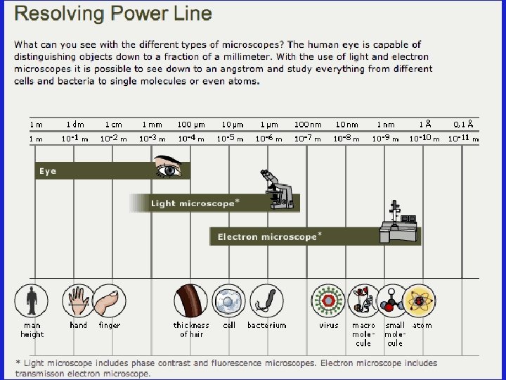
Microscopes l Magnification: refers to the microscope’s power to increase an object’s apparent size l Resolution: refers to the microscope’s power to show detail clearly
Light Microscope
Light Microscope Elodea - Aquatic Plant 40 X 400 X
Transmission Electron Microscope (TEM)
Transmission Electron Microscope (TEM) Herpes Virus Plant Root Cell
Scanning Electron Microscope (SEM)
Scanning Electron Microscope (SEM)
Scanning Electron Microscope (SEM) Mosquito Head 200 X 2000 X
Scanning Electron Microscope (SEM) Fly Eye
Scanning Electron Microscope (SEM) Surface of Tongue Neuron Inside of Stomach
Scanning Electron Microscope (SEM) Pollen Yeast Red Blood Cell, Platelet, and White Blood Cell
The Cell Theory l Who developed the cell theory? – Matthias Schleiden (1838): concluded that all plants are composed of cells – Theodor Schwann (1839): concluded that all animals are composed of cells – Rudolph Virchow (1855): determined that cells come only from other cells
The Cell Theory l What is the cell theory? 1. All living things are composed of one or more cells. 2. Cells are organisms’ basic units of structure and function. 3. Cells come only from existing cells.
Cell Diversity l Size l Shape l Internal Organization
Smallest Cells: Cell Diversity- Size Longest Cells: Biggest Cells: 6 inches long, 5 inches wide, 3 pounds Ostrich Egg
Cell Diversity- Shape l Cells differ widely in shape. l Most cells are roughly cuboidal or spherical.
Cell Diversity- Internal Organization l Nucleus: contains DNA which directs the activity of the cell l Organelle: a cell component that performs specific functions in the cell l Eukaryotes: cells that contain a nucleus and membrane-bound organelles l Prokaryotes: cells that lack nuclei and membrane-bound organelles
Eukaryotes vs. Prokaryotes l Eukaryotes (animals, plants, fungi, protists) and prokaryotes (bacteria) differ greatly in structure.
Prokaryotic Cell
Structural Organization of Eukaryotic and Prokaryotic Cells
Section 2: Parts the Cell
The Parts of the Cell Each living cell carries out the tasks of taking food, transforming food into energy, getting rid of wastes, and reproducing. l Most eukaryotic cells have three main components: – Cell Membrane – Cytoskeleton – Nucleus l
Structure and Function of Organelles l The Structure and Function of the following organelles will be discussed: – – – Cell Membrane Nucleus Cell Wall Cytoplasm Cytoskeleton – Mitochondria – Cilia and Flagella – Vacuoles
Cell Membrane Structure: phospholipid bilayer with proteins that function as channels, markers, and receptors -also contains cholesterol which provides rigidity l Function: selectively permeable boundary between the cell and the external environment l
Nucleus Structure: the nucleus is a sphere that contains another sphere called a nucleolus l Function: -storage center of cell’s DNA -manages cell functions l
Cell Wall Structure: rigid wall made up of cellulose, proteins, and carbohydrates l Function: boundary around the plant cell outside of the cell membrane that provides structure and support l
Cytoplasm Structure: gelatin-like fluid that lies inside the cell membrane l Function: -contains salts, minerals and organic molecules -surrounds the organelles l
Cytoskeleton Structure: a network of thin, fibrous elements made up of microtubules (hollow tubes) and microfilaments (threads made out of actin) l Function: -acts as a support system for organelles -maintains cell shape l
Endoplasmic Reticulum Structure: a system of membranous tubules and sacs l Function: intercellular highway (a path along which molecules move from one part of the cell to another) l Two types: – Rough Endoplasmic Reticulum – Smooth Endoplasmic Reticulum l
Golgi Apparatus Structure: stacked flat sacs l Function: receives proteins from the r. ER and distributes them to other organelles or out of the cell (receiving, processing, packaging, and shipping) l
Mitochondria Structure: folded membrane within an outer membrane – The folds of the inner membrane are called cristae l Function: -converts energy stored in food into usable energy for work – cellular respiration l
Cilia and Flagella Structure: hair-like organelles that extend from the surface of cells – When they are present in large numbers on a cell they are called cilia – When they are less numerous and longer they are called flagella – Both organelles are composed of nine pairs of microtubules arranged around a central pair. l Function: cell motility l
Cillia and Flagella
Vacuoles Structure: a sac of fluid surrounded by a membrane – Very large in plants l Function: used for temporary storage of wastes, nutrients, and water l
Plastids l There are three types of plastids in plant cells: – Chloroplasts (discussed on next slide) – Chromoplasts: synthesize and store pigments – Leucoplasts: store food such as starches, proteins, and lipids Chromoplasts Red Pepper Flower Leucoplasts
Chloroplasts Structure: stacked sacs (thylakoids) that contain chlorophyll surrounded by a double membrane l Function: photosynthesis (conversion of light energy to chemical energy stored in the bonds of glucose) l
Secretory Pathway
Secretory Pathway
Plant Cells vs. Animal Cells l Animal cells are very similar to plant cells except for the following major differences: – Animal cells do not contain chloroplasts – Animal cells are not surrounded by cell walls – The vacuoles in plants are much larger than those of animals
Animal Cell
Plant Cell
Microscope Pictures of a Plant Cell and an Animal Cell Elodea Human Cheek Cells
Hierarchy of Biological Order
THE END!