CELLS LESSON 2 VIEWING CELLS WITH A MICROSCOPE


- Slides: 18
CELLS LESSON 2 – VIEWING CELLS WITH A MICROSCOPE
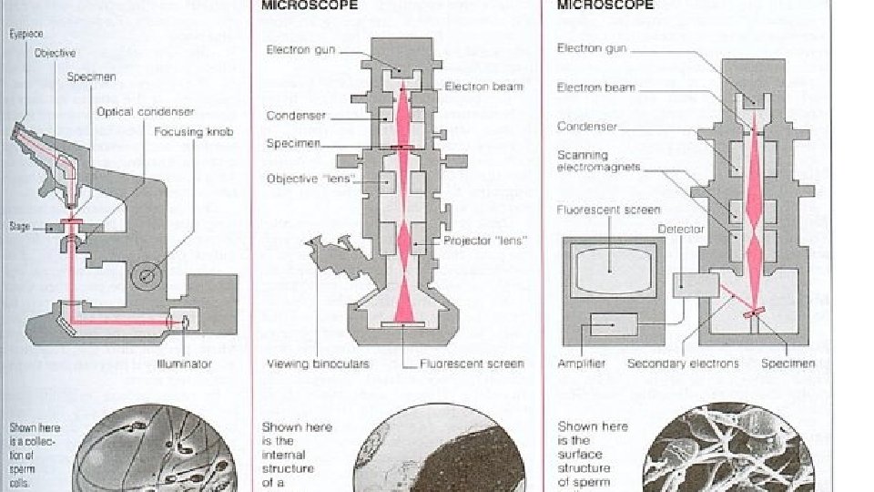
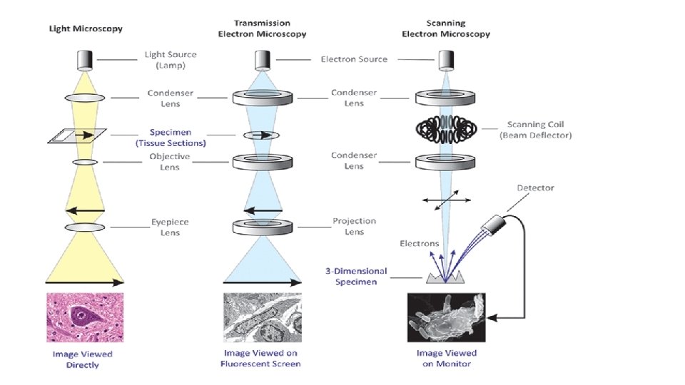
OBJECTIVES 1. Explain the basic functions of a light microscope 2. Compare light and electron microscopes in terms of equipment itself and specimens viewed 3. List the parts of the light microscope
INTRODUCTION • The cell is the basic structural and functional unit of all living things – plants, animals, bacteria and fungi. • They are so small – microscopic that they cannot be seen with the naked eye but only with the aid of a microscope. • What can you see with the different types of microscopes? • The human eye is capable of distinguishing objects down to a fraction of a millimeter. • With the use of light and electron microscopes it is possible to see down to an angstrom and study everything from different cells and bacteria to single molecules or even atoms.
TYPES OF MICROSCOPES There are two major types of microscopes • The light microscope or compound microscope • The electron microscope (of which there are different types) • Visit the following sites for more information • http: //www. microbehunter. com/electron-microscopes-vs-opticallight-microscopes/ • http: //universe-review. ca/R 11 -13 -microscopes. htm
USING THE LIGHT MICROSCOPE • Go online and try out the https: //www. brainpop. co m/games/virtuallabsusing themicroscope/ - virtual microscope use
MICROQUIZ • Using the microquiz given, label the parts of the light microscope • Keep and stick the paper into your notebooks
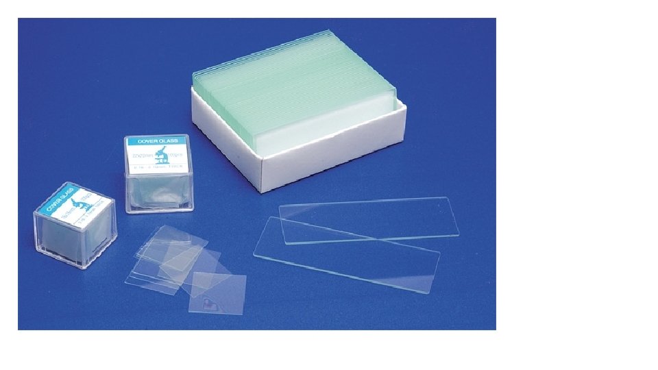
SLIDES • In order to view specimens with the microscope slides are made • A blank slide is a thin, flat piece of glass • A thin slice of a specimen is placed (mounted) on the slide to view • Usually a little square called a cover-slip is placed over the specimen, forming a sandwich – this is so the specimen will not move or dry out. • Cover slips are smaller and thinner than the blank slide. • Slides can be readymade permanent or you can make your own temporary ones.
SLIDE 1 • Look at the first slide • Write down in your book a description as follows • Name of slide: pumpkin stem at x 4 • Shapes of cells: • Colours of cells: • Plant/ Animal specimen?
SLIDE 2 • Look at the first slide • Write down in your book a description as follows • Name of slide: • Shapes of cells: • Colours of cells: • Plant/ Animal specimen?
SLIDE 3 • Look at the first slide • Write down in your book a description as follows • Name of slide: • Shapes of cells: • Colours of cells: • Plant/ Animal specimen?
SLIDE 4 • Look at the first slide • Write down in your book a description as follows • Name of slide: • Shapes of cells: • Colours of cells: • Plant/ Animal specimen?
SLIDE 5 • Look at the first slide • Write down in your book a description as follows • Name of slide: • Shapes of cells: • Colours of cells: • Plant/ Animal specimen? http: //medcell. med. yale. edu/histology/urinary_system_lab/kid ney. php
ELECTRON MICROSCOPE • Electron microscopes are used to view specimens in even more details. It can be used to see inside cells, not just the outline • There are more than one types of electron microscope • The Transmission Electron Microscope (TEM) was developed in 1931. • The first Scanning Electron Microscope (SEM) debuted in 1942
PROJECT - ASSIGNMENT Use your laptop to do research and type up the answers to the following questions: Use Microsoft word. Make sure to add a cover page with your name, class and subject. 1. Compare a light and an electron microscope in a table format. 2. How do microscopes help scientists and doctors? Provide an example of how the microscope has greatly enhanced scientific knowledge. 3. How might the SEMs help diagnose and treat diseases? 4. Can you determine a cell’s function from its size and shape? 5. Which images were most interesting and why? 6. Were there any differences between plant and animal cells? Describe or draw them. Adapted from http: //www. discoveryeducation. com/teachers/free-lesson-plans/virtual-electron-microscope. cfm
Slides online! - Histology • http: //www. histologyguide. org/01_Introduction/Chapter. html#The_ Cell – scroll down to the section marked Morphology • Click on MH 002 Cells and Tissues – there is a clickable picture that zooms in step by stem. – May need a fast speed of connection for the webpage to load properly.