Cells I Basic History Every living thing from

Cells

I. Basic History _______ Every living thing, from the tiniest bacterium to the largest whale, are made of one or more cells! Before the seventeenth century, no CELL one knew that _______ existed. S

I. Basic History small to be cells are too _______ seen with the _______. unaided eye Not discovered until after the invention of the ______ microscope in the early 17 th century. Most
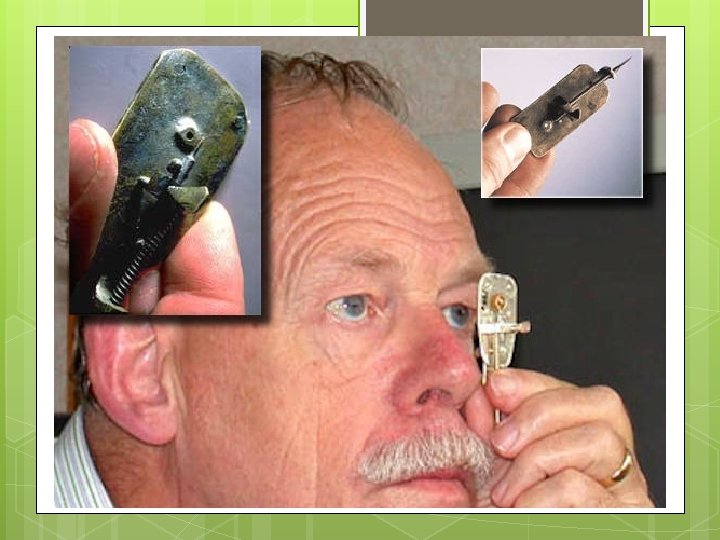

II. Important Scientists A Dutch drapery storeowner ____________, Anton von Leeuwenhoek became the _______ person to FIRS OBSERV _____ and _____ TDESCRIB E E MICROSCOPIC ORGANISMS and LIVING CELLS.

II. Important Scientists 1665: the English scientist Robert Hooke used a ________ microscope to examine a thin slice of ______ and described it as cork consisting of “a great many little boxes”. It was after his observation that Hooke called what he saw “_______”. They looked cellslike “little boxes” and reminded him of the small rooms in which monks lived. So he called them “_______”. cells
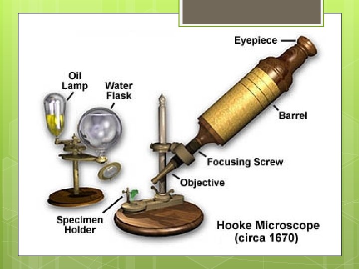

Illustration of Cork drawn by Robert Hooke

II. Important Scientists 1824: the French scientist Henri Dutrochet, concluded ______ and ____ plant animal were always made 1831: Robert Brown named the _____ nucleus that tissue up of cells.

II. Important Scientists 1838: German botanist Matthias Schleiden concluded that all ____ are made of cells. plants 1839: German zoologist Theodor Schwann animals reported that _____ are also made of cells.

II. Important Scientists 1845: Felix Dujardin studied the living cell and noted it contained a material called _______. protoplasm 1855: German physician Rudolf Virchow induced that ALL cells come from _______ cells. preexisting

II. Important Scientists The COMBINED work of Schleiden, Schwann, and Virchow makeup what is now known as the modern _______. cell theory

1. 2. 3. III. The Cell Theory Consists of 3 Principles All living things are ______ composed of one or more _______. cells Cells are the basic units of ___________ structure and _____ function in an organism. Cells come ______ ONLYfrom the ________ cells. reproduction of existing

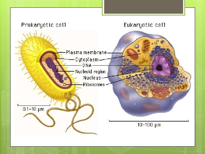
IV. Two Types of Cells 1. EUKARYOT ________ = cell that E contains a _____ other nucleus and ___________ membrane-bound organelles __________ plants Ex: ____, fish, insects mammals, _____ humans and ____

IV. Two Types of Cells 1. PROKARYOT ________ = cell that E _______ lacks a _____ nucleus andother __________ membrane-bound organelles _____. unicellular Ex: _______ bacteria organisms such as _____ and their relatives

V. Cell Diversity o Not all cells are _______. alike o Cells within the same organism show size enormous diversity in ____, ______, and __________. shape internal organization o Your body contains at least 200 different cell types! _____

VI. Cell Size: o. A few types of cells are large enough to be seen by the _____ unaided eye. Female egg o ________ is thelargest _____ cell in the body and can be seen without the aid of a microscope. o Most cells are visible only with a ______. microscope

VI. Cell Size: Most cells are small for 2 reasons o _______________ ___: RATIO area Cells are limited outer in size surface by the _______ volume between their ___________ 1. and their ____. • • As a cell’s size increases, its volume increases much faster than its surface area. (see picture on the next slide!)
 can only control a certain")
VI. Cell Size: 2. The cell’s nucleus (the brain) can only control a certain amount of living, active cytoplasm.

VII. Cell Shape: Variety o _____ of shapes shape o The _______ of the cell depends on the _____. function

VII. Cell Shape: cells o Ex: Nerve ______ that carry information from your toes to your brain are long and threadlike. o Ex: ______ are Blood shaped cells like round discs that can squeeze through tiny blood vessels.

VIII: Cellular Organization Multicellular organisms o ____________ are made up of many cells, each of which is specialized to perform a distinct function. o Digestion, movement, respiration, filtering, etc. o _________ DO NOT Individual cells carry out ALL life functions, but rather depend on each other.

VIII: Cellular Organization o ____ Tissu = a group of cells functioning together to perform an activity. e o o Ex: muscle and nerve tissues Ex: Plant tissues = stem and root o Organs ____ = groups of two or more tissues that function together. o o Stomach, leaf of a plant Cooperation among organs makes life functions within an organism efficient.

VIII: Cellular Organization o ____ Tissu = a group of cells functioning together to perform an activity. e o o Ex: muscle and nerve tissues Ex: Plant tissues = stem and root o Organs ____ = groups of two or more tissues that function together. o o Stomach, leaf of a plant Cooperation among organs makes life functions within an organism efficient.

VIII: Cellular Organization Summary Cells Tissues Organs

Microscope Basics

Always carry a microscope with one hand holding the arm and one had under the base.

What’s my Power? o To calculate the power of magnification, _______ of the multiply thepower _______ of the ocular lens by thepower ______. objective O Objectives (4 X, 10 X, 40 X) O O Ocular lens (10 X)

What’s my Power? o Low Power o Ocular lens = 10 X o Objective = 4 X o TOTAL magnification for LOW power 40 X = _____

What’s my Power? o Medium Power o Ocular lens = 10 X o Objective = 10 X o TOTAL magnification for MEDIUM 100 X power = _____

What’s my Power? o High Power o Ocular lens = 10 X o Objective = 40 X o TOTAL magnification for HIGH power 400 X = _____

Comparing Powers of Magnification: o We can see better details with ____ HIGHER powers of magnification, but we can’t see as much of the image.

Which of these images would be higher power of viewed at a _______ magnification?

Microscope Pictures

Compound Light Microscopes o You will be using a compound light microscope in several labs. o These microscopes have a maximum magnification of 400 X CANNOT see most of the o So you _____ organelles like ribosomes, Golgi bodies, lysosomes, etc. o More powerful microscopes are needed (2000 X plus)

Common Problem. . . AIR BUBBLES

Stained Onion Cells o Can you identify the cell walls? o Can you identify any other organelles?

Stained Cheek Cells

Elodea – Typical Plant Cells seen with the light microscope as

Animal Cell

Nucleus

Rough ER

Golgi Body

Mitochondria

Chloroplasts

Ribosome

Cytoskeleton

How to Make a Wet-Mount Slide 1. Get a clean slide and cover slip from your teacher. 2. Place ONE drop of water/iodine in the middle of the slide. Don’t use too much or the water will run off the edge and make a mess! 3. Place the edge of the cover slip on one side of the water/iodine drop. 4. Slowly lower the cover slip on top of the drop. 5. Place the slide on the stage and view it first with the LOW power objective. Once you see the image, you can rotate the nosepiece to view the slide with the different objectives.

Let’s give it a try. . . 1. 2. 3. Turn on the microscope and then rotate the nosepiece to click the LOW power objective into place. Place a slide on the stage and secure it using the stage clips. Use the coarse adjustment knob (large knob) to get the image into view and then use the fine adjustment knob (small knob) to make it clearer. Once you have the image in view, rotate the nosepiece to view it under different powers. Draw what you see on a piece of paper. BE CAREFUL WITH THE LARGEST OBJECTIVE! Sometimes there is not enough room and you will not be able to use it!

Organelles of the Cell

Nucleus Appearance: Location: Function: Large oval varies control center for all cell functions

Cytoplasm Appearance: Location: Function: clear fluid inside the cell membrane suspends organelles site of chemical reactions

Nucleolus Appearance: Round structure inside the nucleus Location: inside the nucleus Function: Site of RNA synthesis Produces ribosomes
 Membrane Appearance: surrounds cell Location: Plant: in cell wall Animal: outer layer")
Plasma (Cell) Membrane Appearance: surrounds cell Location: Plant: in cell wall Animal: outer layer Semipermeable Composed of lipids & proteins Function: controls materials in and out of the cell

Smooth Endoplasmic Reticulum Appearance: mesh of hollow sheets Location: connected to the nucleus and plasma membrane Function: Smooth: produces lipids

Ribosomes Appearance: small, dense granules Location: Free in the cytoplasm; attached to the rough ER Function: Synthesize proteins

Golgi Body Appearance: Location: Flattened sacs Near the ER Function: Temporary storage, packaging and secretion of proteins and fats Produces lysosomes

Mitochondria Appearance: usually bean shaped with folded membranes (greater surface area – hence more energy) Location: many mitochondria in a cell Function: Powerhouse of the cell (energy production ATP)

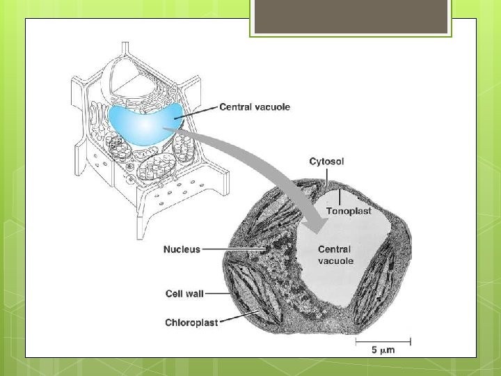
Vacuoles Appearance: cavities filled with fluid Location: Plant: usually 1 large waterfilled vacuole (maintains structure) Animal: many tiny vacuoles Function: storage of water, starch fats, etc. Two types: Contractile vacuole: removes water and wastes Food vacuole: breaks down food

Water Vacuole

Lysosomes Appearance: egg shaped, membranebound structure Location: ONLY found in animal cells Function: Contain digestive enzymes that break down molecules aid in digestion of nutrients break down destructive cells (bacteria)
 Location: entire cell Function:")
Cytoskeleton Appearance: network fibrous (microtubules & of thin, proteins microfilaments) Location: entire cell Function: acts as sort of a scaffold to provide support for organelles helps maintain cell shape

Microfilaments Appearance: long, threadlike proteins Location: a part of cytoskeleton Function: associated with muscle contractions in large organisms associated with cell movement

Microtubules Appearance: thin, hollow cylinders of proteins Location: a part of cytoskeleton Function: provide shape and rigidity to the cell Assist organelles to move from to place within the cell place

Cilia Appearance: thin hair-like projections Location: formed from specialized microtubules Attached to outside of cell Function: aid in movement and locomotion (lungs and intestinal cells)

Flagella Appearance: whip-like tails Location: formed from specialized microtubules Attached to outside of cell Function: aid in movement and locomotion (sperm)

Chromatin Appearance: strings of “spaghetti” Location: inside nucleus Function: uncoiled DNA; involved in duplicating cells. Coils into chromosomes during cell division.
")
Chromosomes Appearance: coiled chromatin Location: inside nucleus Function: contains genetic information (DNA)
")
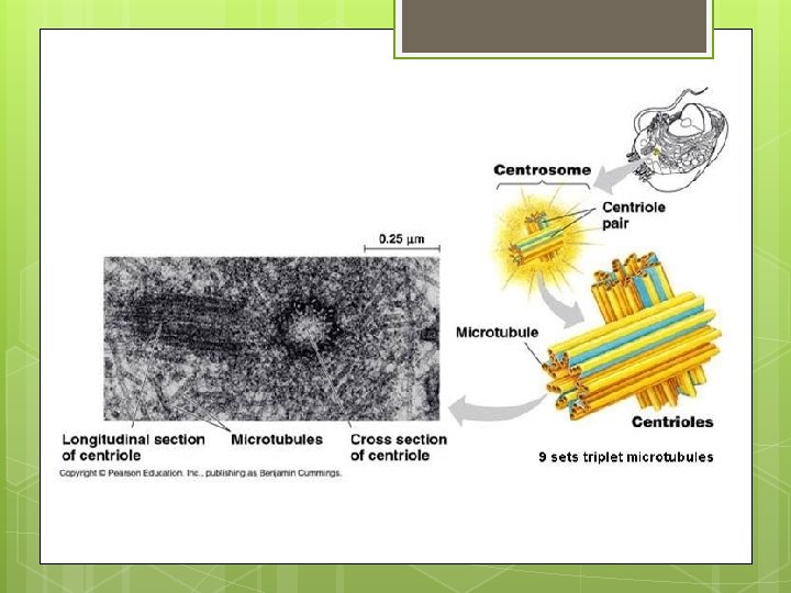
Centrioles Appearance: two small structures Location: found inside the centrosome (only in animal cells) Function: moves chromosomes during cell division
 Appearance: varies; have own DNA Location: ONLY in plants Function: based")
Plastids (plants only) Appearance: varies; have own DNA Location: ONLY in plants Function: based on type: Leucoplast (store starch), chromoplast (store pigments), chloroplast
 Appearance: small, circular, green (contains chlorophyll-green pigment) Location: ONLY in plants")
Chloropast (plants only) Appearance: small, circular, green (contains chlorophyll-green pigment) Location: ONLY in plants Function: site of photosynthesis
 Appearance: made of cellulose; rigid, strong, stiff structure Location: surrounds")
Cell Wall (plants only) Appearance: made of cellulose; rigid, strong, stiff structure Location: surrounds cell membrane (ONLY in plants) Function: support and protection Allows of cell H 2 O, O 2, CO 2 to pass into and out

NOTE: All the ______ organelles work together! For example, after some proteins are made by the ______, ribosomes the rough ER transports these proteins to the __________, then the Golgi apparatus makes vesicles that can fuse with the cell’s plasma membrane to release proteins to the ________ environments outside the cell or used within the cell.
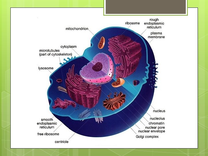
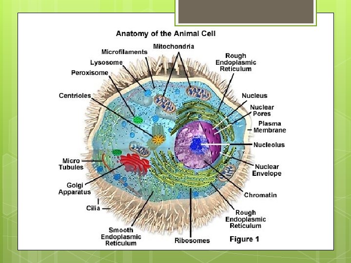

Microfilaments Golgi body Smooth ER Ribosomes Nucleolus *Centrioles *Lysosome Mitochondria Chromatin Microtubule Rough ER Nucleus Plasma Membrane *Vacuole many small ones *ANIMAL CELL

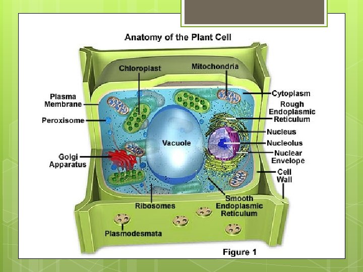
Ribosomes Microtubule *Plastid *Water Vacuole Microfilaments Nucleolus Mitochondria Plasma Membrane Golgi body Chromatin Smooth ER *Chloroplast Nucleus Rough ER *Cell Wall

Plasma Membrane

I. Maintaining Balance How do cells maintain balance o _______________ __? o Cells need to maintain a balance by controlling material that move in and HOMEOSTASI out of the cell ________. S

I. Maintaining Balance Small o _______ molecules like water, oxygen, and carbon dioxide can move in and out of the cell _______. freely o _______ molecules like proteins and Large carbohydrates ____ cannot o __________ Eliminating wastes

I. Maintaining Balance
 Membrane o All cells are surrounded by a plasma")
Structure of the Plasma (Cell) Membrane o All cells are surrounded by a plasma membrane __________. o Functions like a GATE, controlling what _______ LEAVE the cell. ENTER and _______ S membrane S o The cell is semipermeable ________ or selectively permeable.
 Membrane o. A ______________ semipermeable membrane only allows certain")
Structure of the Plasma (Cell) Membrane o. A ______________ semipermeable membrane only allows certain molecules to pass through. o Some substances easily cross the membrane, while others cannot cross at all.
 Membrane o Made of a thin layer of ____")
Structure of the Plasma (Cell) Membrane o Made of a thin layer of ____ lipids and _____ proteins o Made mostly of ________ phospholipid molecules (Phosphate + Lipid) o Phospholipids are a kind of lipid that consists of 2 FATTY ACIDS (_______) and tails PHOSPHATE GROUP (_______) heads
 Membrane o Cell membranes consist of TWO phospholipid layers")
Structure of the Plasma (Cell) Membrane o Cell membranes consist of TWO phospholipid layers called a LIPID ________ BILAYER
 Membrane")
Structure of the Plasma (Cell) Membrane
 Membrane o Water molecules surround ______ both sides of")
Structure of the Plasma (Cell) Membrane o Water molecules surround ______ both sides of the cell membrane. o Polar _________ phosphate heads sticking TOWARD the water (______) hydrophilic o Nonpolar _____ lipid tails point AWAY from the water (_______) hydrophobic
 Membrane o The cell membrane is constantly being _______")
Structure of the Plasma (Cell) Membrane o The cell membrane is constantly being _______ formed and broken down _______ in living cells.

III. Lipid Bilayer o Moving with and among the phospholipids are cholesterol, proteins, and carbohydrates. o _______ Cholesterol o Nonpolar, found among the phospholipids to help prevent the fatty acid tails from _____ together. sticking o Helps with structure and homeostasis

III. Lipid Bilayer

III. Lipid Bilayer o _____: Protein Found on the surface of the plasma membrane = _____ transmit signals to the inside of cell. o Embedded in the plasma membrane = structure and support of cells shape, and move _______ large substances in and out of the cell. o

III. Lipid Bilayer

III. Lipid Bilayer o ________: Carbohydrates Attached to proteins, helps cells identify ___________ chemical signals o Ex: help disease fighting cells recognize and attack a potentially harmful cell o

Cellular Transport
")
Cellular Transport o All particles move and have _______ energy kinetic (energy of motion) o Movement is ____ random and usually in a ________ water solution o Cells are mostly made of water and there is a constant flow of ions and particles.

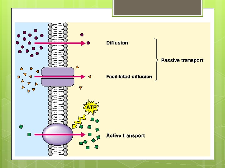
IV. 2 Types of Cellular Transport 1. 2. __________ = Passive transport movement of molecules across the membrane by using the molecules kinetic The cell _____ energy. NO exerts ___ energy! _________ = transport Active transport of materials against the concentration gradient and requires _________. cellular energy

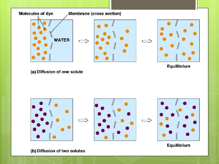
V. Passive Transport o 3 1. types of passive transport: Diffusion = the net movement of ______ HIGHE ______ particles from an area of _____ concentration of R particles to an LOWE area of _____ concentration R of particles

Diffusion. . . o Molecules move _____ randomly until equally distributed. they are _____ o Diffusion continues until the concentration of substances is uniform throughout.

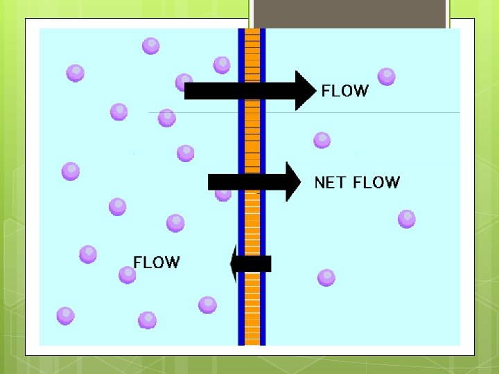
Diffusion. . . o ___________ Dynamic equilibrium = continual movement but no overall change in concentration. o Movement of materials into and out of the cell at equal rates maintains its dynamic equilibrium with its environment. E C N A L A B

Diffusion. . . o Diffusion depends on the Concentration gradient ____________ o o _____________ is the Concentration gradient difference between the concentration of a particular molecule in one area and the concentration in an adjacent area. Ex. Gas exchange in the lungs (oxygen from air to blood and carbon dioxide from blood to air)

V. Passive Transport 2. Facilitated diffusion ____________ = type of passive transport that increases the rate of diffusion with the use of _________ carrier proteins o Ex: Facilitated diffusion of glucose

Facilitated Diffusion. . .

V. Passive Transport 3. _____ Osmosi = the diffusion of w _______ s ater molecules from an area of HIGH water concentration to an area of LOW water concentration

V. Passive Transport: Osmosis o Occurs in response to the concentration of solutes dissolved in water! o Solutes _____ are dissolved substances in a solution water containing is mostly _______ many dissolved _____. solutes o Cytoplasm

V. Passive Transport: Osmosis o Because ___ no TWO molecules can occupy the same space at the same time, the MORE solutes there are in a certain volume of water, the FEWER water molecules there can be in the same volume.

V. Passive Transport: Osmosis o Plant and animal cells behave differently because plant cells have a large water vacuole ____ and a cell wall _____
 cell H")
V. Passive Transport: Osmosis o Ex. Osmosis occurring in a slug (animal) cell H 2 O Na. Cl - water leaves the cell - cells shrivel, slug dies

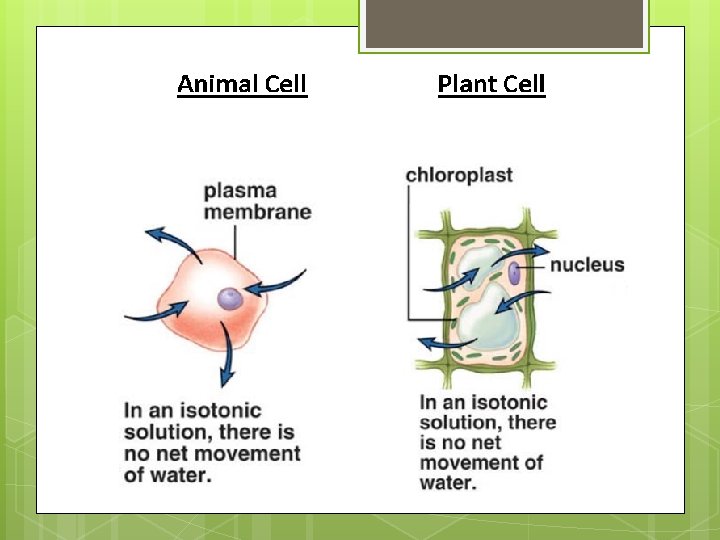
V. Passive Transport: Osmosis A. Isotonic solution __________ = a solution in which the concentration of dissolved substances (solutes) is the _______ SAME as the concentration of solutes inside the cell. o Osmosis ______ DOES NOT occur since a concentration gradient is not established!

What happens to cells when placed in an isotonic solution? o _____ Plant cell flaccid (limp) becomes ____ o plant wilts because no net tendency water to enter for

What happens to cells when placed in an isotonic solution? Animal cell o _______ - normal

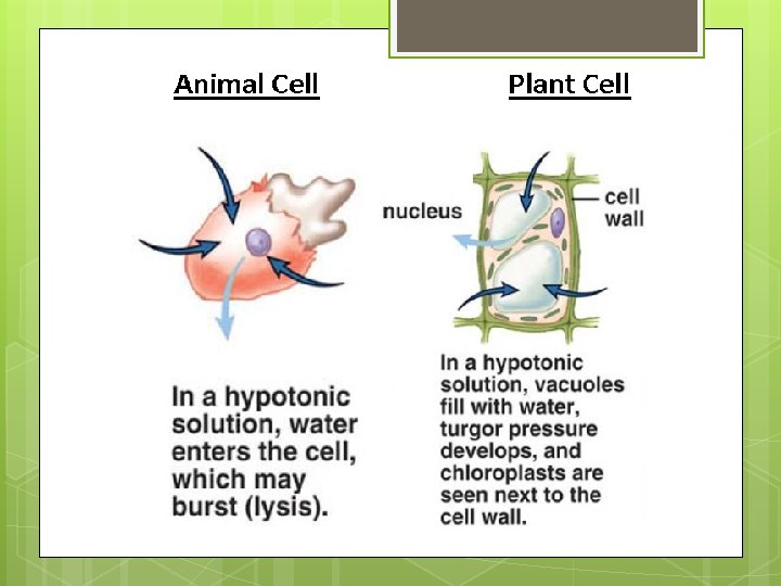
V. Passive Transport: Osmosis B. Hypotonic solution ___________ = a solution in which the concentration of solute is LOWE _____ than R the concentration of solutes inside the cell.

What happens to cells when placed in an hypotonic solution? Animal cell o _______ - water will move through plasma membrane into the cell. This causes the cell to swell and internal pressure increase. o Cell _______ lyses (bursts) Drinking too much water

What happens to cells when placed in an hypotonic solution? o _____ Plant cell - normal The vacuole & cytoplasm increase in volume o The cell membrane is pushed harder against the cell wall causing it to stretch a little o The plant tissue becomes stiffer (turgid) o Why they spray fruit in the grocery store.

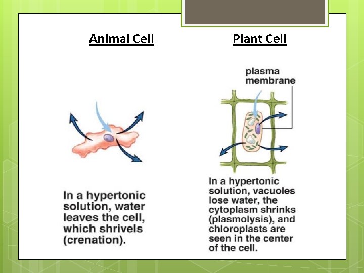
V. Passive Transport: Osmosis B. Hypertonic solution = ___________ a solution in which the concentration of dissolved substances is _____ HIGHE than the R concentration inside the cell.

What happens to when placed in an hypertonic solution? cells Animal cell o _______ - will _____ shrivel because decreased turgor pressure Drinking salt water on a deserted island of

What happens to when placed in an hypertonic solution? cells Plant cell o _____ - will lose water from _____ vacuole and a decrease in turgor pressure will occur; so it is plasmolyzed ________ Plants die when salt is spread on icy roads.

What happens to when placed in an hypertonic solution? Turgor o _________ = internal pressure of a cell due to water Pressure held there by osmotic pressure Plasmolysis = the o _______ loss of turgor pressure causing the plasma membrane to pull away from the cell wall o causes the plant to wilt cells

Summary of Cell Behavior in Different Environments:

VI. Active Transport o Movement of molecules from an area of ______LOW to an area of _______ concentration (opposite of passive HIGH transport) o REQUIRES cellular _________! energy o Moves large, complex molecules such as proteins across the cell membrane

VI. Active Transport o Large molecules, food, or fluid droplets are packaged in membrane-bound sacs called _____. vesicles

2 Types of Active Transport: Endocytosis = process by _______ which a cell surrounds and takes in material from its environment. 1. • Used by amoeba to feed and white blood cells to kill bacteria.

Endocytosis Outside of Cell

2 Types of Active Transport: 2. _______ Exocytosis = expels materials out of the cell reverse of endocytosis wastes, mucus & o Used to remove __________ cell products ________ o Proteins made by ribosomes in a cell are packaged into transport vesicles by the Golgi Apparatus o Transport vesicles fuse with the cell membrane and then the proteins are secreted out of the cell (ex. Insulin)

Exocytosis Outside of Cell
- Slides: 135