Cells A typical cell is only about 0

Cells • A typical cell is only about 0. 1 mm in diameter. The period on your paper is 13 times as large as our smallest cell and 2/3 rds the size of an ovum. • 1665 Robert Hooke inspected thin slices of cork and said they consisted of millions of small irregular units he called a CELL. (similar to a room or cell of a monastery or prison. ) • Cells are the building block of all plants and animals. • Two types: Eukaryotic – Cells that have a membrane bound nucleus, human cells Prokaryotic – Cells don’t have a membrane bound nucleus like bacteria.

The Cellular Level of Organization • Basic, living, structural and functional unit of the body – Two classes of cells 1) Sex cells or gametes 2) Somatic cells – Sex cells: Sperm & oocytes ( the fusion of sperm and an oocyte at fertilization is the first step in creation of new human (zygote). – Somatic cells: Include all other cells in the body. • Cytology = study of cellular structure • Cell physiology = study of cellular function

Cells • Cell is surrounded by Extracellular Fluid, also known as Interstitial Fluid. • Cell membrane (Plasma membrane) separates cell contents (cytoplasm) from the extracellular fluid • It has nucleus (genetic materials of the cell; DNA) and the cytoplasm inside the plasma membrane • The cytoplasm has the i) cytosol – fluid part ii) organelles (tiny organs)

Mitochondrion Microbody Nuclear pore complex Nuclear envelope Nucleus Chromatin Nucleolus Rough ER Ribosome Centrioles Ribosome Lysosome Endoplasmic reticulum Smooth ER Microtubules Microfilaments Vesicle Plasma membrane Golgi complex Cytosol

Plasma membrane • Isolates cell from outside environment • Regulates movement of molecules in and out of cell. • Permeable to small molecules and non-polar molecules; impermeable to polar molecules and ions.

Integral proteins Carbohydrate groups Outside cell Integral proteins Glycolipid Plasma membrane Gate Integral protein (gated channel protein) Integral Cholesterol protein (transport Glycoprotein) Peripheral proteins Cytosol Peripheral Microfilament protein of cytoskeleton (linking microtubule Peripheral to membrane) protein
 Phosphate group Glycerol Nonpolar end (hydrophobic)")
A. Phospholipid molecule Polar alcohol Polar end (hydrophilic) Phosphate group Glycerol Nonpolar end (hydrophobic) Hydrophobic tail

Functions of Membranes: Passive and Active Transport • Transport is the controlled movement of specific ions and molecules across a membrane by membrane proteins • Passive transport moves ions and molecules with the concentration gradient – from the side with a higher concentration to the side with a lower concentration • Active transport moves ions or molecules against the concentration gradient; from lower to higher concentration – uses energy directly or indirectly from ATP
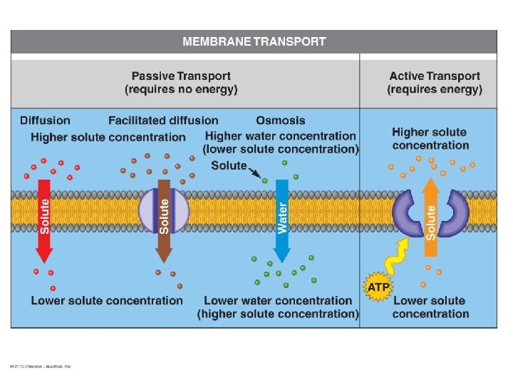

Types of passive transport • • • Simple Diffusion Facilitated Diffusion Osmosis Filtration Dialysis

Principles of Diffusion • Random mixing of particles in a solution as a result of the particle’s kinetic energy – more molecules move away from an area of high concentration to an area of low concentration • When the molecules are evenly distributed, equilibrium has been reached • • • Crystal of dye placed in a cylinder of water Net diffusion from the higher dye concentration to the region of lower dye Equilibrium has been reached in the far right cylinder

Osmosis • Diffusion of free water across membrane • Moves from low concentration of salts to high concentration – think slugs • Osmosis controlled to maintain cell size and shape • Net movement of water through a selectively permeable membrane from an area of high water concentration to an area of lower water concentration
 –")
Effects of Tonicity on Cell Membranes • Isotonic solution (same as the cell) – water concentration the same inside & outside of cell results in no net movement of water across cell membrane • Hypotonic solution (lower than that of the cell) – Red Blood Cells, in a hypotonic solution will swell and eventually burst; the phenomenon called hemolysis • Hypertonic solution (higher than that of the cell) – Red Blood Cells, in a hypertonic solution will shrink; the phenomenon known as crenation.

Facilitated Diffusion • Substance binds to specific transporter protein • Transporter protein conformational change moves substance across cell membrane • Facilitated diffusion occurs down the concentration gradient only – if no concentration difference exists, no net movement across membrane occurs • Rate of movement depends upon – steepness of concentration gradient – number of transporter proteins (transport maximum)

Facilitated Diffusion of Glucose

Active Transport • Movement of polar or charged substances against their concentration gradient – energy-requiring process • energy from hydrolysis of ATP (primary active transport) • energy stored in an ionic concentration gradient (secondary active transport) • Na+, K+, H+, Ca+2, and Cl-, amino acids and monosaccharides

Primary Active Transport • Transporter protein called a pump – works against concentration gradient – requires 40% of cellular ATP • Na+/K+ ATPase pump – all cells have 1000 s of them – maintains low concentration of Na+ and a high concentration of K+ in the cytosol – operates continually

Vesicular Transport • Endocytosis = bringing something into cell – phagocytosis = cell eating by macrophages & WBCs • particle binds to receptor protein • whole bacteria or viruses are engulfed & later digested – pinocytosis = cell drinking • Exocytosis = release something from cell • Vesicles form inside cell, fuse to cell membrane • Release their contents – digestive enzymes, hormones, neurotransmitters or waste products • Receptor mediated Endocytosis = target molecules bind to the receptor protein and then taken in.

Vesicular transport

Organelles • Ribosomes • Endoplasmic Reticulum – Rough ER – Smooth ER • • Golgi Bodies Lysosomes Peroxisomes Mitochondria Centrosome Cytoskeleton Cilia and Flagella

Ribosomes • Packages of Ribosomal RNA & protein. Site of protein synthesis. “Protein factories”. They are molecular machines that translate the code to make proteins. ( structural and functional) • Free ribosomes are loose in cytosol – synthesize proteins found inside the cell • Membrane-bound ribosomes – attached to endoplasmic reticulum or nuclear membrane (rough ER) – synthesize proteins needed for plasma membrane or for export

Endoplasmic Reticulum and Golgi bodies • Microscopic circulatory system of cell • Rough ER – continuous with nuclear envelope & covered with attached ribosomes – synthesizes, processes & packages proteins for export to their next destination, the golgi bodies or apparatus (Camillo Golgi). (Fed. Ex) • Smooth ER – No attached ribosomes. Well developed in liver cells (hepatocytes) – synthesizes phospholipids, cholesterol, steroids for maintenance of plasma membrane. – detoxifies harmful substances (alcohol)

Smooth & Rough Endoplasmic Reticulum Rough ER is covered with fixed ribosomes.

Packaging by Golgi Complex • Proteins pass from rough ER to golgi complex in transport vesicles • Finished proteins exit golgi as secretory (exocytosis), membrane renewal or storage vesicle (lysosome- digestive enzymes)

Lysosomes • Membranous vesicles – formed in Golgi complex – filled with digestive enzymes • Functions – digest foreign substances and release usable substances such as sugars and amino acids. – autophagy(autophagosome forms) • recycles own organelles. Clean up and recycling. – autolysis • lysosomal damage after death

Lysosomes • Membrane bound enzymes • Digest food from extracellular fluid or damaged organelles

Peroxisomes • Membranous vesicles – smaller than lysosomes – form by division of preexisting peroxisomes • Function – part of normal metabolic breakdown of amino acids and fatty acids – oxidizes toxic substances such as alcohol and formaldehyde – Found in kidney and liver – Contain enzymes, which are important in detoxification reactions involving hydrogen peroxide.

Mitochondria • Double membrane organelle – central cavity known as matrix – inner membrane folds known as cristae • surface area for chemical reactions of cellular respiration • Function – generation of ATP – powerhouse of cell • Mitochondria self-replicate – increases with need for ATP

The Cytoskeleton • The cytoskeleton is an interconnected system of protein fibers and tubes that extends throughout the cytoplasm • The cytoskeleton maintains a cell’s characteristic shape and internal organization, and functions in movement • The cytoskeleton of animal cells contains microtubules, intermediate filaments, and microfilaments
 – Interact with myosin to")
The Cytoskeleton Filaments • Microfilaments – thinnest filaments (actin) – Interact with myosin to produce movement of a cell • Intermediate filaments – Structurally reinforce cell – Keratin of superficial layers of skin make this layer strong and resistant to stretch – Microtubules – large cylindrical structures (composed of tubulin) – Acts like a monorail system. – Form the spindle apparatus which distributes duplicated chromosomes to opposite ends of the dividing cell. – Form the structural components of cilia & centrioles

Flagella and Cilia • Flagella and cilia are elongated, motile structures that extend from the cell surface – cilia are shorter than flagella and occur in greater numbers • Movements of a flagellum propel a cell through a watery medium – the tail of a sperm cell is a flagellum • Cilia move fluids over the cell surface – cilia line the air passages of the lungs and sweep out mucus containing bacteria, dust particles, and other contaminants

Flagellum and Cilia • • Used for locomotion, moving particles Made of protein filaments Cilia – many “hairs” Flagellum – Usually a single undulating “tail”

Nucleus • The control center of the cell. • Contains the genetic material of the cell DNA • DNA of a cell codes for specific protein produced by the cell • Proteins have specific structure which gives them specific function. • Proteins do the work of the cell.

Nucleus • Large organelle with double membrane nuclear envelope – outer membrane continuous with rough ER – The control center – Skeletal Muscle cells have many nuclei but RBC’s have none. • Nucleolus – spherical, dark bodies within the nucleus (no membrane) – site of ribosome assembly. • Normal Human nucleus has 46 chromosomes
- Slides: 34