CellMediated Effector Responses Chapter 14 Cell Mediated Immune


")


")


 • Cells Capable of Cytotoxicity Express Fc Receptors")
- Slides: 27
Cell-Mediated Effector Responses Chapter 14
Cell Mediated Immune Responses • Primary Function Of Cell Mediated Response – Eliminate Intracellular Pathogens – Eliminate Tumor Cells • Both Ag Specific And Non-specific cells Are Involved – Ag Specific: CD 8+ Cells (TC) And TH (DTH) – Non-specific: M , Neutrophils, NK • Both Specific And Non-specific Require Cytokines • Humoral And Cell Mediated Do Collaborate – Ex. M Use Abs As Receptors To Recognize Target Cells
Importance Of Cell Mediated Immunity • Di. George Syndrome Proves The Importance – No Thymus, No T-cell Mediated Immunity – Extracellular Infections Are Effectively Addressed – Intracellular Infections Are NOT (viruses, intracellular bacteria) • Cell Mediated Immunity Can Be Divided Into 2 Major Categories – Effectors lyse target • 2 groups of cells: CTLs (specific) and NK, M (non-specific) – Effectors which are CD 4+ and mediate DTH
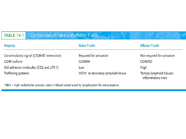
What Are Effector T Cells • 3 Types: CD 4+ (TH 1 and TH 2) and CD 8 (CTLs) • Differences Between Naïve and Effectors – Easy to activate – Increased expression of adhesion molecules – Production of soluble and membrane bound effectors molecules • Easy To Activate – Naïve require co-stimulation, Effectors minimal Co-stimulation – Reason for this is expression of CD 45 Isoforms • CD 45 RO (expressed on effectors), CD 45 RA (naïve) – RO associates much better to CD 4/CD 8, TCR complex (dephosphorylates Lck and Fyn) – Association results in more efficient signaling • Cell Adhesion Molecules – CD 2 and LFA-1 2 -4 fold higher on Effectors – These molecules adhere to LFA-3 and ICAMs on APCs
What Are Effector T Cells • Membrane Bound Molecules – Ex. FASL (CD 8+) • Soluble Molecules – CTLs secrete cytotoxins • perforins and granzymes – Also secret IFN- and TNF-
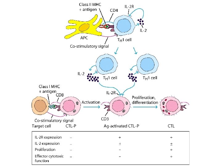
Cytotoxic T Cells • CTLs Recognize Cells That Have Been infected – Virus – Transformed to tumor • CTL Activation Is Divided Into 2 Phases – Activation and differentiation of naïve CTL – Effector recognizes Class I MHC/peptide and destroys target • Naïve CTLs Cannot Kill – Referred to as CTL-Ps (precursors) – 3 signals needed for activation • Ag specific signal thru TCR/MHC I+Ag • Co-stimulatory signal CD 28(CTL)/B 7 (APC) • IL-2 signaling inducing proliferation (CTL-P do not express IL-2 R) – IL-2 is provided by TH 1 or CTL-P itself – IL-2 R is expressed only after activation
Cytotoxic T Cells • TH 1 And CTL Collaborate To Induce Effector CTL – IL-2 seems to be crucial (knock out data) – Lack of IL-2 R In CTL-P Ensures Ag Specificity • Upon Clearance Of Antigen CTLs Undergo Apoptosis • TH 1 Induce Up-regulation Of Co-stimulatory Molecules On APCs Enhancing APC/CTL Costimulation
Memory CTL-P vs Naïve CTL-P
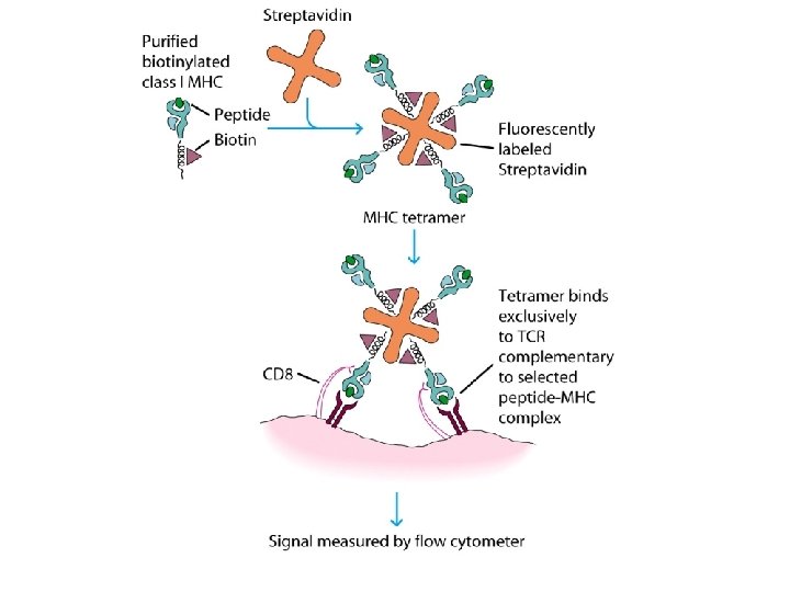
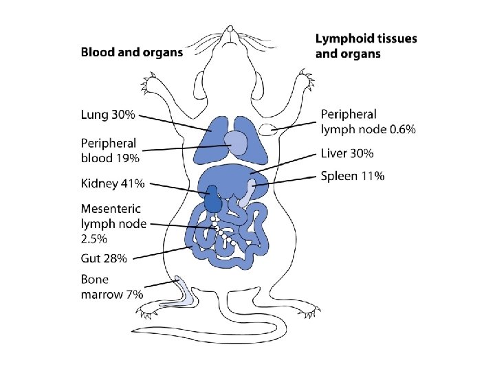
Tracking CTLs With Tetramers • MHC Tetramers Are Synthetic – 4 MHC I molecules bound together by Sv – Sv is conjugated with fluorescence – Tetramer is bound to peptide • Tetramer Recognizes CTLs With A TCR Capable Of Binding Peptide/Tetramer • Flow Cytometry Is Used To Detect Fluorescent Cells • Very Sensitive Technique, 0. 1% • Comparison Between Pathogen Exposed Animals and Naïve • Large Populations Of Ag Specific CTLs Found In VSV Infected Mice In Liver, Kidney, Lung and Gut (low levels in peripheral lymph nodes!!)
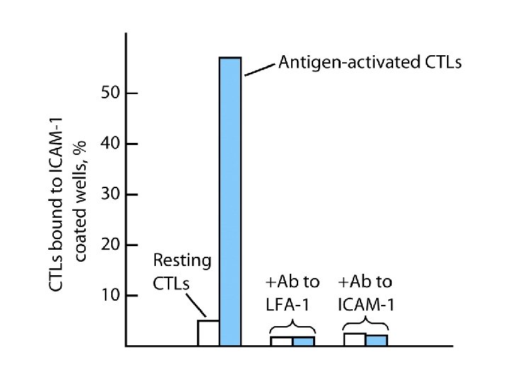
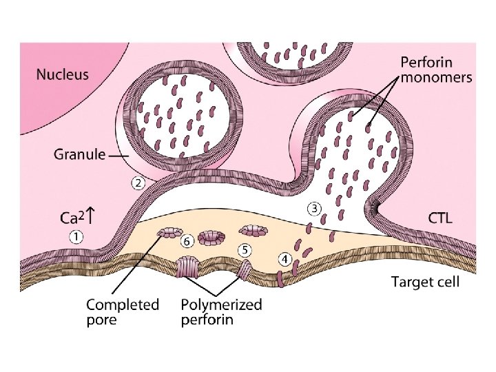
How CTLs Kill • 4 Phases In CTL Killing – Conjugate formation • LFA-1 (CTL) binds ICAMs (Target) – LFA-1 changes to high avidity if Ag Is Recognized – Activated LFA-1 persists for 5 -10 mins – Membrane attack • Requires Ca 2+ and energy – Granules release Perforins (65 k. Da) and Granzymes (serine proteases) at the junctional space – Perforins polymerize forming cylindrical pores (5 -20 nm), Ca 2+ is needed – Granzymes enter target cell – Granzyme B can enter thru mannose-6 -phosphate receptor in a vesicle – DNA fragmentation – CTL dissociation – Target cell destruction • Apoptotic death within a few hours
CTL Making Contact With Tumor Cell (small cell)
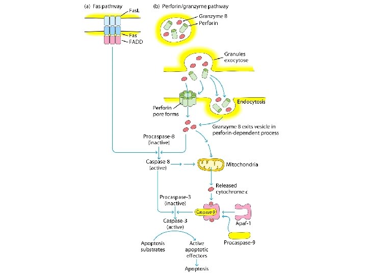
Fas. L Mediated Cytotoxicity • Some CTLs Lack Granzyme And Perforin • They Kill Using Fas. L-Fas Interaction – Fas. L is found on CTLs – Fas is found on target cell – Fas. L-Fas interaction induces apoptosis • 2 Mechanisms Are Responsible For CTL Induced Apoptosis – Fas. L-Fas (FADD Activation leading to pro-caspase 8 activation) – Perforin and granzyme – During apoptosis caspases (cysteine proteases that cleave aspartic acid) are activated – Family of more than 12 caspases exist – Activation of caspases results in orderly destruction of target cell
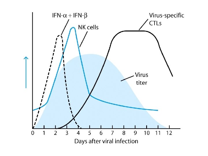
Natural Killer Cells • NK Make Up 5 -10% Of Circulating Lymphocytes – Major Producers of IFN – Thru IFN they influence innate immunity (M ) – They also influence adaptive, favor TH 1 – Eliminate viruses and tumor cells • Early Responders To Viral Infections – IFN and IFN produced by p. DCs Stimulates NK activity – IFN production induces M To Make IL-12 – IL-12 Results In More IFN Pushing Towards TH 1 – TH 1 Thru IL-2 Induces CTL activation
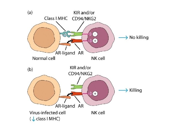
Natural Killer Cells • NK Eliminate Target Cells Same Way CTLs – Thru perforin/granzyme and Fas. L/Fas • However They Are Different From CTLs – No Ag Specific TCR – No CD 3 – No MHC restriction – No memory, same intensity regardless of repeated exposure • How Do They Recognize The Target?
Target Recognition • Balance Between Activating and Inhibiting Molecules Allows NK Cells To Differentiate Normal From Altered • Still Not Clear What The Activating Receptors Are – C-type lectins Are Candidates • • NKR-P 1 c CD 2 (Receptor for adhesion molecule LFA-3) CD 16 (Fc RIII, Involved In Antibody Mediated Recognition) NKp 30, NKp 44 and NKp 46 • Inhibitory Receptors – – – MHC Molecules CD 94/NKG 2 Recognize HLA-E If HLA-E is present –ve signal, no killing No HLA-E (during viral infection), no –ve signal, killing KIRs recognize specific MHC molecules, -ve signal, no killing
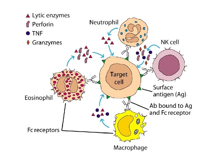
Antibody Dependent Cell Mediated Cytotoxicity (ADCC) • Cells Capable of Cytotoxicity Express Fc Receptors • Antibody Binds Target Cell, Cytotoxic Cells Bind Fc Portion Of Ab • Antibody Provides The Specificity • Examples Of Cells Capable Of ADCC – M , NK, Neutrophils, eosinophils • Killing Of Target Is Accomplished – Thru perforin, granzyme (NK, Eosinophils) – TNF (M , NK) – Lytic enzymes (M , Neutrophils, Eosinophils, NK)