Cell Wall Chemical Composition Structure Growth Dr Amit


- Slides: 12
Cell Wall, Chemical Composition, Structure & Growth Dr. Amit Saha Assistant Prof. in Botany(Stage III) Surendranath College, Kolkata.
Introduction • A strong, rigid, extra protoplasmic layer in plant cells whose growth is directed from within the cell is called cell wall. • Most plant cells except reproductive cells are provided with a rigid cell wall. • It provides a skeletal support to the whole plant and act as a barrier against injury and infection.
Chemical Constituents • Cellulose- the most common compound in the cell wall. The common carbohydrate apart from it are non cellulosic polysaccharides, organic compounds and mineral substances are also present. • It is hydrophilic crystalline compound. It is a polysaccharide hexan and closely related to starch. The molecules of cellulose are chain like structure with 1000 or more of the glucose residues connected together by oxygen bridges with beta 1, 4 glycosidic bond.
• Hemicellulose- these are cellulose molecules. They are not made up of glucose molecules but of other sugars. e. g. xylans, mannans, galactans, glucans, etc. • Pectic substances- they occur in 3 forms- protopectin, pectin and pectic acid. Pectic substances belong to polyuronides (polymer of uronic acids). Pectic compounds are amorphous, colloidal, plastic and highly hydrophilic. It is associated with cellulose in the primary cell wall. • Gums & Mucilage- these are compound carbohydrate related to pectic compounds and have the property of swelling in water. • Gums exude as a result of physiological or pathological disturbances that induce a breakdown of walls and cell contents. • Mucilages occur in gelatinous and mucilaginous type of cell walls.
• Mineral substances- Silica, calcium carbonate, and organic compounds like tannins, resins, fatty substances, volatile oils, acids, crystalline pigments etc. • Lignin- It is found in all 3 wall layers. • It is an organic compound containing high carbon contents, complex, cross linked polymer comprising of phenylpropane units, chemically inert. • Cutin & suberin – They are fatty substances which do not melt and show solubility in fat solvents. They are closely related, highly polymerized compounds consisting of fatty acids. Waxes- It is associated with the above two and are present in the surface of cuticle in various forms. It is melt able and soluble in fat solvents. • Water- Most imp. and variable compound. Part of it occurs in micro capillaries and relatively free, the rest is associated with hydrophilic substances.
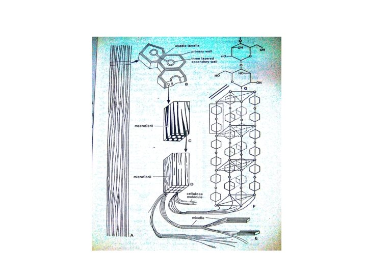
Gross structure • Cell wall is complex and consists of 3 layers • 1. Middle lamella- it is amorphous, colloidal and optically inactive(isotropic). Composed of pectic compounds(calcium & magnesium pectate) 2. Primary Cell wall- it is the first true cell wall to be formed by the developing cell. It is formed on either side of middle lamella. It contains cellulose, hemicelluloses and pectins, may be lignified. • It grows both in surface and thickness and such changes are reversible.
3. Secondary cell wall- it is formed on the inner surface of the primary wall and is laid down as a supplementary wall after the primary wall when it ceases to increase in surface area. Consists of cellulose, hemicellulose, lignin and other substances. In tracheids & fibres secondary walls are 3 layered(outer, middle-thickest and inner layer/tertiary wall) Secondary wall is present in cells which are non living at maturity e. g. sclerenchyma fibers, tracheary elements
Ultrastructure • The cell wall under electron microscope shows 2 parts • 1. Matrix- non cellulosic part • 2. Fibrils- embedded in matrix and made up of cellulose. • The largest fibril seen by a light microscope is called macro fibril and smaller fibrils under electron microscope are called micro fibrils. • Smaller fibrils visualized as subunits of micro fibrils are called micelles which in turn are further divided to form cellulose molecules.
Growth of cell wall • The cell wall increases both in size and thickness. • The growth in thickness is due to the formation of new layers by protoplasm on the inner surface. This method is called growth by apposition(Strasburger). • On both sides of middle lamella primary walls develop. The secondary walls are developed later outside the primary walls.
• According to Nageli the growth of cell wall takes place by intussusception. • In this method some gaps are formed in the cell wall due to force of stretching. These gaps are filled by new particles of cell wall formed by the cytoplasm. • The cytoplasm of the 2 neighboring cells is interconnected through fine protoplasmic threads called plasmodesmata.
Suggested References • Plant Anatomy by B. P. Pandey • Plant Anatomy by Pijush Roy • A Textbook of Botany Vol II by Bhattacharya, Hait, Ghosh • Studies in Botany Vol I by Mitra, Choudhury. • Plant Anatomy by Fahn