Cell to Cell Communication AP Biology Chapter 11

Cell to Cell Communication AP Biology Chapter 11

Objectives Explain why and how cells communicate. � Explain the common features shared among cell communication processes. � Compare the purpose cell communication in unicellular and multicellular organisms. � Describe the major features of signal transduction pathways in cells. � Connect cellular signaling pathways to specific examples. � Discuss evolutionary/adaptive considerations of cellular signaling pathways. �

Introduction � Cell-to-cell communication is essential for multicellular organisms and many unicellular organisms. • Cells must communicate to coordinate their activities.

Introduction � Biologists have discovered some universal mechanisms of cellular regulation, involving the same small set of cell-signaling mechanisms. � Cells may receive a variety of signals, chemical signals, electromagnetic signals, and mechanical signals. • Signal-transduction pathway: The process by which a signal on a cell’s surface is converted into a specific cellular response.

Talking to cells, both near and far… � Multicellular organisms can release signaling molecules that target other cells. � Local Signaling: • Some transmitting cells release local regulators that influence cells in local vicinity. • In synaptic signaling, a nerve cell produces a neurotransmitter that diffuses to a single cell that is almost touching the sender. the

Talking to cells, both near and far… � Long-distance Signaling: • Plants and animals use hormones to signal at greater distances. • Cells may communicate by direct contact.

How do Cells Communicate? � The process must involve three stages. • Reception: a chemical signal binds to a cellular protein, typically at the cell’s surface. • Transduction: binding leads to a change in the receptor that triggers a series of changes along a signal-transduction pathway. • Response: the transduced signal triggers a specific cellular activity.

Signal molecules and Receptor Proteins � A cell targeted by a particular chemical signal has a receptor protein that recognizes the signal molecule. • Recognition occurs when the signal binds to a specific site on the receptor because it is complementary in shape. � When ligands (small molecules that bind specifically to a larger molecule) attach to the receptor protein, the receptor typically undergoes a change in shape. • This may activate the receptor so that it can interact with other molecules. • For other receptors this leads to the collection of receptors.

Signal Molecules � Most signal molecules are water-soluble and too large to pass through the plasma membrane. � They influence cell activities by binding to receptor proteins on the plasma membrane. • Binding leads to change in the shape or the receptor or to aggregation of receptors. • These trigger changes in the intracellular environment. � Three major types of receptors: • G-protein-linked receptors • Tyrosine-kinase receptors • Ion-channel receptors.

G-Protein-Linked Receptor � Consists of a receptor protein associated with a G-protein on the cytoplasmic side. • The receptor consists of seven alpha helices spanning the membrane. • Effective signal molecules include: �yeast mating factors �epinephrine, other hormones �neurotransmitters
 is")
�The G protein acts as an on-off switch. • If GDP (guanine diphosphate) is bound, the G protein is inactive. • If GTP (guaninetriphosphate) is bound, the G protein is active.

The G-protein system cycles between on and off. When a G-protein-linked receptor is activated by binding with an extracellular signal molecule, the receptor binds to an inactive G protein in membrane. � This leads the G protein to substitute GTP for GDP. � The G protein then binds with another membrane protein, often an enzyme, altering its activity and leading to a cellular response. �

� G-protein receptor systems are extremely widespread and diverse in their functions. • In addition to functions already mentioned, they play an important role during embryonic development and sensory systems. � Similarities among G proteins and G-proteinlinked receptors suggest that this signaling system evolved very early. � Several human diseases are the results of activities, including bacterial infections (e. g. cholera and botulism), that interfere with Gprotein function.

Tyrosine-kinase Receptors �Effective when the cell needs to regulate and coordinate a variety of activities and trigger several signal pathways at once. �A tyrosine-kinase is an enzyme that transfers phosphate groups from ATP to the amino acid tyrosine on a protein.

Tyrosine-kinase Receptors � Individual tyrosinekinase receptors consists of several parts: • an extracellular signal- binding site, • a single alpha helix spanning the membrane, and • an intracellular tail with several tyrosines.

Tyrosine-kinase Receptors � When ligands bind to both receptor polypeptides, the polypeptides bind, forming a dimer. � This activates the tyrosine-kinase sections of both. � These add phosphates to the tyrosine tails of the other polypeptide.

� The fully-activated receptor proteins initiate a variety of specific relay proteins that bind to specific phosphorylated tyrosine molecules. • One tyrosine-kinase receptor dimer may activate ten or more different intracellular proteins simultaneously. � These activated relay proteins trigger many different transduction pathways and responses.

Ligand-gated Ion Channels � Protein pores that open or close in response to a chemical signal. • This allows or blocks ion flow, such as Na+ or Ca 2+. • Binding by a ligand to the extracellular side changes the protein’s shape and opens the channel. • Ion flow changes the concentration inside the cell. • When the ligand dissociates, the channel closes. • Very important in the nervous system

Intracellular Receptors � Other signal receptors are dissolved in the cytosol or nucleus of target cells. � The signals pass through the plasma membrane. � These chemical messengers include the hydrophobic steroid and thyroid hormones of animals. � Also in this group is nitric oxide (NO), a gas whose small size allows it to slide between membrane phospholipids.

Testosterone � Testosterone, like other hormones, travels through the blood and enters cells throughout the body. � In the cytosol, they bind activate receptor proteins. � These activated proteins enter the nucleus and turn on genes that control male sex characteristics.

Turning Genes On �These activated proteins act as transcription factors. • Transcription factors control which genes are turned on - that is, which genes are transcribed into messenger RNA (m. RNA). �The m. RNA molecules leave the nucleus and carry information that directs the synthesis (translation) of specific proteins at the ribosome.

Transduction � The transduction stage of signaling is usually a multistep pathway. � These pathways often greatly amplify the signal. • If some molecules in a pathway transmit a signal to multiple molecules of the next component, the result can be large numbers of activated molecules at the end of the pathway. � A small number of signal molecules can produce a large cellular response. � Also, multistep pathways provide more opportunities for coordination and regulation than do simpler systems.

Signal Transduction Pathways � Signal transduction pathways act like falling dominoes. • The signal-activated receptor activates another protein, which activates another and so on, until the protein that produces the final cellular response is activated. � The original signal molecule is not passed along the pathway, it may not even enter the cell. • Its information is passed on. • At each step the signal is transduced into a different form, often by a conformational change in a protein.
 � The phosphorylation of proteins by a specific enzyme (protein")
Phosphorylation (adding on Phosphates) � The phosphorylation of proteins by a specific enzyme (protein kinase) is a mechanism for regulating protein activity. • Most protein kinases act on other substrate proteins, unlike the tyrosine kinases that act on themselves. � � � Most phosphorylation occurs at either serine or threonine amino acids in the substrate protein. Many of the relay molecules in a signal-transduction pathway are protein kinases that lead to a “phosphorylation cascade”. Each protein phosphorylation leads to a shape change because of the interaction between the phosphate group and charged or polar amino acids. http: //images-mediawikisites. thefullwiki. org/07/9/4/0/569439340965241. jpg
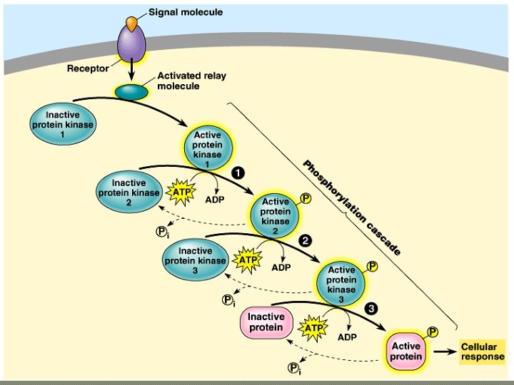

Phosphorylation � Phosphorylation of a protein typically converts it from an inactive form to an active form. • The reverse (inactivation) is possible too for some proteins. � A single cell may have hundreds of different protein kinases, each specific for a different substrate protein. • Fully 1% of our genes may code for protein kinases. � Abnormal activity of protein kinases can cause abnormal cell growth and contribute to the development of cancer.

Protein Phosphatase � The responsibility for turning off a signal-transduction pathway belongs to protein phosphatases. • These enzymes rapidly remove phosphate groups from proteins. • The activity of a protein regulated by phosphorylation depends on the balance of active kinase molecules and active phosphatase molecules. � When an extracellular signal molecule is absent, active phosphatase molecules predominate, and the signaling pathway and cellular response are shut down. Inactive Form Active Form http: //kinasephos. mbc. nctu. edu. tw/image/phosphorylation. jpg

Second Messengers � Many signaling pathways involve small, nonprotein, water-soluble molecules or ions, called second messengers. • These molecules rapidly diffuse throughout the cell. � Second messengers participate in pathways initiated by both G-protein-linked receptors and tyrosine-kinase receptors. • Two of the most important are cyclic AMP and Ca 2+.

Pathway involving c. AMP as a secondary messenger. Pathway using Ca 2+ as a secondary messenger.

The Response �Ultimately, a signal-transduction pathway leads to the regulation of one or more cellular activities. • This may be a change in an ion channel or a change in cell metabolism. • For example, epinephrine helps regulate cellular energy metabolism by activating enzymes that catalyze the breakdown of glycogen.

Regulation of Transcription � Some signaling pathways do not regulate the activity of enzymes but the synthesis of enzymes or other proteins. � Activated receptors may act as transcription factors that turn specific genes on or off in the nucleus.

Benefits of Multiple Steps � Signaling pathways with multiple steps have two benefits. • They amplify the response to a signal. • They contribute to the specificity of the response. � At each catalytic step in a cascade, the number of activated products is much greater than in the preceding step. • A small number of epinephrine molecules can lead to the release of hundreds of millions of glucose molecules.

Specificity of Cell Signaling �Various types of cells may receive the same signal but produce very different responses. �These differences result from a basic observation: • Different kinds of cells have different collections of proteins.

�The response of a particular cell to a signal depends on its particular collection of receptor proteins, relay proteins, and proteins needed to carry out the response.

Scaffolding � Rather than relying on diffusion of large relay molecules like proteins, many signal pathways are linked together physically by scaffolding proteins. • Scaffolding proteins may themselves be relay proteins to which several other relay proteins attach. • This hardwiring enhances the speed and accuracy of signal transfer between cells.

Relay Proteins � The importance of relay proteins that serve as branch or intersection points is underscored when these proteins are defective or missing. • The inherited disorder, Wiskott-Aldrich syndrome (WAS), is due to the absence of a single relay protein. • It leads to abnormal bleeding, eczema, and a predisposition to infections and leukemia. • The WAS protein interacts with the microfilaments of the cytoskeleton and several signaling pathways, including those that regulate immune cell proliferation. • When the WAS protein is absent, the cytoskeleton is not properly organized and signaling pathways are disrupted.

Termination of the Signal � As important as activating mechanisms are inactivating mechanisms. • For a cell to remain alert and capable of responding to incoming signals, each molecular change in its signaling pathways must last only a short time. • If signaling pathway components become locked into one state, the proper function of the cell can be disrupted. • Binding of signal molecules to receptors must be reversible, allowing the receptors to return to their inactive state when the signal is released. • Similarly, activated signals (c. AMP and phosphorylated proteins) must be inactivated by appropriate enzymes to prepare the cell for a fresh signal.
- Slides: 37