Cell Theory The following theory was proposed following


![Intravenous therapy: Normal saline = 0. 9% Na. Cl Contains 154 mmol/L [Na+] and](https://slidetodoc.com/presentation_image/789948f3ff05bcf8c49fef06cc2c0a97/image-9.jpg "Intravenous therapy: Normal saline = 0. 9% Na. Cl Contains 154 mmol/L [Na+] and")


. RNA contains four nitrogenous bases (ACGU).")


")


.")


 must")


")


 • Specific hormonereceptor interactions. • Transmembrane signalling. •")

. Prednisolone")


- Slides: 59
Cell Theory The following theory was proposed following the development of microscopy in the 16 th century – when cells could be directly visualized. • All living things are composed of one or more cells. • Cells are the basic units of structure and function in living things. • New cells are produced from existing cells. (Hooke, Leeuwenhoek, Schleiden, Schwann, Virchow).
Why do you, potential doctors, need to know so much about cells? • Because disease is generally about either cell damage or cell death. • Because, the same injurious stimuli that kill the cell, also kill the organism. • Save the cell, the organism.
Injurious Stimuli: The cell, like the body, can adapt to and survive small injurious stimuli. However if the extent of the stimulus is too great, then cell death occurs. The term for such cell death is “necrosis. ” • • Examples: Physical - Temperature/Pressure/Radiation/Electric shock/Mechanical Trauma Chemical - Small molecules/poisons/drugs etc. Infectious - Viruses, Bacteria, Fungi Immunological - Autoimmune attack, Allergic response
Cell Structure - Key points • Cells have membranes. • Cells are compartmentalized. • The compartments represent different functions = organelles. • For the cell to function and survive, this compartmentation must be maintained.
The cell
Organelle Major function Mitochondrion Generation of cell’s energy currency. Ribosome Protein synthesis. Rough Endoplasmic Reticulum Protein synthesis, processing and transport in the cell. Smooth Endoplasmic Reticulum Range of metabolic processes lipid, carbohydrate and detoxification. Golgi Body Cell’s post office - packages and labels molecules for distribution throughout the cell. Nucleus Store and handling of genetic material; controlling cell function. Lysosome Enzymatic digestion of intracellular or extracellular waste material.
Cells are charged • Cell membrane = Plasmamembrane • The unequal distribution of ions across the membrane causes the cell to be charged. The charge ranges from -9 to -100 m. V depending on the cell. This is termed the membrane potential.
• In mammalian spinal neurons this membrane potential is -70 m. V. Ion Na+ K+ Cl- Concentration (mmol/L H 20) Inside Outside Cell 15. 0 150. 0 5. 5 9. 0 125. 0
Intravenous therapy: Normal saline = 0. 9% Na. Cl Contains 154 mmol/L [Na+] and 154 mmol/L [Cl-] 5% dextrose Contains 278 mmol/L [Glucose]
Why you need to know some genetics: • To understand drug action and prescribe the right drugs. • To genetically counsel your patients. • To engage in medical research. • To understand disease.
Central Dogma DNA Transcription RNA Translation Protein
Structure of DNA and RNA are polymers of ribonucleotides. Nucleotide = base + sugar + phosphate. DNA contains deoxyribose, RNA contains ribose. DNA – deoxyribose nucleic acid. RNA – ribose nucleic acid. DNA = H RNA = OH
Simple sugars have the suffix …ose. e. g. Glucose Fructose Sucrose Maltose Mannose Ribose
Chemical Bonds I - Hydrogen Bond The hydrogen bond is a weak polar bond, between a relatively positive hydrogen atom ( +) and an electronegative atom ( -), (N, O or F). It is responsible for the solubility of sugars. It is responsible for the double helical structure of DNA and explains how DNA can be unzipped for replication or transcription. It accounts for the denaturation of proteins at high and low temperatures, or acidic or alkaline conditions. If the physical or chemical conditions do not allow the hydrogen bonds to maintain integrity, then the organism will die.
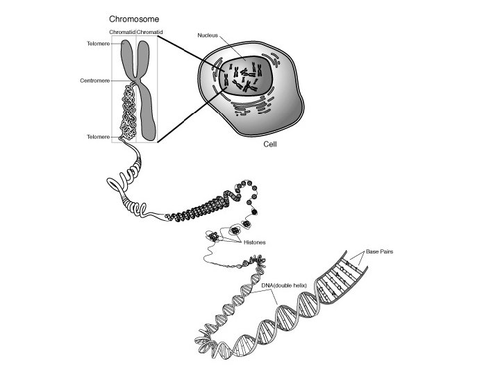
Structure of DNA contains four nitrogenous bases (ACGT). RNA contains four nitrogenous bases (ACGU). DNA RNA
DNA backbone
Chromosomes Men 22+XY Women 22+XX
Making more DNA; Copies are made by Replication DNA replication Both parental strands are replicated. Direction of replication is 5’-to-3’. Replication requires DNA polymerase.
Reading the instructions in the DNA code ; Transcription RNA polymerase DNA is unwound m. RNA
Transcription is controlled by specific DNA sequences Only ‘coding regions’ of the DNA are transcribed into RNA. Start and Stop sites are defined by specific DNA sequences. Protein factors regulate transcription. Signals and factors regulate: Transcription rates - how fast the m. RNA is made. Tissue specificity - which tissues make the m. RNA. Inducible expression - switching on m. RNA synthesis.
Translation is the conversion of m. RNA information into protein Transfer RNA (t. RNA) molecules act as the translators. The dictionary is the genetic code. The words of the code are based on 3 -letter codons. The code is degenerate.
Exercise - 2 minutes List all the protein functions that you can think of, for example: Enzymes/catalysts
Functions of Proteins; what can they do? • Enzymes – Kinases, phosphatases - Metabolic reactions • Structures – Muscles, cytoskeleton • Messengers - Signalling molecules, Hormones • Transport - Transporters • Carriers – Haemoglobin, Myoglobin • Make other proteins - Protein synthesis, • Make DNA, RNA • Make Lipids • Make Carbohydrate
How are proteins able carry out these functions? Because of their structure: Primary structure Amino acid sequence Secondary structure Primary folds Alpha helix Beta pleat/sheet Tertiary structure Secondary folds Globular protein (enzyme) Fibrous protein (collagen) Quaternary structure Multiple subunits Subunit structure (haemoglobin) ------
Protein synthesis Synthesis occurs on ribosomes (in the cytoplasm).
Examples of Antibiotics that interfere with protein synthesis in bacteria: Aminoglycosides - Gentamicin, Streptomycin Aminoglycosides antibiotics work by binding to the bacterial 30 S ribosomal subunit, inhibiting the translocation of the peptidyl-t. RNA from the A-site to the P-site and also causing misreading of m. RNA, leaving the bacterium unable to synthesize proteins vital to its growth. Used for serious systemic bacterial infections “septicaemia. ” Chloramphenicol This works by binding to the 50 S ribosomal subunit and inhibiting the peptidyl-transferase. Used for eye infections.
Membranes
Why do you need to know about membranes? • The cell membrane protects the cell from its external environment. • Proteins that mediate diffusion and active transport are housed in the membrane. The membrane is selectively permeable. • It is the letterbox through which messages are received from the outside world. Hence specific signals to grow, metabolize, die etc. , are transmitted through the membrane. • A range of receptors, and molecules that they interact with, are situated in the cell membrane. • The glycoproteins that mediate tissue rejection are based in the cell membrane. • Many of the cell’s organelles are compartmentalized by membranes.
How does the membrane achieve all its functions? • By having a range of proteins floating in a sea of lipids. The proteins and lipids can carry out a range of activities. • The major lipid component is phospholipid. * Proteins Lipids Integral proteins Peripheral proteins Cholesterol Phosphatidylserine* Phosphatidylcholine* Phosphatidylethanolamine* Phosphatidylinositol* Glycolipids
Cell Membrane
Illustrations of a glycerophospholipid Most contain one saturated and one unsaturated fatty acid.
Saturated fatty acids have no C=C double bonds. OH C H 2 C H 2 C H 3 C O CH 2 CH 2 Laurate = CH 3(CH 2)10 COOH No of carbons Common name 12 Laurate 14 Myristate 16 Palmitate 18 Stearate 20 Arachidate 24 Lignocerate Systematic name Dodecanoate Tetradecanoate Hexadecanoate Octadecanoate Eicosanoate Tetracosanoate
Unsaturated fatty acids contain at least one C=C double bond. C=C introduces a kink in the chain (causes irregular packing). OH C H 2 C H 2 C CH 2 CH CH H 2 C O Palmitate CH 3(CH 2)14 COOH CH 2 H 2 C H 2 C CH 2 CH 3 H 2 C Palmitoleate CH 3(CH 2)5 CH=CH(CH 2)7 COOH cis-D 9 -hexadecenoic acid H 3 C OH C O CH 2 CH 2
Steroids have a common structure of four hydrocarbon rings. Cholesterol is the only steroid in human membranes. Cholesterol has important effects on membrane fluidity. Steroid rings Hydrophobic Hydrophilic HO Cholesterol Has a small hydrophilic group. Has a longer hydrophobic group.
Crossing the membrane; Diffusion and Transport If the membrane is effectively a water - oil - water sandwich, how do substances cross the membrane? How is the asymmetric charge across the membrane maintained?
Three processes must be considered: • Simple diffusion • Facilitated transport (i. e. facilitated diffusion) • Active transport
Simple diffusion For effective diffusion, the molecule trying to cross the membrane (“solute”) must be appreciably soluble in the hydrophobic core of the membrane. The diffusion rate depends on the size and hydrophobicity of the solute. Small, hydrophobic solutes (O 2, N 2) can diffuse across. Small, uncharged, polar solutes (CO 2, urea, ethanol) can diffuse across. (Water itself is a small uncharged molecule that can diffuse across. ) Solutes that are charged (ions) or large (glucose) cannot diffuse across without help. Rate & direction of transport depends on concentration gradient. Transport is down the concentration gradient, from [High] to [Low]. Transport continues to a (dynamic) equilibrium. (Examples - Oxygen in the lungs, alcohol in the stomach).
Facilitated Diffusion / Facilitated Transporters are proteins called permeases. The solutes go from areas of high to low concentrations. The solutes cannot be concentrated in one compartment as no energy is used to power the transport. (Examples of facilitated diffusion - glucose and amino acids. )
Active versus Passive Transport Passive transporters do not use metabolic energy. Transport is by facilitated diffusion from [High] to [Low]. Active transporters use metabolic energy. Energy is usually derived from the hydrolysis of ATP. Transport can be against a concentration gradient. Simple diffusion Facilitated diffusion Active transport Energy
An important example of Active Transport; The Sodium/Potassium Pump (Na+/K+-ATPase)
The Sodium-Potassium Pump has the following functions: • Maintains cell membrane charge by pumping out 3 sodium ions and taking in 2 potassium ions in each cycle. This is important in all cells - but is particularly so in electrically excitable cells such as heart and skeletal muscle. • The sodium concentration gradient it creates is used to power a range of transporters e. g. the sodium and glucose cotransporter in the gastrointestinal tract. The pump is important in nutrient absorption. • Pumping sodium out of the cell, tends to cause water to leave with the sodium. This maintains cell volume. In the absence of the pump, oncotic considerations would cause the cell to burst and die.
Clinical Example - Digoxin and Cardiac Arrhythmias Irregular heartbeats are dangerous and can be fatal. A number of drugs have been developed that help to correct these abnormal rhythms, one of which is digoxin. A normal part of muscle contraction is the depolarisation (loss of charge from the cell membrane). As we know, the sodiumpotassium pump is responsible for returning that charge, so that the muscle can contract again. The sodium potassium pump is important in repolarisation of the cell membrane. Digoxin binds to the extracellular part of the sodium-potassium pump and inhibits action. The consequent slowing of repolarisation, helps to correct the abnormally rapid pace of heart contractions.
Oncotic Pressure - A special kind of transport issue
Clinical examples of disorders of oncotic pressure • Renal impairment due to nephritic or nephrotic syndrome; Hypoalbuminaemia causing periorbital oedema and swelling of the lower legs. • Cirrhotic liver causing pulmonary oedema; decreased protein synthesis.
Signal Transduction - How hormones tell cells what to do The cells in a human body are usually a symphony of perfectly orchestrated biochemistry. In contrast, if each cell in the body behaved independently, there would be chemical chaos and the organism (us!) would die. None of the 7 characteristics of life can occur unless the cells are coordinated by some sort of extracellular signal. 7 characteristics of life: Growth, Nutrition, Excretion, Respiration, Movement, Reproduction, Sensitivity. Examples of signals: Insulin - tells cells to take in glucose for growth. (Nor)Adrenalin - tells muscle cells to prepare to move.
Moderating control signals can cure cancer; “Gleevec” is designed to kill cancer cells. • Several signal transduction pathways use tyrosine kinase enzymes to Intracellularly transmit the normal growth signal. In chronic myelogenous leukemia, the Philadelphia chromosome translocation leads to a mutant fusion protein. This aberrant protein is a continuously active tyrosine kinase. Gleevec is a drug that is used to decrease the aberrant tyrosine kinase activity without affecting the cell’s normal tyrosine kinase activity. The cancer cells lose their growth signal and die. Gleevec is one of the earliest of a new family of STI (signal transduction Inhibitor drugs), that are believed to offer hope against cancer.
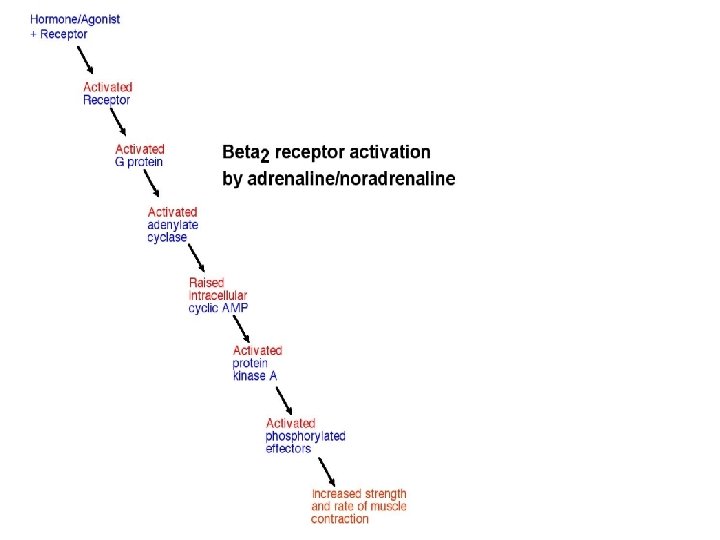
Principles of Cell Signalling (Signal Transduction) • Specific hormonereceptor interactions. • Transmembrane signalling. • Intracellular magnification of the signal to trigger specific proteins. • Secondary messengers usually act at this level. • Effector proteins are activated. • The pattern of activated proteins leads to a specific cell response.
Receptors - Cellular aerials • • • Receptors are usually transmembrane proteins. They bind the signal molecule and transmit its message. Receptors are membrane-bound. Receptors pass on the message into the cell. They do this by activating other proteins.
There are five types of receptor 1. Intracellular receptors (transcription factors or enzymes). Prednisolone (an immunosuppressant steroid). 2. Receptors that are ion channels. Acetylcholine at nicotinic acetylcholine receptors. 3. Receptors with built-in enzyme activity. Insulin receptor. 4. Receptors linked to soluble protein kinases. Adrenaline receptors. 5. Receptors coupled to target proteins via a G protein.
Neurotransmission at nicotinic acetylcholine receptors Agonist = acetylcholine
Second messengers help to transmit the extracellular message into a magnified intracellular signal. The concentration of second messenger changes after stimulation. Second messengers regulate the activity of target proteins. Class of secondary messenger Hydrophobic membrane bound. Hydrophilic - water soluble and cytosolic. Gases - diffuse through membrane and cytosol. Examples Diacylglycerol, Phosphatidylinositols c. AMP, c. GMP, IP 3, Ca 2+/Calmodulin Nitric oxide
Signalling pathways use three mechanisms to change cell behaviour: 1. Alter gene transcription. 2. Alter ion balance across the plasma membrane. 3. Alter the activity level of existing enzymes. Whatever the mechanism, the signal transduction process achieves altering protein function / enzyme activity to achieve the cell response.
Steroid hormones use intracellular receptors
Receptor-G protein interaction
G proteins - A family of proteins • Gαs stimulates adenylate cyclase to produce c. AMP from ATP. c. AMP acts as a second messenger that goes on to interact with and activate protein kinase A that phosphorylates a myriad of downstream targets. • Gαi inhibits the production of c. AMP from ATP. • Gαq/11 stimulates membrane-bound phospholipase C beta, which then cleaves PIP 2 into IP 3 and diacylglycerol (DAG). • Gα 12/13 control cell cytoskeleton remodeling, thus regulating cell migration. • Gβγ sometimes also have active functions, e. g. , coupling to Ca 2+ channels and K+ channels.
Signalling cascade for growth factors Growth factor 1 2 P Receptors 3 P Membrane P P adaptor GDP GTP 4 GTP GDP Binding of growth factor induces receptor dimerisation. Dimerisation triggers phosphorylation of receptors. Adaptor and Ras-GDP bind to phosphorylated receptors. Nucleotide exchange generates activate Ras-GTP. Ras
Signalling cascade for inhibitory growth factors Inhibitory growth factor Binding of inhibitory growth factor induces receptor dimerisation Receptor dimers phosphorylate SMAD protein SMAD P co. SMAD-P forms complex with co. SMAD (SMAD 4) protein SMAD P co. SMAD-co. SMAD complex migrates to nucleus. Complex activates transcription factors. Transcription of target genes.