Cell Theory Mathias SchleidenBotanist 1838 Theodor Schwann Zoologist
 1838 Theodor Schwann (Zoologist) 1838 1. All organisms are made")
Cell Theory Mathias Schleiden(Botanist) 1838 Theodor Schwann (Zoologist) 1838 1. All organisms are made of cells 2. Cells are the smallest unit of living things. 3. Cells give rise to other cells.

The Simple Microscope
")
Anton Von Leeweunhoeke (1630’s)
")
Robert Hooke (1665)

Compound Microscope

Italian Compound Microscope, signed by François Baillou, Milan, c. 1700.
")
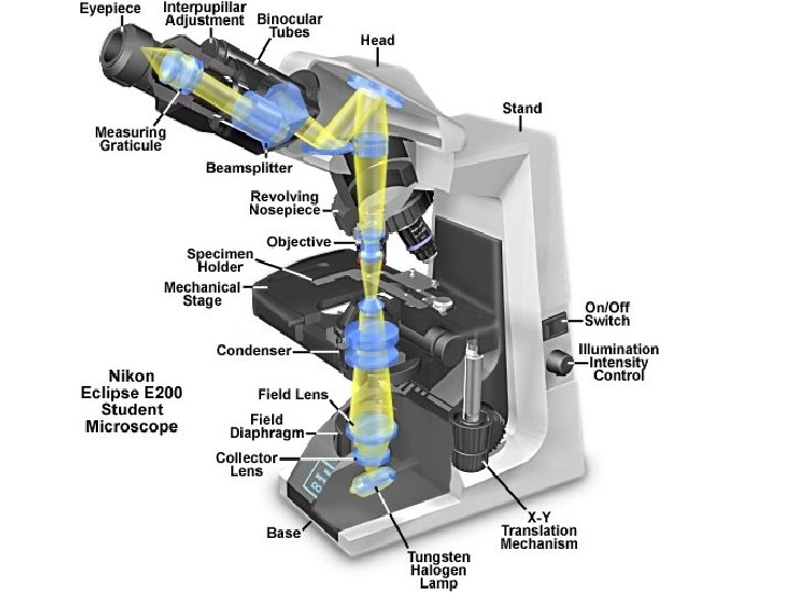
Compound Microscope (Modern)

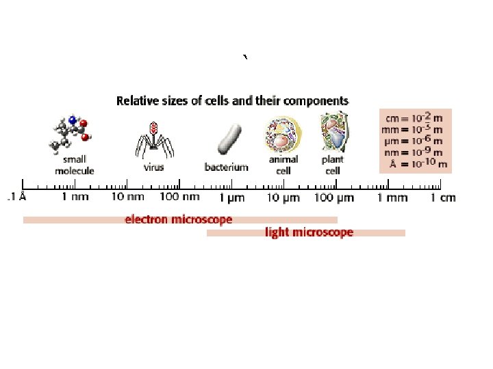
Images Stomata of a plant

Ameoba
 • The ocular")
Compound Microscope: Key Points • Two lenses ( ocular and objective) • The ocular magnifies the image from the objective. *2 -D images *can use live samples *light must pass through sample to see it *light source needed: reflected by a mirror (old) or electric power • 1000 x at the most; we have 400 x as the highest mag. at school. • Resolution and clarity is ok

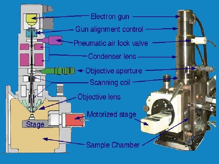
Electron Microscope First one around 1930

Two coccolithophores viewed under a scanning electron microscope. Top: Helicosphaera hyalina. Bottom: Discosphaera tubifera (Photos by Vita Pariente)
 DNA Golgi body")
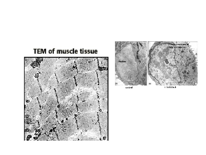
Transmission Electron (TEM) DNA Golgi body
")
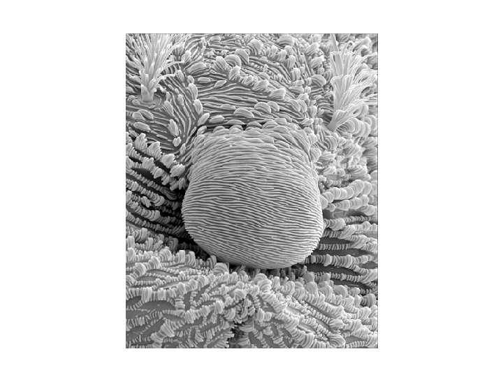
Scanning Electron(SEM)

Pollen Hookworm

Weevil head

Fern leaf cross section

Black Widow spider claw

Dentist Drill

Electron Microscope: Key points • Electrons as a source to view sample; image captured by computer…not light. *very specific magnification ( 1 million x) • Computer needed for precision and accuracy.

SEM • Sample coated with a metal ion • Electrons shot over sample and scan across • Electrons bounce off the surface and provide a 3 -D image • Can view whole samples

TEM • Samples must be thin…very thin for electrons to pass through the sample. • Can’t be used to view whole or live samples • Very good clarity and resolution • 1 million x • 2 -D images; very detailed
- Slides: 27