CELL THEORY History of Cell Theory In 1655


 • Electrons are passed through a specimen to a fluorescent")
 • Directs electrons over the surface of the specimen and")
- Slides: 18
CELL THEORY
History of Cell Theory • In 1655, Robert Hooke made a simple microscope and looked at a piece of cork. • He saw small, box-like structures in the magnified cork and called them Cellulae. • It is from Hooke’s work that we have the term cell.
• A cell is the basic structural and functional unit of all living organisms. • In the late 1600 s, Anton van Leeuwenhoek designed a microscope and found that he was able to view living organisms in pond water, milk and other various substances.
• In 1838, Matthias Schleiden studied plant tissues and concluded that all plants are composed of cells. • In 1839, Theodor Schwann reported that animal tissues also consisted of individual cells. • In 1855, Rudolph Virchow proposed that all cells are produced from the division of existing cells.
The Cell Theory • The cell theory is one of the fundamental ideas of modern biology and includes the following three principles:
• 1. All living organisms are composed of one or more cells. • 2. Cells are the basic unit of structure and organization of all living organisms. • 3. Cells arise only from previously existing cells, with cells passing copies of their genetic material onto their daughter cells.
Microscope Technology • The discovery of cells and the development of cell theory would not have been possible without microscopes. • Developments in microscope technology have given scientists the ability to study cells in greater detail than early scientists ever thought possible.
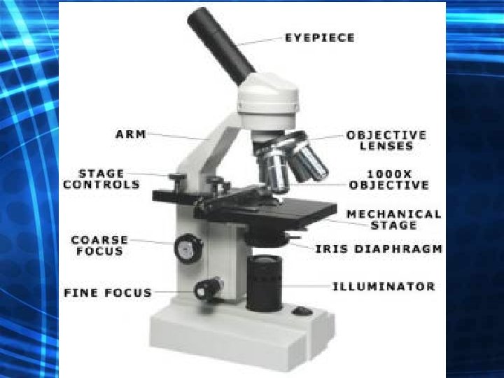
Compound Light Microscopes • Consists of a series of glass lenses and uses visible light to produce a magnified image. • Each lens in the series magnifies the image of the previous lens. • The maximum magnification without blurring is around 1000 x.
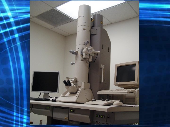
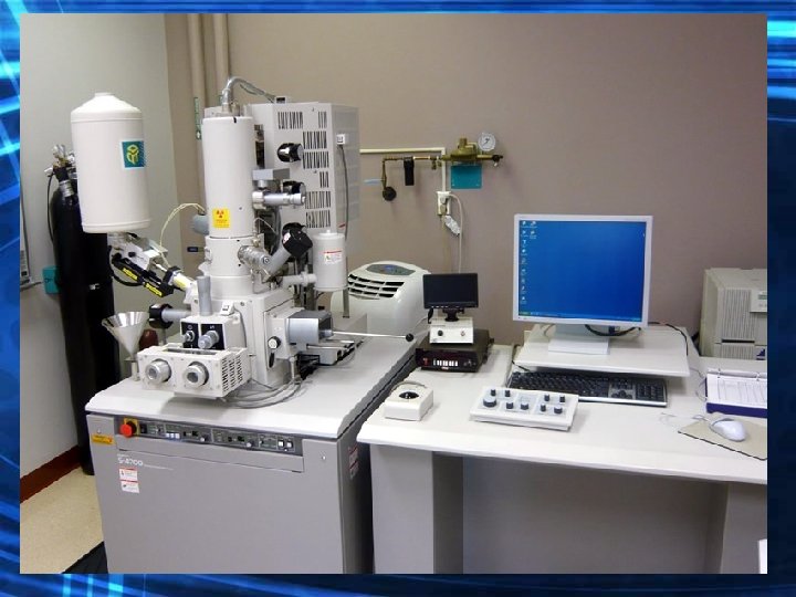
Electron Microscopes • Instead of lenses, the Electron Microscope uses magnets to aim a beam of electrons at thin slices of cells.
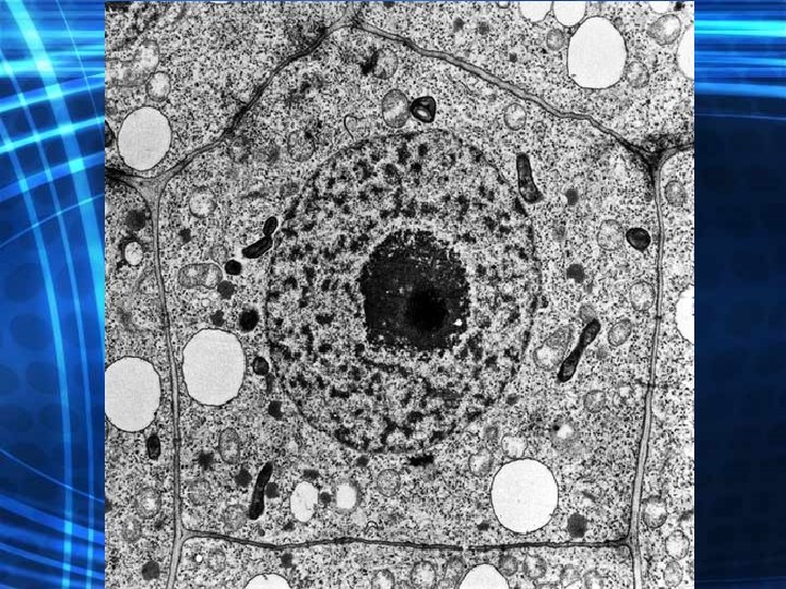
Transmission Electron Microscope (TEM) • Electrons are passed through a specimen to a fluorescent screen. • Thick parts of the specimen absorb more electrons than thin parts, forming a black and white shaded image of the specimen. • Can magnify up to 500, 000 x. • The specimen must be dead, sliced very thin, and stained with heavy metals.
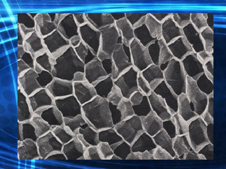
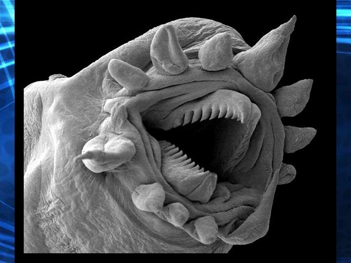
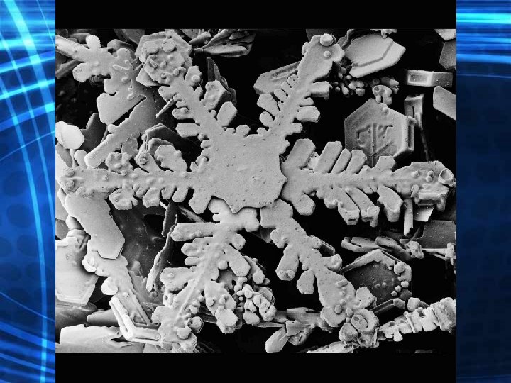
Scanning Electron Microscope (SEM) • Directs electrons over the surface of the specimen and produces a 3 D image. • One disadvantage of using a TEM and SEM is that only nonliving cells and tissues can be observed.