CELL THEORY ENDOSYMBIOTIC THEORY Slide Show by Kelly

CELL THEORY ENDOSYMBIOTIC THEORY Slide Show by Kelly Riedell/Brookings Biology http: //www. parlament-berlin. de/Galeriecopy. nsf/0/8 ABC 720262898739 C 1256 A 480037 F 869? Open. Documen http: //www. ncu. edu. tw/~ls/graph/faculty_pictures/whole_time/SLC_lab-1. jpg

CELL THEORY 1. All living things are ____________. MADE OF CELLS 2. Cells are the basic unit of STRUCTURE & _______ FUNCTION ______ in an organism. life (cell = basic unit of _______) 3. Cells come from the reproduction of ______ cells existing Cell image: http: //waynesword. palomar. edu/lmexer 1 a. htm

1970 American Biologist ___________ Lynn Margulis Provided evidence for the idea that ancestors of ______ Mitochondria + _______ chloroplasts were at one time free-living prokaryotes that were engulfed by other cells and stayed to live symbiotically inside Endosymbiotic Theory = _____________ http: //en. wikipedia. org/wiki/Lynn_Margulis

ENDOSYMBIOTIC THEORY See a movie about ENDOSYMBIOTIC THEORY http: //www. mun. ca/biology/scarr/Endosymbiosis_theory. gif

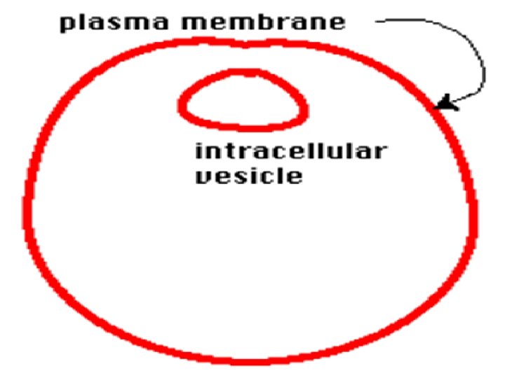

Bacterial plasma membrane Vesicle

Bacterial DNA Bacterial ribosomes

Remember host cell is a eukaryote. Add a nucleus and host DNA How is this different from bacterial DNA? Host Nucleus HOST DNA

Add some eukaryotic host ribosomes. How are these different from bacterial ribosomes? Host RIBOSOMES

Evolved to become mitochondrion
 Vesicle OUTER MITOCHONDRIAL MEMBRANE")
Bacterial cell membrane Evolved to become mitochondrion (CRISTAE) Vesicle OUTER MITOCHONDRIAL MEMBRANE

WHAT’s the EVIDENCE? Look at the colors in your picture. Which parts of mitochondria/chloroplasts have a prokaryotic bacterial origin? Which have a eukaryotic host origin? Make a list of the evidence for the ENDOSYMBIOTIC THEORY? Find these in your picture!

CELL THEORY ENDOSYMBIOTIC THEORY LYNN MARGULIS ENDOSYMBIOTIC THEORY Proposed by ___________ Ancient prokaryotes were taken in by eukaryotic cells and stayed to live inside them in a symbiotic relationship; eventually lead to mitochondria and chloroplast organelles MITOCHONDRIA and __________ CHLOROPLASTS Explains origins of _________ EVIDENCE: MITOCHONDRIA/CHLOROPLASTS OWN DNA 1. are only cell parts with DOUBLE _________ MEMBRANES and ______ RIBOSOMES 2. have __________like bacteria PHOSPHOLIPIDS 3. have _________ in their inner membranes like bacteria BINARY FISSION like bacteria 4. divide using __________ 5. have a SINGLE, CIRCULAR loop of DNA like bacteria

All living things made of cells BUT… organisms can be very different. Image from: http: //www. agen. ufl. edu/~chyn/age 2062/lect_06/bacsiz. GIF UNICELLULAR MULTICELLULAR http: //www. angelbabygifts. com/ http: //www. inclusive. co. uk/downloads/images/pics 2/tree. gif

CELL SIZE http: //facstaff. bloomu. edu/gdavis/links%20100. htm Typical cells range from: 5 – 50 micrometers (microns) in diameter
 ? http: //www. talentteacher. com/pics/005 cb.")
How big is a micron ( µm ) ? http: //www. talentteacher. com/pics/005 cb. jpg 1 cm = 10, 000 microns 1” = 25, 000 microns

MULTICELLULAR ORGANISM don’t just contain MANY CELLS. They have different kinds of cells doing different jobs Image from: http: //www. isscr. org/images/ES-cell-Fig-2. jpg

Cells in a multi-cellular organism become SPECIALIZED by turning different genes on and off Image from: http: //www. ncu. edu. tw/~ls/graph/faculty_pictures/whole_time/SLC_lab-1. jpg Cell Specialization =DIFFERENTIATION

SPECIALIZED ANIMAL CELLS Muscle cells Red blood cells http: //www. biologycorner. com/bio 3/images/bloodcells 3 D. jpg Cheek cells http: //www. mlms. logan. k 12. ut. us/~ajohnson/Cells. html

Specialized Plant cells Guard cells Xylem cells Pollen Guard cells: http: //botit. botany. wisc. edu/courses/img/Botany_130/Diversity/Bryophytes/Anthoceros/Guard_cells. jpg Xylem: http: //botit. botany. wisc. edu/images/130/Secondary_Growth/Woody_Stems/Tilia_Stem/Secondary_Growth/One_Year_Stem/Primary_xylem_MC. j Pollen: http: //www. uic. edu/classes/bios 100/labs/pollen. jpg

How Do Cell Compare? Plant cells Animal cells Bacterial cells _________> _____ http: //slideplayer. com/slide/3524544/12/images/16/Bacterial+cell+Animal+cell+Plant+cell. jpg

CELL THEORY CELLS 1. All living things are made of _______ 2. Cells are the basic unit of structure & function in an organism. (= basic unit of ______) life existing 3. New cells are produced from _______cells. Cell image: http: //waynesword. palomar. edu/lmexer 1 a. htm

____ ATOMS MOLECULES _____ ORGANELLES ______ IMAGE SOURCES: see last slide

CELLS TISSUES ____________ Similar cells working together IMAGE SOURCES: see last slide

ORGANS SYSTEMS ______ Different tissues working together ORGANISM ______ Different organs working together IMAGE SOURCES: see last slide

SOUTH DAKOTA SCIENCE STANDARDS Students will be able to: • explain the process of specialization 9 -12. L. 1. 3. A (ADVANCED) • describe the relationships between the levels of organization in multi-cellular organisms (cells, tissues, organ systems, and organism) (PROFICIENT) • explain how gene expression regulates cell growth and differentiation 9 -12. L. 1. 3. A (Tissue formation, development of new cells from original stem cells (ADVANCED)

NATURE OF SCIENCE: Indicator 1: Understand the nature and origin of scientific knowledge. 9 -12. N. 1. 1. Students are able to evaluate a scientific discovery to determine and describe how societal, cultural, and personal beliefs influence scientific investigations and interpretations. • Recognize scientific knowledge is not merely a set of static facts but is dynamic and affords the best current explanations.

SOUTH DAKOTA ADVANCED SCIENCE STANDARDS 9 -12. L. 1. 3 A. Students are able to explain how gene expression regulates cell growth and differentiation. Examples: Tissue formation Development of new cells from original stem cells

Core High School Life Science Performance Descriptors High school students performing at the PROFICIENT level: Describe the relationship between structure and function (cells, tissues, organ systems, and organisms);

IMAGE BIBLIOGRAPHY http: //www. uic. edu/classes/bios 100/summer 2004/lect 02. htm Paint image by Riedell http: //www. emc. maricopa. edu/faculty/farabee/BIOBK/Bio. Book. CHEM 2. html#Organic%20 molecules http: //evolution. berkeley. edu/evosite/evo 101/images/dna_bases. gif

http: //bioweb. wku. edu/courses/BIOL 115/Wyatt/Biochem/Carbos/Carb_poly. gif http: //vilenski. org/science/safari/cellstructure/golgi. html http: //www. science. siu. edu/plant-biology/PLB 117/JPEGs%20 CD/0076. JPG http: //classes. kumc. edu/som/bioc 801/lectures/images/mem 01 -08. gif http: //www. biology 4 kids. com/files/cell_nucleus. html

http: //www. biologyclass. net/mitochondria. jpe http: //www. ncu. edu. tw/~ls/graph/faculty_pictures/whole_time/SLC_lab-1. jpg http: //www. kufm. kagoshima-u. ac. jp/~anatomy 2/BON/1016 A 03. jpg http: //www. carolguze. com/text/102 -19 -tissuesorgansystems. shtml http: //academic. pg. cc. md. us/~aimholtz/Aand. P/206_ONLINE/Immune/Innate_Images/cilia. jpg http: //www. emc. maricopa. edu/faculty/farabee/BIOBK/Bio. Book. Animal. TS. html http: //www. agen. ufl. edu/~chyn/age 2062/lect_19/147 b. gif

http: //www. proctitispages. force 9. co. uk/ http: //vilenski. org/science/safari/fungus. html http: //www. harrythecat. com/graphics/ http: //bestanimations. com http: //www. inclusive. co. uk/downloads/images/pics 2/tree. gif http: //people. eku. edu/ritchisong/homepage. htm http: //sps. k 12. ar. us/massengale/animal%20 dissections. htm
- Slides: 34