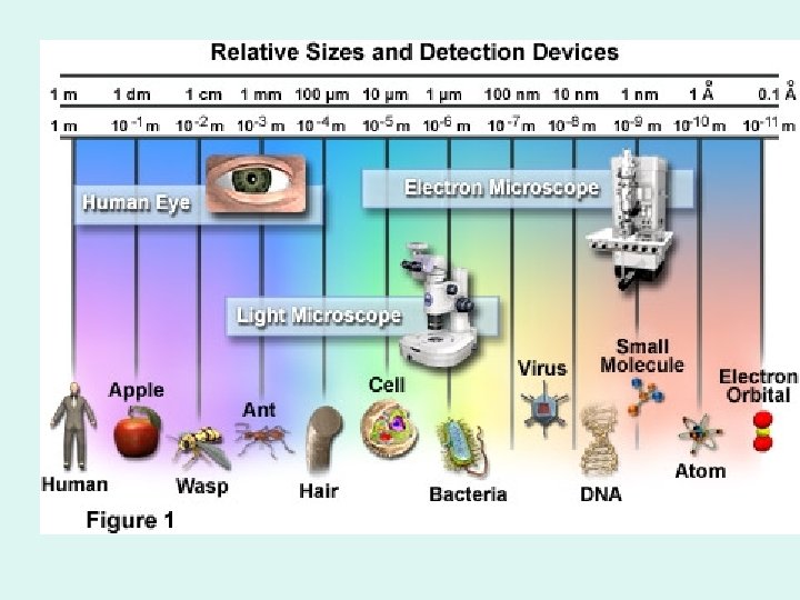
Cell Structure Function Robert Hooke 1600 s named


Cell Structure & Function
 named the cell after viewing cork under m’scope")
• Robert Hooke (1600 s) named the cell after viewing cork under m’scope

At 40 x tattoo ink in dermis of skin

Comparing Prokaryotic and Eukaryotic Cells • Basic features of all cells: – Plasma membrane – Semifluid substance called cytosol – Chromosomes (carry genes) – Ribosomes (make proteins) Copyright © 2008 Pearson Education, Inc. , publishing as Pearson Benjamin Cummings

2 Types of Cells • Prokaryotes: earliest cells; Have NO NUCLEUS • Eukaryotes: modern cells/most cells( all but bacteria) HAVE A NUCLEUS
 A typical rod-shaped")
Fig. 6 -6 Fimbriae Nucleoid Ribosomes Plasma membrane Bacterial chromosome (a) A typical rod-shaped bacterium Cell wall Capsule Flagella 0. 5 µm (b) A thin section through the bacterium Bacillus coagulans (TEM)

Sickle Cell Anemia *note misshapen RBC

Cell Theory 1. All living things are made of cells. 2. Cells are the basic unit of life 3. New cells come from existing cells -Schleiden, Schwann, Virchow

Functions of Organelles
 • Thick, clear gel-like substance found throughout cell • Supports the organelles")
Cytoplasm (Cytosol) • Thick, clear gel-like substance found throughout cell • Supports the organelles
")
Nucleus • “control center” of cell • Contains the chromosomes (genetic info. = DNA*) • Has all instructions to make new proteins • *DNA from both parents found here
")
Nucleolus • Center of nucleus • Site of ribosome synthesis (ribosomes are made here)

Fig. 6 -UN 1 a Structure Cell Component Concept 6. 3 The eukaryotic cell’s genetic instructions are housed in the nucleus and carried out by the ribosomes Nucleus Function Surrounded by nuclear envelope (double membrane) perforated by nuclear pores. The nuclear envelope is continuous with the endoplasmic reticulum (ER). Houses chromosomes, made of chromatin (DNA, the genetic material, and proteins); contains nucleoli, where ribosomal subunits are made. Pores regulate entry and exit os materials. Two subunits made of ribosomal RNA and proteins; can be free in cytosol or bound to ER Protein synthesis (ER) Ribosome

Fig. 6 -10 Nucleus 1 µm Nucleolus Chromatin Nuclear envelope: Inner membrane Outer membrane Nuclear pore Pore complex Surface of nuclear envelope Rough ER Ribosome 1 µm 0. 25 µm Close-up of nuclear envelope Pore complexes (TEM) Nuclear lamina (TEM)

Chromosomes • Contain genetic information/DNA • Chromatin combines to form • Humans have 46 chromosomes or 23 pairs

Fig. 15 -1

Fig. 15 -5 X Y

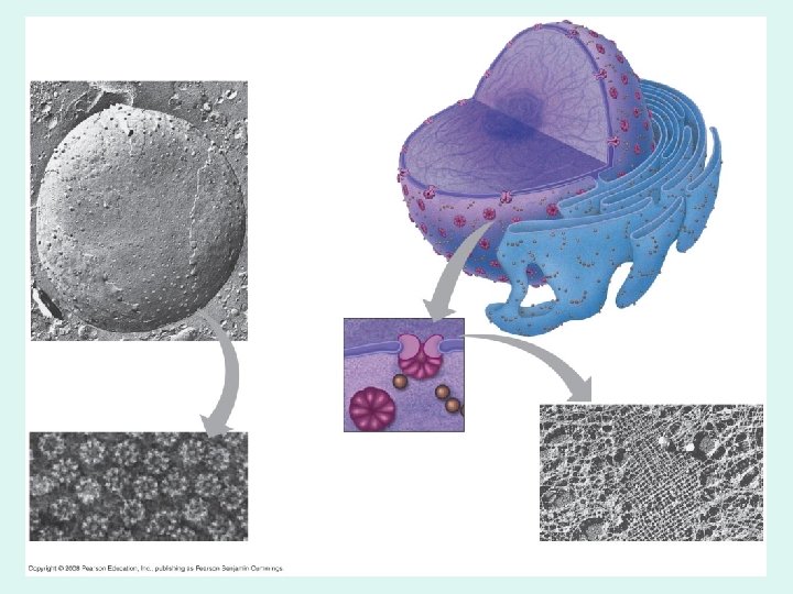
Endoplasmic Reticulum • Extensive system of tubules and membranes – 2 Types: 1. Smooth ER 2. Rough ER
 • Breaks down/metabolizes carbohydrates • Packages enzymes")
Smooth ER • Synthesis of lipids (cholestrol) • Breaks down/metabolizes carbohydrates • Packages enzymes for secretion • De-toxification of alcohol in liver ER

Fig. 6 -12 Smooth ER Rough ER ER lumen Cisternae Ribosomes Transport vesicle Smooth ER Nuclear envelope Transitional ER Rough ER 200 nm
 Free ribosomes Bound ribosomes Large subunit 0.")
Fig. 6 -11 Cytosol Endoplasmic reticulum (ER) Free ribosomes Bound ribosomes Large subunit 0. 5 µm TEM showing ER and ribosomes Small subunit Diagram of a ribosome

Rough ER • Has ribosomes attached • One of the sites of protein assembly

Ribosomes • Site of protein synthesis • Made of RNA + protein • Means: proteins are made here • Free Ribosomes: NOT attached to ER • Bound Ribosomes: attached to ER
 • Support • Protection • Regulates which substances enter &")
Cell Membrane (plasma membrane) • Support • Protection • Regulates which substances enter & exit = Selectively permeable
 TEM")
Fig. 6 -7 Outside of cell Inside of cell 0. 1 µm (a) TEM of a plasma membrane Carbohydrate side chain Hydrophilic region Hydrophobic region Hydrophilic region Phospholipid Proteins (b) Structure of the plasma membrane

Fig. 6 -30 Collagen Proteoglycan complex EXTRACELLULAR FLUID Polysaccharide molecule Carbohydrates Fibronectin Core protein Integrins Proteoglycan molecule Plasma membrane Proteoglycan complex Microfilaments CYTOPLASM

What might ENTER a cell? • Oxygen • Dissolved nutrients • Potassium and other ions • water

Vacuoles • Storage of –Water –Dissolved nutrients –Even Waste **animals have few, very small

Fig. 6 -15 Central vacuole Cytosol Nucleus Central vacuole Cell wall Chloroplast 5 µm

Central Vacuole • Plants have a large central vacuole. • Takes up most of plant cell –Supports –Turgor Pressure –“Wilting” process: how?
 • Has Nuclear Pores: holes to")
Nuclear Membrane • Protects nucleus (why necessary? ) • Has Nuclear Pores: holes to allow substances to enter/exit

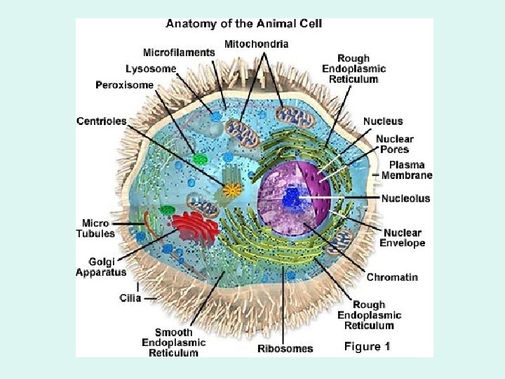
Lysosomes • Animal Cells • Bags of hydrolytic enzymes • Digests old cell organelles
 1. Gives cell shape 2.")
Cytoskeleton • System of protein fibers • (Microtubules, microfilaments) 1. Gives cell shape 2. Supports cell 3. Helps move organelles

Fig. 6 -1

Golgi Apparatus Golgi Body • Proteins are modified and packaged here for secretion • “warehouse/UPS” of cell • Lysosomes are made here
 0. 1 µm Cisternae")
Fig. 6 -13 cis face (“receiving” side of Golgi apparatus) 0. 1 µm Cisternae trans face (“shipping” side of Golgi apparatus) TEM of Golgi apparatus

Fig. 6 -16 -3 Nucleus Rough ER Smooth ER cis Golgi trans Golgi Plasma membrane
 respiration")
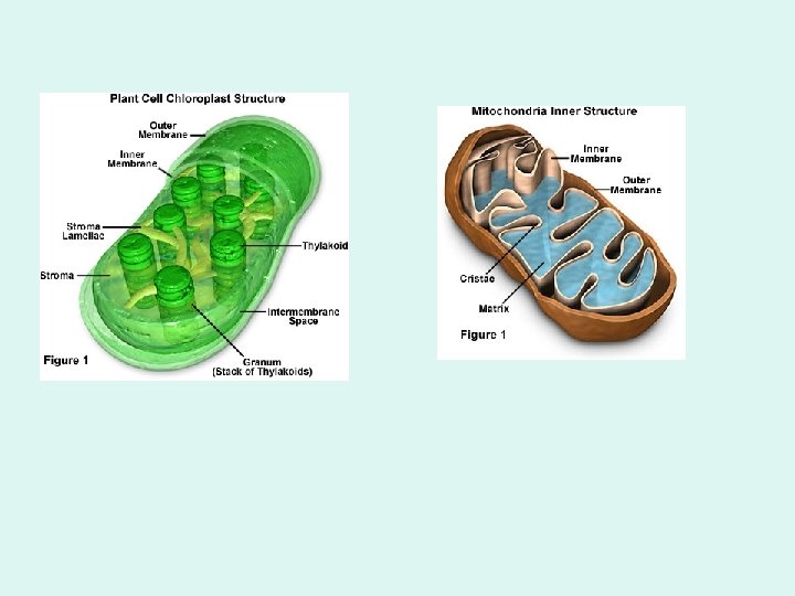
Mitochondria • “powerhouse of cell”= energy is produced • Site of cellular (aerobic) respiration (ATP is made) • Was once an independent, free-living organism

Fig. 6 -17 Intermembrane space Outer membrane Free ribosomes in the mitochondrial matrix Inner membrane Cristae Matrix 0. 1 µm

• More active cells have more mitochondria- WHY? Ex: muscle cells have more • Has a membrane surrounding And DNA of its own! mt. DNA- inherited from mother/materlineal Used in forensics (sometimes)

Endosymbiont Theory: Idea that…. . 1. Chloroplasts & mitochondria were once free-living 2. Moved into eukaryotic cell 3. Became an organelle of cell
 Outer membrane b)")
Why do we think this? Both chloroplasts & mitochondria have: a) Outer membrane b) Energy source/function c) Both have bits of genetic material

Fig. 6 -19 Chloroplast Peroxisome Mitochondrion 1 µm

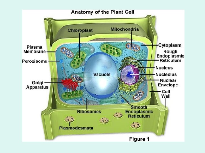
Chloroplast • Plant Cells • Located in middle of leaf tissue • Site of photosynthesis • green- chlorophyll • Was once free-living, independent • *has maternal DNA (interesting!)

Fig. 6 -18 Ribosomes Stroma Inner and outer membranes Granum Thylakoid 1 µm

Cell Wall • Plants only • Support • Protection • Made of cellulose: strong carbohydrate

Fig. 6 -28 Secondary cell wall Primary cell wall Middle lamella 1 µm Central vacuole Cytosol Plasma membrane Plant cell walls Plasmodesmata

Centrioles • Animal cells • Helps move chromosomes apart during mitosis

Cilia and Flagella • Protein fibers • Cilia- short fibers; all over • Flagella- long fibers; 1 or 2 • purpose: locomotion (movement) • Ex: paramecium, spermatozoa
 Motion of flagella 5 µm Direction of")
Fig. 6 -23 Direction of swimming (a) Motion of flagella 5 µm Direction of organism’s movement Power stroke Recovery stroke (b) Motion of cilia 15 µm

How Are Plant Cells and Animal Cells Different? Plants: • Chloroplasts • Cell walls • Central vacuole • Green: chlorophyll • None • Rectangular shape Animals • None • Few, smaller • None • Lysosomes • Different shapes • More mitochondria
- Slides: 57