Cell Structure and Microscope The Microscope Learning Objectives

Cell Structure and Microscope

The Microscope

Learning Objectives By the end of this topic, you will be able to: 1. Identify the parts of a light microscope 2. Give the function of each part of the light microscope 3. Describe how to use a light microscope 4. Distinguish between the light and the Electron Microscope 5. Calculate magnification

There are 2 types of microscope 1. Light microscope 2. Electron microscope

Light Microscopes 1. Simple microscope: 1 lense 2. Compound microscope: 2 lenses. These are the microscopes used in our lab a. The Eyepiece Lens is the lens that you look through. b. The objective lens is the lens that is immediately above the specimen.

Parts of the Microscope
 Magnification Stage To place")
Parts of the Microscope Part Function Lenses (eyepiece & objective) Magnification Stage To place the slide on Clips Hold the slide in place Diaphragm To control the amount of light reaching the object Coarse Focus Knob To focus the image Fine Focus Knob To precisely focus the image Light source To supply light to the object Nose piece Revolves to move desired lens into place Stage height adjustment Allows longest lens to fit over slide

Remember Microscope magnifications Eyepiece lens Objective lens X 5 X 10 X 40 Total Exam Qs 2007 OL True or False, if the eyepiece lens of a microscope is marked X 10 and the objective lens is marked X 4, the total magnification is X 14

Recording what you can see: Cheek Cells Onion Cells

Recording what you can see: Animal Cell membrane Cytoplasm Nucleus Plant Cell membrane Cytoplasm Vacuole Nucleus Cell wall Chloroplast

How are we going to look at an animal cell? 1. Collect your equipment 2. Carefully wipe the side of your cheek with a cotton bud 3. Gently wipe the sample onto the slide 4. Stain the sample with methylene blue 5. Cover the sample with a cover slip 6. Have a look at your cells.

Preparing Onion Slides

Now Fill out your workbooks

Recording what you can see: Cheek Cells Things outlined in black are probably air bubbles! Get us to help you if you can’t see!

Recording what you can see: Animal Cell membrane Cytoplasm Nucleus
")
What do all the bits do? Controls what happens in the cell (contains DNA) Jelly-like substance where reactions happen Keeps substances in the cell and controls what goes in and out What do the organelles do? • Nucleus • Cell membrane • Cytoplasm • Chloroplast • Cell wall • Vacuole Contain chlorophyll – where photosynthesis happens Contains cell sap – keeps the cell firm Made of cellulose and supports the cell

ELECTRON MICROSCOPE

Electron Microscope • Uses Electrons instead of light • Resolution is much better • Magnification is much greater • Allows us to see the cell ultrastructure (organelles of the cell)

Images from Electron Microscope Bacteria Platelet Cytoplasm

Learning Check What is the function of each of these parts of the Microscope? Eyepiece Lens Stage Diaphragm Fine focus Knob Objective Lens Clips Coarse Focus Knob Light

What have you learned? Can you? 1. Name the parts of the microscope and their functions. 2. Outline the differences between the light microscope and the electron microscope
 Name the parts of the light microscope labelled A and B.")
2004 OL (a) Name the parts of the light microscope labelled A and B. (b) Answer the following in relation to preparing a slide of stained plant cells and viewing them under the microscope. (i) From what plant did you obtain the cells? (ii) Describe how you obtained a thin piece of a sample of the cells (iii)What stain did you use for the cells on the slide? (a) Describe how you applied this stain (b) What did you do before placing the slide with
 (b) (ii) A = eye piece B = objective or lens or")
7. (a) (b) (ii) A = eye piece B = objective or lens or high power (allow lens for A or B but not for both) X 400 2 2 name of plant 3 2 description – peel off thin film of plant tissue with forceps / cut thin section of plant tissue with blade (or microtome) or any other correct method i. e. How = 3 plus instrument = 3 2(3) name of stain 3 application of stain – use dropper to place stain on tissue on slide or place tissue in stain or any other correct method. 3 put on cover slip or remove excess stain one 3 any cell wall/ chloroplasts or chlorophyll/ (large) vacuoles/ (starch) granules/ leucoplasts/ chromoplasts / shape any two 2(3)

Can you…. . 1. Identify the parts of a light microscope 2. Give the function of each part of the light microscope 3. Describe how to use a light microscope 4. Distinguish between the light and the Electron Microscope 5. Calculate magnification

Txt Msg Write a txt msg explaining your learning Back to Plenaries

Aidhm 1. Identify the parts of a plant cell as seen under light microscope 2. Identify the parts of an animal cell as seen under light microscope 3. Give the function of each of parts of a cell under light microscope 4. Identify the ultra structure and give the function of part of cell 6. Draw the ultra structure of the mitochondrion and the chloroplast 7. Definition of prokaryotic and eukaryotic cells (HL)

Anton von Leeuwenhoek with his hand-held microscope, was the first person to observe and describe Living Cells in the early 17 th century

Robert Hooke looked a thin slices of cork under a microscope. He thought the spaces he saw reminded him of Monks Cells Hence the name cells

Plant Cell as seen under a light microscope

Cells • All living things are composed of one or more cells. • Cells are the basic units of structure and function in an organism. • Cells come only from reproduction of existing cells.

Organelles • Cells contain ORGANELLES. • An organelle has a SPECIFIC FUNCTION FOR THE CELL.

Organelles we need to know • • Cell membrane Nucleus Mitochondria Chloroplast Ribosomes DNA Cell Wall Vacuole

Learning Check What are Cells? What are organelles? Can you name 8 organelles?

Video Intro to animal cells • https: //www. youtube. com/watch? v=Fzj 6 TR n. Xmps&feature=player_embedded

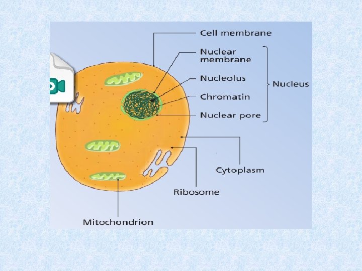
Animal Cells contain the following structures Cell Membranes Nucleus can be seen under the light microscope Cytoplasm Mitochondrian Parts of nucleus Ribosomes can be seen under electron microscope

• Protoplasm: All the living parts of the cell • Cytoplasm: The living parts outside the nucleus

Cell Membrane

Cell Membranes are made up of phospholipids and proteins The phospholipids and proteins are in constant motion. Membranes are fluid.

Functions of Cell Membranes • • • Separate the cell organelles and cytoplasm from the outside Semi permeable which means it only allows certain substances through the membrane. It allows movement of water by osmosis Support the cell

Cytoplasm • Watery substance that contains all the organelles within the cell

Nucleus • Contains DNA which make up genes. Genes are found on thread like structures called chromosomes • Genes contain information to make proteins. Proteins control the function of the cell

Ultra Structure of The Nucleus Nuclear pore: Controls movement of substances into and out of nucleus Chromatin contains DNA, arranged into chromosomes which are not dividing Nucleolus contains RNA, DNA, and proteins. Makes Ribosomes

Ultra Structure of the Mitochondrion

Ultra Structure of the Mitochondrion • Powerhouses of the cell • Composed of inner and outer membranes • Site of energy release (through respiration). Enzymes are attached to inner membrane. In respiration makes molecules of ATP (chemical energy) • Cells with lots of mitochondria produce a lot of energy

Ribosomes: make proteins Ribosomes

Ribosomes • Each cell contains thousands • Found in cytoplasm of cell Use To make proteins Made from Rna and Protein


A typical animal cell

The parts of a typical animal cell

Learning Check 1. All cells have a cell membrane. What are its 3 functions? 2. Label the picture 3. What is the function of a ribosome? 4. What is the cytoplasm? 5. What is the function of the cytoplasm

Learning Check What is this organelle? Why are they known as powerhouses? What type of cells would have these organelles in large numbers?
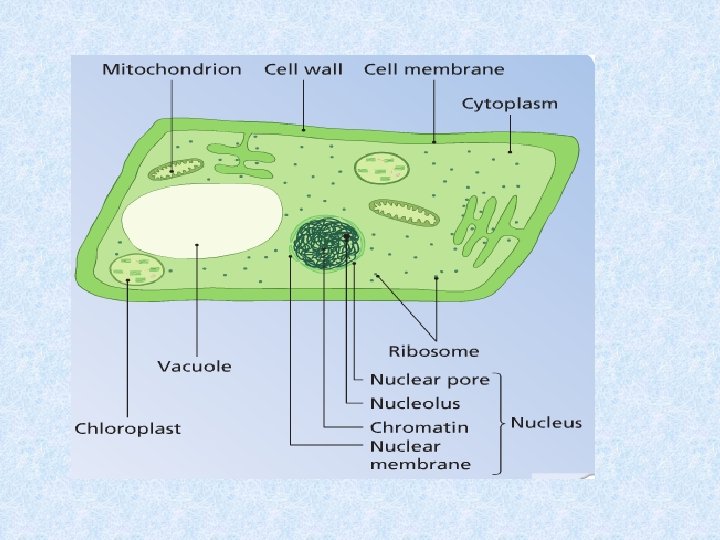

Plant Cells contain the following structures Cell Membranes Mitochondrian Nucleus Cytoplasm Cell Wall Vacuole Chloroplasts can be seen under the light microscope Parts of nucleus Ribosomes Parts of Chloroplasts can be seen under electron microscope

Ultra structure of an plant cell Draw
")
Cell wall • Gives shape, support and strength • Made of cellulose (structural polysaccharides) • Fully permeable: allows all substances into and out of the cell

Vacuoles • Provides support • Stores water, salts, sugars/sap. • Plant cells have large vacuoles (Animal cells have either small or no vacuoles. )

Chloroplasts: The function of chloroplasts is photosynthesis. Contain chlorophyll.
 • Have a double membrane • The membrane")
Ultra structure of the Chloroplast (draw) • Have a double membrane • The membrane stacks (grana) contain the chlorophyll which traps the sun’s energy • Have loops of DNA

A typical plant cell

The parts of a typical plant cell

http: //www. youtube. com/watch? v=r. ABKB 5 a. S 2 Zg&feature=related

Learning check 1. What organelle carries out photosynthesis? 2. What type of cells have large vacuoles and cell walls? 3. What is the function of vacuoles? 4. What is the function of cell walls? 5. What makes cells walls rigid?

Eukaryotes and Prokaryotes

Eukaryotes and Prokaryotes • Prokaryotic cells: contain no true nucleus and no membrane-bound organelles. Found as bacteria • Eukaryotic cells: contain a nucleus and other membrane-bound organelles. Found as plant and animal cells, fungi and amoeba

Comparing animal and plant cells

Can you…. 1. Identify the parts of a plant cell as seen under light microscope 2. Identify the parts of an animal cell as seen under light microscope 3. Give the function of each of the following parts: Cell wall, cell membrane, nucleus, cytoplasm, vacuole and chloroplast 4. Identify the ultra structure and give the function of each of the following cell parts: Cell membrane, mitochondrion, chloroplast, nucleus, nuclear pores, ribosome and DNA 6. Draw the ultra structure of the mitochondrion and the chloroplast 7. Definition of prokaryotic and eukaryotic cells (HL)

END
- Slides: 69