Cell ReproductionCell Cycle DNA DNA deoxyribonucleic acid a

Cell Reproduction/Cell Cycle

DNA • DNA deoxyribonucleic acid – a long, thin molecule that contains info to run cell • DNA is organized into genes • Gene segment of DNA that contains information; passed from parent to offspring – Genes code for proteins • Proteins determine traits

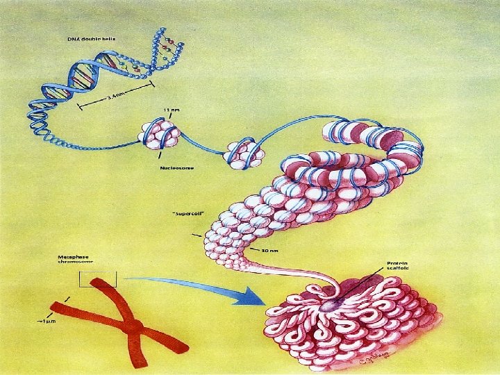
Chromosomes • Chromosome a rod-shaped structure that forms when a single DNA molecule is coiled tightly before cell division. • Chromatid a copy of a chromosome • Centromere protein disk that attaches two chromatids around the middle. – Attached chromatids are called sister chromatids

Chromosomes • The DNA in eukaryotic cells wraps around proteins called histones. – They aid in shaping and maintaining the tight packing of DNA

I. Prokaryotic Cell Cycle A. Bacterial cells contain single, simple, circular strands of “naked” DNA. A. Binary Fission- asexual reproduction of prokaryotic cells. 1. COPY- Cell replicates DNA 2. SLIDE- Each copy of DNA attaches to opposite sides of the cell membrane 3. SPLIT- Cell membrane pinches in creating 2 identical cells.

Binary Fission

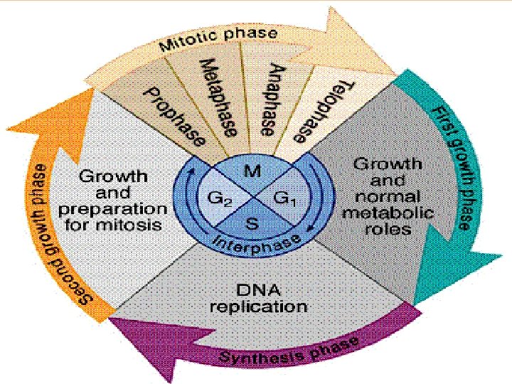
II. Eukaryotic Cell Cycle Life cycle of a cell that begins when a cell is formed and ends when it divides. G 1 growth phase of the cell -cell grows and carries out major functions -majority of cell life. S When the DNA is copied G 2 Mitochondria and other organelles replicate-prepare for nuclear division M Mitosis occurs-nucleus of cell divides into two nuclei C cytoplasm divides-called cytokinesis

HW Questions #1 Write out questions and answer in complete sentences. • In binary fission, how does the offspring’s genetic code compare to the parent’s? Explain. • Using your knowledge of prokaryotic and eukaryotic cells, why do you think the eukaryotic cell cycle is longer/has more steps than the prokaryotic cell cycle?

A. Interphase – normal growth phase of the cell. - the majority of the cell cycle is spent in interphase 1. G 1 phase- growth of the cell immediately following cell division 2. S phase- DNA replication 3. G 2 phase- replication of organelles

• Cells that don’t reproduce don’t have an S or G 2 phase. (ex. Red blood cells) • Genetic material is in loose tangles of DNA and associated proteins (Histones) called Chromatin. • Chromatin is invisible to most light microscopes.
 a. Chromatin")
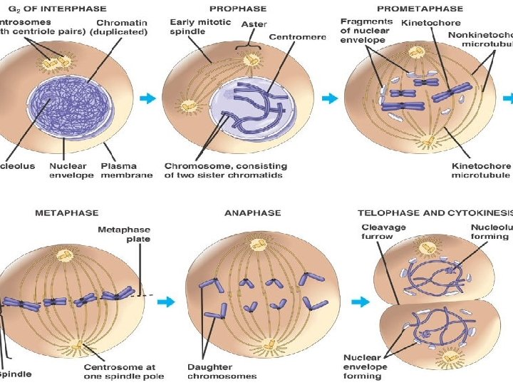
B. Mitosis- the production of 2 new, identical nuclei. 1. Prophase (pairing) a. Chromatin coils to form thick, compact structures called Chromosomes. b. Each chromosome is composed of two identical copies of DNA (Sister Chromatids) held together by a centromere. c. Nuclear membrane and nucleolus begin to break down. d. Centrosomes containing bundles of microtubules called centrioles begin to migrate to opposite poles of the cell. e. Long microtubules called Spindle fibers extend from the centrioles across the cell

Chromosome

Prophase
 a. Spindles fully form and attach to the centromeres of each")
2. Metaphase (midline) a. Spindles fully form and attach to the centromeres of each chromosome. b. Chromosomes are pulled to the equator of the cell forming a line. -half of chromatids on each side ensures equal split
 a. Centromeres are split and sister chromatids are pulled towards opposite")
3. Anaphase (away) a. Centromeres are split and sister chromatids are pulled towards opposite poles of the cell. ** At this point each chromatid is now called a Chromosome.
 a. Identical chromosomes sets reach opposite poles of the cell.")
4. Telophase (two nuclei) a. Identical chromosomes sets reach opposite poles of the cell. b. Spindle fibers break down. c. Two new nuclear membranes begin to form around the chromosome sets. d. Chromosomes begin to uncoil into chromatin. ** This ends mitosis, but not the cell cycle.

Telophase

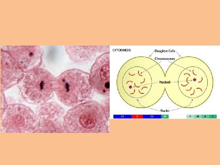
C. Cytokinesis- the splitting of the cytoplasm into two identical daughter cells. - In animal cells, the cell membrane is pinched in by constricting microtubules creating a cleavage furrow. - In plant cells, secretion vesicles form a cell plate between the chromosome sets splitting the cell membrane and cell wall. - Can begin as early as Anaphase - Each new cell immediately begins the G 1 phase of Interphase.

Mitosis Rap • https: //www. youtube. com/watch? v=p. Os. Ab. Ti 9 t. Hw

Animal cell mitosis 4 2 6 1 3 5 7 9 10 8 11 12

Plant cell mitosis
 Homologous chromosomes A. matching pairs of")
III. Meiosis- production of haploid gametes (sex cells) Homologous chromosomes A. matching pairs of chromosomes that are the same size, shape and code for the same hereditary traits B. but may not be exactly alike.

Chromosome numbers • Sex chromosomes – are chromosomes that determine the sex of an organism. – XX – female (mom gives X, dad gives X) – XY – male (mom gives X, dad gives Y) • Autosomes – All the other chromosomes other than the sex chromosomes (humans have 22 pairs).

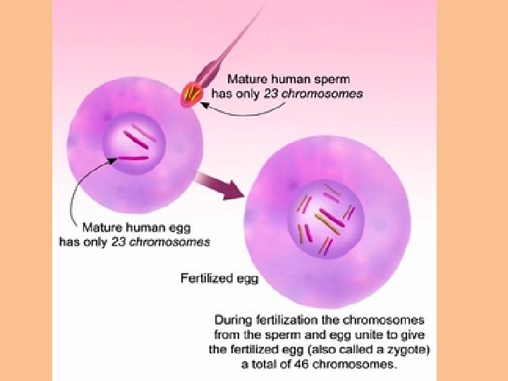
Human Chromosome Number • Humans have two sex chromosomes – One from mother – One from father • Human # Sex Chromosomes – 1 pair 2 chromosomes

Human Chromosome Number • Humans have two copies of each autosome. – One set from mother – One set from father • Human # Autosomes – 22 pair 44 chromosomes • Total #= 23 pair 46 chromosomes
 cells- contain a full set of homologous pairs - 1")
1. Diploid (2 n) cells- contain a full set of homologous pairs - 1 set are maternal - 1 set are paternal a. Diploid # is the total # of chromosomes in a normal body cell. ex. Human diploid # is 46; Chimps is 48
 cells- contain a half set of homologous pairs or 1 of")
2. Haploid (n) cells- contain a half set of homologous pairs or 1 of each homologous pair. a. Haploid # is half of the diploid #. ex. Human haploid # is 23; chimps is 24 b. Gametes (sex cells) are haploid. - male sperm - female egg - fusion of egg and sperm produces a diploid (2 n) cell called a zygote

Overview of Meiosis • Interphase occurs normally – G 1 –S – G 2 • Goal of meiosis is similar to mitosis – now the goal is production of haploid cells (instead of diploid) • There are two rounds of division in meiosis – Split into Meiosis I and Meiosis II – Phase names are the same (Pro-, Meta-, Ana-, Telo-)

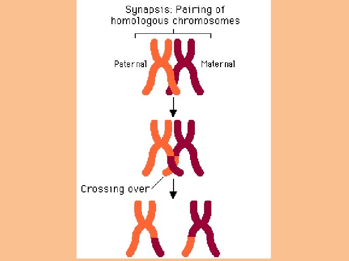
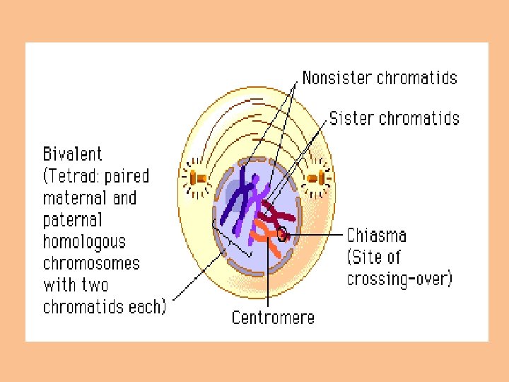
B. Meiosis I *All DNA is replicated 1. Prophase I a. Chromatin coils to form chromosomes. b. Nucleus disappears *** Special to Meiosis I: c. Homologous chromosomes pair up to form tetrads. d. The chromatid arms of the homologous pairs can become intermingled and exchange genetic material. (Crossing Over) e. The random exchange of genes caused by crossing over creates chromosomes w/ a unique combination of genes. (Genetic Recombination)

2. Metaphase I a. Homologous pairs of chromosomes line up at the equator of the cell. -Homologous chromosomes are randomly lined up on midline. (Independent Assortment) b. Spindle fibers attach to the centromeres.

When gametes combine, offspring show variation due to independent assortment and crossing over

3. Anaphase I - Half of the chromosomes are pulled in one direction; other half pulled in opposite direction. -Centromeres are not divided, sister chromatids remain attached - Chromosome number is reduced from diploid(2 n) to haploid(n).

4. Telophase I/ Cytokinesis a. New nuclei may form. b. Cell splits into two haploid cells (not genetically identical).

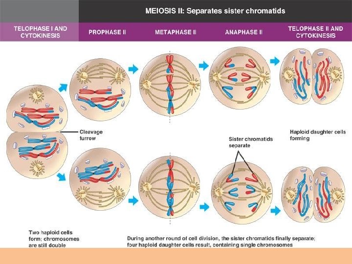
C. Meiosis II - Similar steps to mitosis, but w/ haploid cells. 1. Prophase II- sister chromatids are paired, centrioles migrate to opposite sides of cell 2. Metaphase II- Chromosomes line up on midline 3. Anaphase II- Sister chromatids are separated 4. Telophase II / Cytokinesis- nuclear membranes form around the chromosome sets -produces 4 genetically different, haploid gametes: -Males produce 4 functional sperm. -Females- only one cell becomes egg - other 3 are non-functional gametes (called polar bodies) http: //highered. mcgrawhill. com/sites/0072495855/student_view 0/chapter 28/animation__

Key points of Meiosis ●The process results in 4 daughter cells ●Daughter cells are haploid (n) ●Daughter cells have unique combinations of chromosomes
 Meiosis ensures variability in offspring Gametes combine to")
Meiosis creates gametes (sperm and eggs) Meiosis ensures variability in offspring Gametes combine to create a zygote which is diploid (2 N) process of sexual reproduction

Check for understanding 1. What phase directly follows telophase I? prophase II 2. How many cells are present at the end of meiosis I ? 2 3. A cell has a diploid number of 60, what is the organism's haploid number? 30 4. Meiosis forms what type of cells? gametes 5. In what phase do homologous chromosomes pair up and crossing-over can occur? prophase 1 6. In what phase do the CHROMATIDS separate? anaphase 2

Pg 180 7. Which of these pictures is metaphase I of MEIOSIS and which is metaphase of MITOSIS? Mitosis Meiosis
")
Questions • What are the differences between mitosis and meiosis? (Big picture ideas)

IV. REGULATION OF THE CELL CYCLE AND CANCER

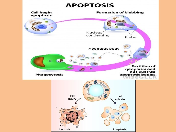
A. Control of the Cell Cycle - G 1 Checkpoint - Check to see if DNA is damaged - G 2 Checkpoint - Check to see if DNA is replicated properly - M Checkpoint - spindle assembly checkpoint, check for alignment of chromosomes Apoptosis - programmed cell death, occurs if any of the checks fail

B. Mutations Can Cause Cancer • Cancer a term used to indicate a disease characterized by abnormal cell growth – Cancer cells are not like normal cells • normal cell growth is a highly regulated process • cancer cells do not respond to stop signals for growth so they continue to divide at fast rates without stopping • Video: From a normal cell to a cancer cell

• Cancer cells that accumulate in an area are called a tumor • Tumors are classified as benign or malignant. – A benign tumor does not invade surrounding tissues and can be surgically removed. – A malignant tumor spreads into other tissues and interferes with organ function. » The most devastating property of malignant tumors is that its cells are able to break free of the tumor and enter the blood and lymph system. » Then these cells are carried to new locations in the body and form new growths » the spread of malignant cells beyond their origins is called metastasis.

1. Cancer Genes • Cells become cancerous when mutations occur in genes that regulate cell growth. • An example of a growth regulating gene is called the ras gene. – The ras gene helps prevent uncontrolled cell division. – When there is a mutation in the ras gene, cell division occurs more rapidly than normal – The ras genes are examples of oncogenes-a gene that when mutated can cause a cell to become cancerous. • It takes time for an individual cell to accumulate the necessary mutations to cause cancer – that is why most cancers occur in people over 40.
- Slides: 53