Cell Reproduction DNA and Inheritance Prokaryotes reproduce by

Cell Reproduction, DNA and Inheritance
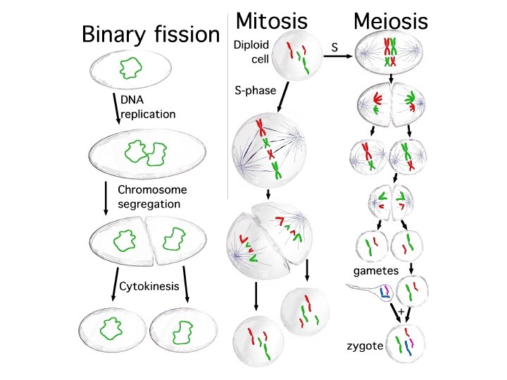

Prokaryotes reproduce by binary fission – Binary fission means “dividing in half” – Occurs in prokaryotic cells – Two identical cells arise from one cell – Steps in the process – A single circular chromosome duplicates, and the copies begin to separate from each other – The cell elongates, and the chromosomal copies separate further – The plasma membrane grows inward at the midpoint to divide the cells Copyright © 2009 Pearson Education, Inc.

Plasma membrane Prokaryotic chromosome Cell wall 1 Duplication of chromosome and separation of copies

Plasma membrane Prokaryotic chromosome Cell wall 1 Duplication of chromosome and separation of copies 2 Continued elongation of the cell and movement of copies

Plasma membrane Prokaryotic chromosome Cell wall 3 1 Duplication of chromosome and separation of copies 2 Continued elongation of the cell and movement of copies Division into two daughter cells

Prokaryotic chromosomes
 Reproductive cells •")
Eukarotes: Two Types of Cells in Body Sex Cells (Germ Cells) Reproductive cells • Male sperm • Female oocyte (a cell that develops into an egg) • undergo Meiosis Somatic Cells Soma = body • All body cells except sex cells • undergo Mitosis

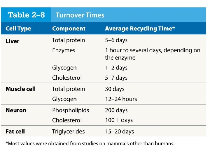
Is the internal epithelium of the bowel the same as it was one month ago? The internal epithelial covering of the intestine acts as protective barrier and also as means of nutrient absorption. The traffic of ingested material inside the intestinal lumen is very intense and the consequent tissue damage requires incessant epithelial renovation through cell division. The tissue renovation is completed in two to three days and is made by mitosis.

The large, complex chromosomes of eukaryotes duplicate with each cell division – Eukaryotic chromosomes are composed of chromatin – Chromatin = DNA + proteins – To prepare for division, the chromatin becomes highly compact, and the chromosomes are visible with a microscope Copyright © 2009 Pearson Education, Inc.
")
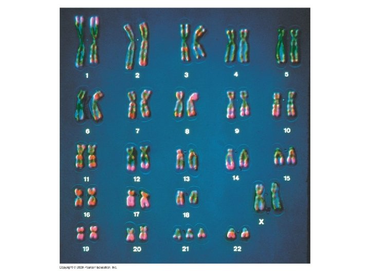
Humans have 46 Chromosomes (23 pairs: One from Mom and One from Dad)
 approximately the same length,")
Homologous Chromosomes • Homologous chromosomes are 2 chromosome strands: 1) approximately the same length, 2) same staining pattern, 3) with genes for the same characteristics at corresponding loci. One homologous chromosome is inherited from the organism's mother; the other from the organism's father.

Before Cell Division, Chromosomes must Replicate

• Next, replicated halves are split during mitosis so that each can go into Centromere a new cell. Chromosome duplication Sister chromatids Chromosome distribution to daughter cells
 cytokinesis metaphase anaphase")
Overview of mitosis interphase prophase I. P. M. A. T. (pro-metaphase) cytokinesis metaphase anaphase telophase

• http: //www. youtube. com/watch? v=Qy-GYYCg 3 Y •

Microscope Lab • Mitosis

Mitosis in whitefish blastula

Mitosis in animal cells

Mitosis in plant cell

onion root tip

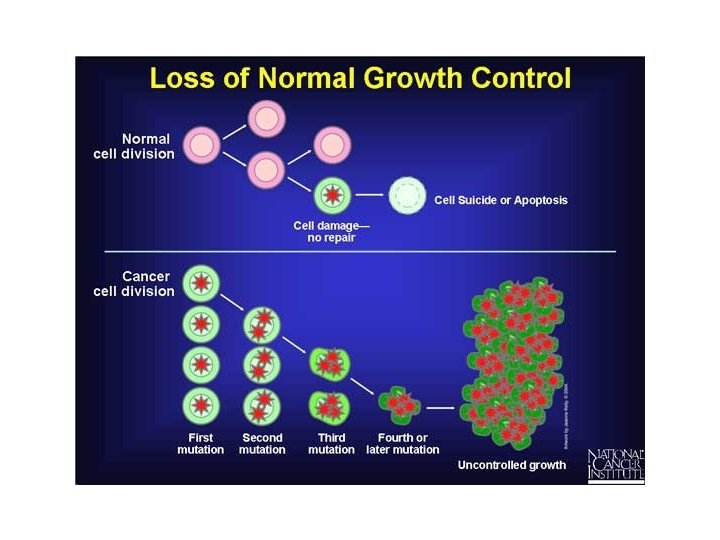
Cells that don’t divide correctly usually undergo APOPTOSIS • Cells that escape this suicide gene go on to Unrestricted cell division and therefor tissue growth. • What is this disorder called?

Breast Cancer Cells Dividing • http: //www. youtube. com/watch? v=Hm 03 r. CU ODqg

Development of Cancer • Cancer develops only after a cell experiences ~6 key mutations (“hits”) – unlimited growth • turn on growth promoter genes – ignore checkpoints • turn off tumor suppressor genes (p 53) – escape apoptosis • turn off suicide genes – immortality = unlimited divisions • turn on chromosome maintenance genes – promotes blood vessel growth • turn on blood vessel growth genes – overcome anchor & density dependence • turn off touch-sensor gene

What causes these Mutations? • Mutations in cells can be triggered by u u UV radiation chemical exposure radiation exposure heat u u cigarette smoke pollution age genetics

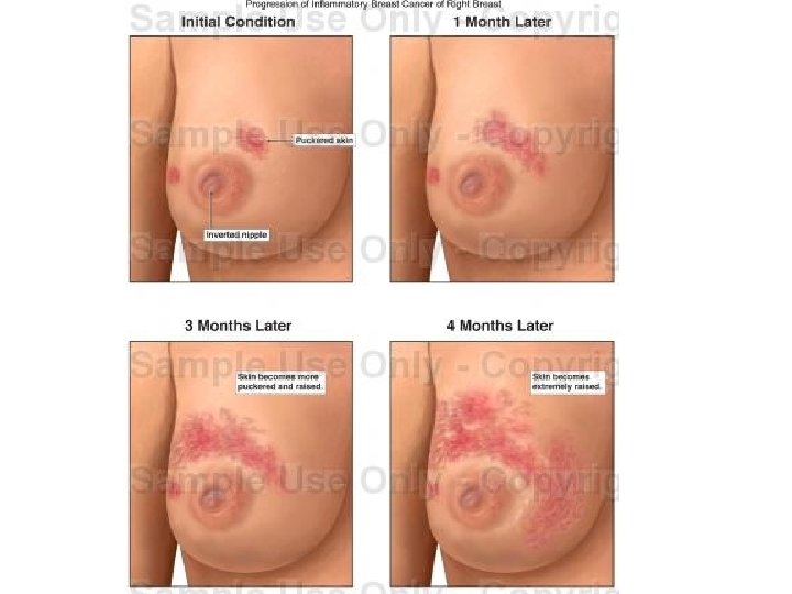
Tumors • Mass of abnormal cells – Benign tumor • abnormal cells remain at original site as a lump – p 53 has halted cell divisions • most do not cause serious problems & can be removed by surgery – Malignant tumor • cells leave original site – lose attachment to nearby cells – carried by blood & lymph system to other tissues – start more tumors = metastasis • impair functions of organs throughout body

Egg and Sperm: Sex Cells • Cannot undergo Mitosis. Why?
 wants to reproduce? – joining of")
What if a complex multicellular organism (like us) wants to reproduce? – joining of egg + sperm • Do we make egg & sperm by mitosis? What if we did, then…. 46 egg + 46 92 sperm zygote
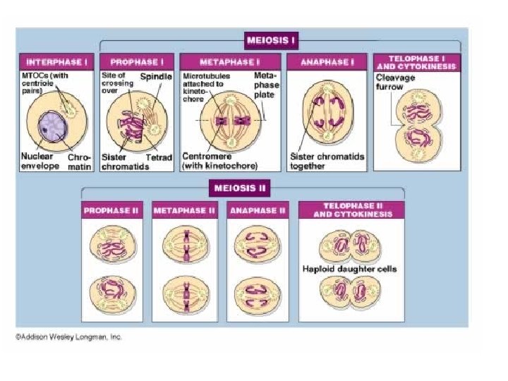

1 st division of meiosis separates homologous pairs MEIOSIS I: Homologous chromosomes separate Centrosomes (with centriole pairs) Nuclear envelope METAPHASE I PROPHASE I INTERPHASE Sites of crossing over Spindle Chromatin Sister chromatids Tetrad Microtubules attached to kinetochore Metaphase plate Centromere (with kinetochore) ANAPHASE I Sister chromatids remain attached Homologous chromosomes separate

2 nd division of meiosis separates sister chromatids MEIOSIS II: Sister chromatids separate TELOPHASE I AND CYTOKINESIS PROPHASE I METAPHASE II ANAPHASE II TELOPHASE II AND CYTOKINESIS Cleavage furrow Sister chromatids separate Haploid daughter cells forming

Trading pieces of DNA • Crossing over – during Prophase 1, sister chromatids intertwine • homologous pairs swap pieces of chromosome – DNA breaks & re-attaches synapsis tetrad prophase 1

Putting it all together… meiosis fertilization mitosis + development gametes 46 23 meiosis 46 egg 23 23 23 zygote fertilization sperm 46 4646 46 mitosis development
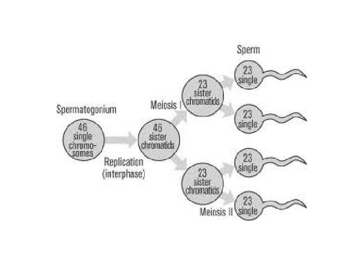
 primary spermatocyte (diploid) MEIOSIS")
Sperm production Epididymis Testis Coiled seminiferous tubules germ cell (diploid) primary spermatocyte (diploid) MEIOSIS I secondary spermatocytes (haploid) Vas deferens spermatids (haploid) spermatozoa • Spermatogenesis – continuous & prolific process Cross-section of seminiferous tubule – each ejaculation = 100 -600 million sperm MEIOSIS II
 fallopian tube fertilization MEIOSIS I primary oocyte (diploid)")
Oogenesis primary follicles germinal cell (diploid) fallopian tube fertilization MEIOSIS I primary oocyte (diploid) secondary oocyte (haploid) MEIOSIS II ovum (haploid) developing follicle mature follicle with secondary oocyte ruptured follicle (ovulation) corpus luteum

The value of sexual reproduction • Sexual reproduction introduces genetic variation – genetic recombination • independent assortment of chromosomes – random alignment of homologous chromosomes in Metaphase 1 – crossing over • mixing of alleles across homologous chromosomes – random fertilization • which sperm fertilizes which egg? • Driving evolution – providing variation for natural selection

What is DNA and How does it Replicate?

DNA and RNA are polymers of nucleotides or polynucleotides= Nucleic Acids – The monomer unit of DNA and RNA is the nucleotide, containing – Nitrogenous base – 5 -carbon sugar (ribose) – Phosphate group What does this look like? Copyright © 2009 Pearson Education, Inc.
 Cytosine (C) Pyrimidines Guanine (G) Adenine")
There are 4 bases in DNA Thymine (T) Cytosine (C) Pyrimidines Guanine (G) Adenine (A) Purines

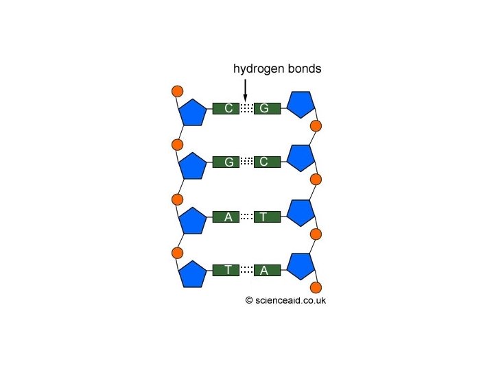
– DNA is composed of two polynucleotide chains joined together by hydrogen bonding between bases, twisted into a helical shape – The sugar-phosphate backbone is on the outside – The nitrogenous bases are perpendicular to the backbone in the interior – Specific pairs of bases give the helix a uniform shape – A pairs with T, forming two hydrogen bonds – G pairs with C, forming three hydrogen bonds

Twist

Hydrogen bond Base pair Ribbon model Partial chemical structure Computer model

Extracting DNA

DNA REPLICATION Copyright © 2009 Pearson Education, Inc.

DNA replication depends on specific base pairing – DNA replication follows a semiconservative model – The two DNA strands separate – Each strand is used as a pattern to produce a complementary strand, using specific base pairing – Each new DNA helix has one old strand with one new strand http: //www. youtube. com/watch? v=zd. Dki. Rw 1 Pd. U&feature=related

Replication: 1 st step • Unwind DNA – helicase enzyme • unwinds part of DNA helix • stabilized by single-stranded binding proteins helicase single-stranded binding proteins replication fork

Replication: 2 nd step § Build daughter DNA strand u DNA Polymerase III add new complementary bases

Editing & proofreading DNA • 1000 bases/second = lots of typos! • DNA polymerase I – proofreads & corrects typos – repairs mismatched bases – removes abnormal bases • repairs damage throughout life – reduces error rate from 1 in 10, 000 to 1 in 100 million bases

Fast & accurate! • It takes E. coli <1 hour to copy 5 million base pairs in its single chromosome – divide to form 2 identical daughter cells • Human cell copies its 6 billion bases & divide into daughter cells in only few hours – remarkably accurate – only ~1 error per 100 million bases – ~30 errors per cell cycle

Parental molecule of DNA

Nucleotides Parental molecule of DNA Both parental strands serve as templates

Nucleotides Parental molecule of DNA Both parental strands serve as templates Two identical daughter molecules of DNA
 DNA double helix (2 -nm diameter) Metaphase chromosome")
Tight helical fiber (30 -nm diameter) DNA double helix (2 -nm diameter) Metaphase chromosome Linker “Beads on a string” Nucleosome (10 -nm diameter) Histones Supercoil (300 -nm diameter) 700 nm

Mutations can change the meaning of genes: the function of proteins – A mutation is a change in the nucleotide sequence of DNA – Base substitutions: replacement of one nucleotide with another – Effect depends on whethere is an amino acid change that alters the function of the protein – Deletions or insertions – Alter the reading frame of the m. RNA, so that nucleotides are grouped into different codons – Lead to significant changes in amino acid sequence downstream of mutation – Cause a nonfunctional polypeptide to be produced Copyright © 2009 Pearson Education, Inc.

Mutations can change the meaning of genes: The function of proteins – Mutations can be – Spontaneous: due to errors in DNA replication or recombination – Induced by mutagens – High-energy radiation – Chemicals (Usually detrimental) Copyright © 2009 Pearson Education, Inc.

Acquired mutations are changes in DNA that develop throughout a person's lifetime.

Hereditary mutations are carried in the DNA of the reproductive cells. When reproductive cells containing mutations combine to produce offspring, the mutation will be in all of the offspring's body cells

Accidents during meiosis can alter chromosome number – Nondisjunction is the failure of chromosomes or chromatids to separate during meiosis – During Meiosis I – Both members of a homologous pair go to one pole – During Meiosis II – Both sister chromatids go to one pole – Fertilization after nondisjunction yields zygotes with altered numbers of chromosomes Copyright © 2009 Pearson Education, Inc.

Nondisjunction in meiosis I

Nondisjunction in meiosis I Normal meiosis II

Nondisjunction in meiosis I Normal meiosis II Gametes n+1 n– 1 Number of chromosomes n– 1

Normal meiosis I

Normal meiosis I Nondisjunction in meiosis II

Normal meiosis I Nondisjunction in meiosis II Gametes n+1 n– 1 n Number of chromosomes n
 90 80 70 60 50 40")
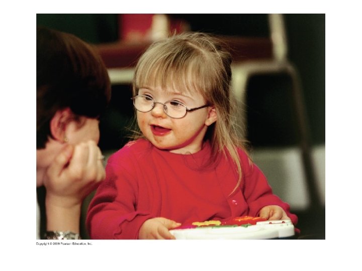
Infants with Down syndrome (per 1, 000 births) 90 80 70 60 50 40 30 20 10 0 20 25 40 30 35 Age of mother 45 50

Abnormal numbers of sex chromosomes do not usually affect survival – Sex chromosome abnormalities tend to be less severe as a result of – Small size of the Y chromosome – X-chromosome inactivation – In each cell of a human female, one of the two X chromosomes becomes tightly coiled and inactive – This is a random process that inactivates either the maternal or paternal chromosome – Inactivation promotes a balance between the number of X chromosomes and autosomes Copyright © 2009 Pearson Education, Inc.
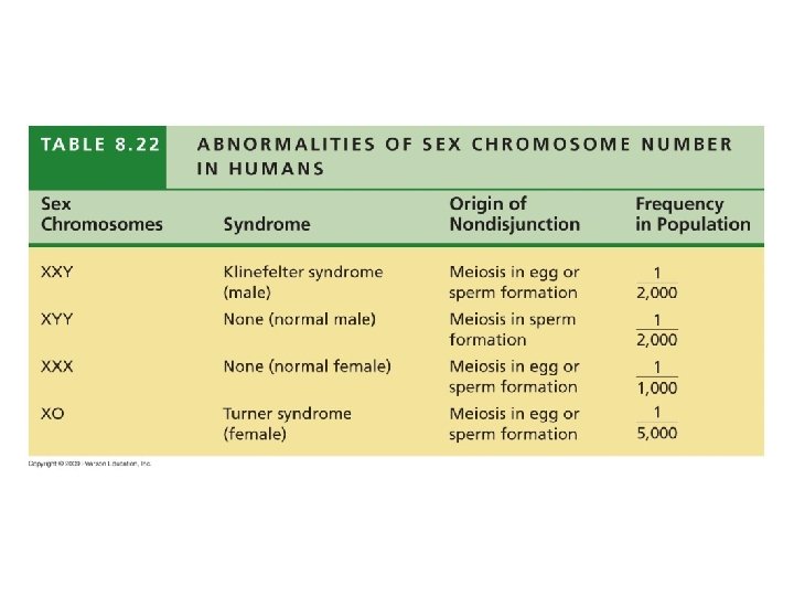

Klinefelters

Turner’s Syndrome
- Slides: 77