Cell Physiology Introduction Organelles Plasma Membrane Transport Mechanisms

Cell Physiology Introduction Organelles Plasma Membrane Transport Mechanisms

I. An Introduction to Cells § Cell Theory § Developed from Robert Hooke’s research § Cells are the building blocks of all plants and animals § All cells come from the division of preexisting cells § Cells are the smallest units that perform all vital physiological functions § Each cell maintains homeostasis at the cellular level Copyright © 2009 Pearson Education, Inc. , publishing as Pearson Benjamin Cummings

Origin of Eukaryotic Cells § Endosymbiotic theory
")
§ Endosymbiotic Theory § Eukaryotes evolved from symbiotic relationships between anaerobic bacteria ( chloroplasts) and intracellular aerobic bacteria ( mitochondria) § Evidence: § Mitochondria and chloroplasts have bacterial traits; they have ribosomes, own DNA, and reproduce by binary fission § We inherit our mitochondrial DNA from our mom
 § All body cells")
An Introduction to Cells § Somatic cells (soma = body) § All body cells except sex cells § Sex cells (germ cells) § Reproductive cells § Male sperm § Female oocyte (a cell that develops into an egg) Copyright © 2009 Pearson Education, Inc. , publishing as Pearson Benjamin Cummings

Cell Functions § Metabolism – Use molecules for cellular functions, to make ATP and heat § Molecule synthesis – Different cells synthesize different molecules. Structural and functional characteristics are based on molecules they produce. § Communication – Cells produce and respond to chemical and electrical signals § � Reproduction and inheritance – Most cells have a complete copy of all of our genetic information. This is passed down from cell to cell and from parent to child

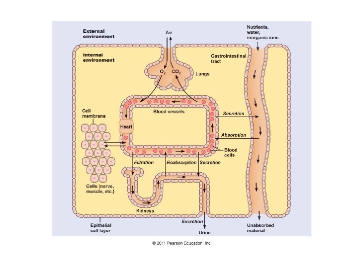
An Introduction to Cells § A cell is surrounded by a watery medium known as the extracellular fluid § Extracellular fluid is interstitial fluid + plasma + cerebrospinal fluid + synovial fluid § The plasma membrane separates cytoplasm (intracellular fluid) from the extracellular fluid (ECF) § Cytoplasm= cytosol + organelles § Cytosol = liquid § Organelles are Intracellular structures

**Components are indicated by blue color

Plasma Membrane
 § Dissolved materials: – nutrients, ions, proteins, and waste")
Cytoplasm § Cytosol (watery matrix) § Dissolved materials: – nutrients, ions, proteins, and waste products § High potassium/low sodium levels compared to ECF § High protein content compared to ECF § High carbohydrate/low amino acid and fat inside cell § Fluids § Organelles=“little organs” § Structures with specific functions
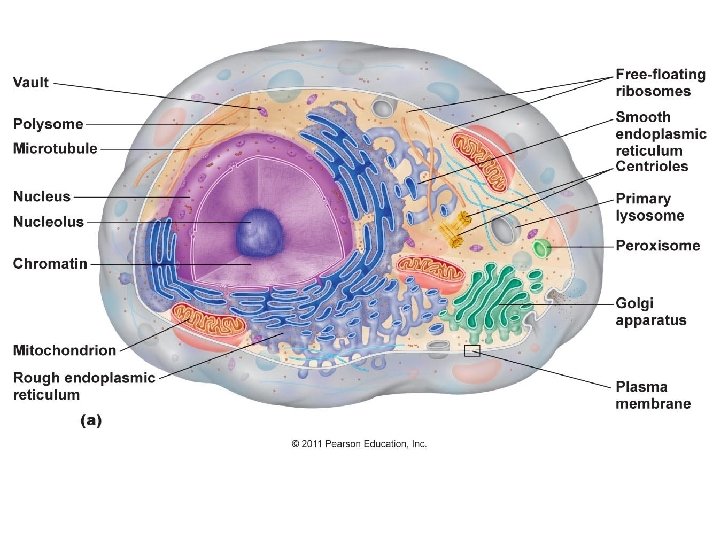

II. Organelles § Nonmembranous organelles § No membrane § Direct contact with cytosol § Includes the cytoskeleton, microvilli, centrioles, cilia, ribosomes, and proteasomes § Membranous organelles § Covered with plasma membrane § Isolated from cytosol § Includes the nucleus, endoplasmic reticulum (ER), the Golgi apparatus, lysosomes, peroxisomes, and mitochondria

Organelles § Nonmembranous Organelles § The Cytoskeleton — structural proteins for shape and strength § Microfilaments § Intermediate filaments § Microtubules Copyright © 2009 Pearson Education, Inc. , publishing as Pearson Benjamin Cummings

Figure 3. 16

Organelles and the Cytoplasm § The Cytoskeleton § Microfilaments—thin filaments composed of the protein actin § Provide additional mechanical strength § Interact with proteins for consistency § Pair with thick filaments of myosin for muscle movement Copyright © 2009 Pearson Education, Inc. , publishing as Pearson Benjamin Cummings

Organelles and the Cytoplasm § The Cytoskeleton § Intermediate filaments—mid-sized between microfilaments and thick filaments § Durable (collagen) § Strengthen cell and maintain shape § Stabilize organelles § Stabilize cell position Copyright © 2009 Pearson Education, Inc. , publishing as Pearson Benjamin Cummings

Organelles and the Cytoplasm § The Cytoskeleton § Microtubules—large, hollow tubes of tubulin protein § Strengthen cell and anchor organelles § Change cell shape § Move vesicles within cell (kinesin and dynein) § During cellular division they form the spindle apparatus that attaches to chromosomes to pull them to opposite ends of the dividing cell

Cytoskeleton

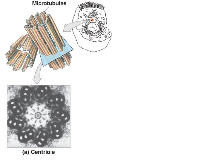
Organelles § Centrioles § Two only and housed in the centromere § Barrel-shaped, composed of nine microtubule triplets § Forms spindle apparatus during cellular division and used in cilia and sperm flagella for movement

Organelles § Microvilli § Extension of the cell to increase surface area of the cell § Found in brush border of small intestine, stereocilia of ear, WBC, and oocyte. § Cilia § Small hair-like extensions § Cilia move fluids across the cell surface § Found in alveoli and fallopian tubes § Flagella § Tail of sperm that consists of microtubules

Microvilli Figure 3– 3 The Cytoskeleton. Copyright © 2009 Pearson Education, Inc. , publishing as Pearson Benjamin Cummings

Microvilli Figure 3– 3 The Cytoskeleton. Copyright © 2009 Pearson Education, Inc. , publishing as Pearson Benjamin Cummings

Cilia

Organelles and the Cytoplasm Figure 3– 4 Centrioles and Cilia. Copyright © 2009 Pearson Education, Inc. , publishing as Pearson Benjamin Cummings

Organelles § Ribosomes § Made in nucleolus and shipped to cytoplasm § Build polypeptides in protein synthesis § Two types § Free ribosomes in cytoplasm: – manufacture proteins for cell § Fixed ribosomes attached to ER: – manufacture proteins for secretion § Proteasomes § Contain enzymes (proteases) § Disassemble damaged proteins for recycling

Figure 3. 12
 § Endoplasmic reticulum (ER)-Rough and Smooth")
Organelles § Membranous Organelles § Nucleus (double membrane) § Endoplasmic reticulum (ER)-Rough and Smooth ER § Golgi apparatus § Lysosomes § Peroxisomes § Mitochondria (double membrane) Copyright © 2009 Pearson Education, Inc. , publishing as Pearson Benjamin Cummings
")
Largest Organelle Nucleus (stained yellow)

Organelles § Nucleus § Houses the DNA § Serves as the cell’s control center § Surrounded by two membranes, together called the nuclear envelope § The nuclear envelope is studded with nuclear pores. § Nuclear pores regulate traffic into and out of the nucleus.

Organelles § Inside the nucleus: § Chromatin – composed of DNA + proteins § Nucleolus – site of ribosome manufacture § Nucleoplasm – fluid inside the nucleus Figure 3. 9
 § Endo- = within, plasm = cytoplasm, reticulum =")
Organelles § Endoplasmic reticulum (ER) § Endo- = within, plasm = cytoplasm, reticulum = network § Has cisternae are storage chambers within membranes § Functions § Synthesis of proteins, carbohydrates, and lipids § Storage of synthesized molecules and materials § Transport of materials within the ER § Detoxification of drugs or toxins

Organelles Figure 3– 5 The Endoplasmic Reticulum. Copyright © 2009 Pearson Education, Inc. , publishing as Pearson Benjamin Cummings
 § Smooth endoplasmic reticulum (SER) § No ribosomes attached")
Organelles § Endoplasmic reticulum (ER) § Smooth endoplasmic reticulum (SER) § No ribosomes attached § Synthesizes lipids and carbohydrates: – phospholipids and cholesterol (membranes) – steroid hormones (reproductive system) – glycerides (storage in liver and fat cells) – glycogen (storage in muscles) Copyright © 2009 Pearson Education, Inc. , publishing as Pearson Benjamin Cummings
 § Rough endoplasmic reticulum (RER) § Surface covered with")
Organelles § Endoplasmic reticulum (ER) § Rough endoplasmic reticulum (RER) § Surface covered with ribosomes: – active in protein and glycoprotein synthesis – folds polypeptides protein structures – encloses products in transport vesicles Copyright © 2009 Pearson Education, Inc. , publishing as Pearson Benjamin Cummings

Organelles Figure 3– 5 Rough Endoplasmic Reticulum. Copyright © 2009 Pearson Education, Inc. , publishing as Pearson Benjamin Cummings

Organelles § Golgi Apparatus § A stack of membranous sacs § Vesicles pinch off from the ER to fuse with the Golgi apparatus and empty their protein contents. § The proteins are then modified, sorted, and sent to their appropriate destination in new vesicles that bud off from the Golgi apparatus.

Organelles Figure 3– 6 The Golgi Apparatus. Copyright © 2009 Pearson Education, Inc. , publishing as Pearson Benjamin Cummings

Organelles Figure 3– 6 The Golgi Apparatus. Copyright © 2009 Pearson Education, Inc. , publishing as Pearson Benjamin Cummings

Organelles Figure 3– 7 Functions of the Golgi Apparatus. Copyright © 2009 Pearson Education, Inc. , publishing as Pearson Benjamin Cummings

Organelles § Lysosomes § Powerful enzyme-containing vesicles: – lyso- = dissolve, soma = body § Primary lysosome: – formed by Golgi apparatus and inactive enzymes § Secondary lysosome: – lysosome fused with damaged organelle – digestive enzymes activated – toxic chemicals isolated Copyright © 2009 Pearson Education, Inc. , publishing as Pearson Benjamin Cummings

Organelles § Functions of Lysosomes § Clean up inside cells § § Break down large molecules Attack bacteria Recycle damaged organelles Eject wastes by exocytosis § Autolysis § Auto- = self, lysis = break § Self-destruction of damaged cells: – – lysosome membranes break down digestive enzymes released cell decomposes cellular materials recycle Copyright © 2009 Pearson Education, Inc. , publishing as Pearson Benjamin Cummings

Organelles and the Cytoplasm Figure 3– 8 Lysosome Functions. Copyright © 2009 Pearson Education, Inc. , publishing as Pearson Benjamin Cummings

Figure 3. 11

Organelles § Peroxisomes § Are enzyme-containing vesicles: § break down lipids and amino acids § produce hydrogen peroxide (H 2 O 2) § replicate by division Copyright © 2009 Pearson Education, Inc. , publishing as Pearson Benjamin Cummings

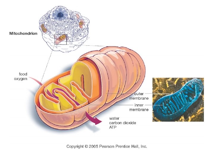
Organelles § Mitochondria § Uses carbs, lipids, and proteins to synthesize ATP § Has outer and inner membranes separated by the intermembrane space § Inner membrane carries proteins involved in ATP production § Matrix is site of reactions that release energy from nutrients

Figure 3. 10

III. Plasma Membrane § Functions of the Plasma Membrane § Physical isolation § Barrier § Regulates exchange with environment § Ions and nutrients enter § Wastes eliminated and cellular products released § Monitors the environment § Extracellular fluid composition § Chemical signals § Structural support § Anchors cells and tissues Copyright © 2009 Pearson Education, Inc. , publishing as Pearson Benjamin Cummings

Plasma Membrane § Comprised of a phospholipid bilayer-double layer of phospholipid molecules § Hydrophilic heads—toward watery environment, both sides § Hydrophobic fatty-acid tails—inside membrane, some are kinked to enhance fluidity of membrane + cholesterol to enhance fluidity § It is selectively permeable (semi-permeable). It is a barrier to large molecules, ions and water soluble compounds.

Plasma Membrane

Plasma Membrane § Fluid Mosaic Model-describes the plasma membrane as fluid, not static. § Movement of plasma membrane due to: § Unsaturated hydrophobic fatty acid tails-kink § Cholesterol

Membrane Fluidity Demonstrated:

Membrane Fluidity

Plasma Membrane § Membrane Proteins § Integral proteins § Span the membrane § They are amphipathic-polar and nonpolar § Peripheral proteins § Bound to inner or outer surface of the membrane Copyright © 2009 Pearson Education, Inc. , publishing as Pearson Benjamin Cummings

Plasma Membrane Figure 3– 2 The Plasma Membrane. Copyright © 2009 Pearson Education, Inc. , publishing as Pearson Benjamin Cummings
 § Attach")
Plasma Membrane § Several Types of Membrane Proteins § Anchoring proteins (stabilizers) § Attach to inside or outside structures § Recognition proteins (identifiers) § Label cells as normal or abnormal § Enzymes § Catalyze reactions § Receptor proteins § Bind and respond to ligands (ions, hormones) § Carrier proteins § Transport specific solutes through membrane § Channels § Regulate water flow and solutes through membrane Copyright © 2009 Pearson Education, Inc. , publishing as Pearson Benjamin Cummings

Plasma Membrane Figure 3– 2 The Plasma Membrane. Copyright © 2009 Pearson Education, Inc. , publishing as Pearson Benjamin Cummings

Plasma Membrane § Membrane Carbohydrates § Proteoglycans, glycoproteins, and glycolipids § Extend outside cell membrane § Form sticky “sugar coat” (glycocalyx) § Functions of the glycocalyx § Lubrication and protection § Anchoring and locomotion § Specificity in binding (receptors) § Recognition (immune response) Copyright © 2009 Pearson Education, Inc. , publishing as Pearson Benjamin Cummings

Plasma Membrane Figure 3– 2 The Plasma Membrane. Copyright © 2009 Pearson Education, Inc. , publishing as Pearson Benjamin Cummings
 membrane is a barrier, but § Nutrients")
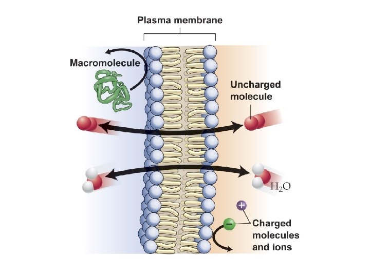
IV. Transport Mechanisms § The plasma (cell) membrane is a barrier, but § Nutrients must get in § Products and wastes must get out § Permeability determines what moves in and out of a cell, and a membrane that § Lets nothing in or out is impermeable § Lets anything pass is freely permeable § Restricts movement is selectively permeable Copyright © 2009 Pearson Education, Inc. , publishing as Pearson Benjamin Cummings

Transport Mechanisms § Plasma membrane is selectively permeable/semipermeable § Allows some materials to move freely § Restricts other materials from crossing over (they may need a transport protein of some type, or ATP) § Selective permeability restricts materials based on § Lipid solubility (lipophilic/ hydrophilic) § Size/ shape (small/ large) § Electrical charge (nonpolar/ polar & charged)

What can diffuse into cell?

Transport Mechanisms § Passive transport-doesn’t require ATP § Diffusion § Osmosis § Facilitated Diffusion § Active transport and secondary active transport § requires ATP § Endocytosis/Exocytosis § Filtration

IVA. Passive Transport § Passive transport – diffusion OR osmosis across the plasma membrane in living organisms § Energy from the cell is not required. § Diffusion involves only very small hydrophobic molecules, small polar molecules, or gasses. § Osmosis is the movement of water. § Both move down their concentration gradient § Rate of movement depends on difference of gradient § What about large molecules or ions? ?
![Diffusion [high] [low]](http://slidetodoc.com/presentation_image/27a813002a457c10a7f3e407f1525563/image-68.jpg "Diffusion [high] [low]")
Diffusion [high] [low]

Figure 3. 20

Osmosis § Osmosis is the passive movement of water across a semipermeable membrane from an area of high water concentration to an area of low concentration of water [high] [low] § OR mvt. of water to the side with more particles. § water wants to dilute the side with excess particles
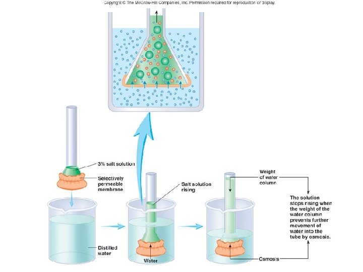

Osmotic Pressure § Osmotic pressure: pressure derived from particles in a solution. These particles influence movement of water. The side with more particles wins. § osmotic pressure activity § The greater the difference in concentration the greater the osmotic pressure will be the greater the pull will be on water § Why is this important for our bodies? ? ?

Osmosis § Comparative terms used to describe osmotic pressures of two solutions: § Both solutions have the same concentrations of solute particles; solution A is isoosmotic to solution § The solution with a greater concentration of solute (A) is hyperosmotic to the other solution (B) § The solution with a lesser concentration of solute (B) is hyposmotic to the other solution (A)

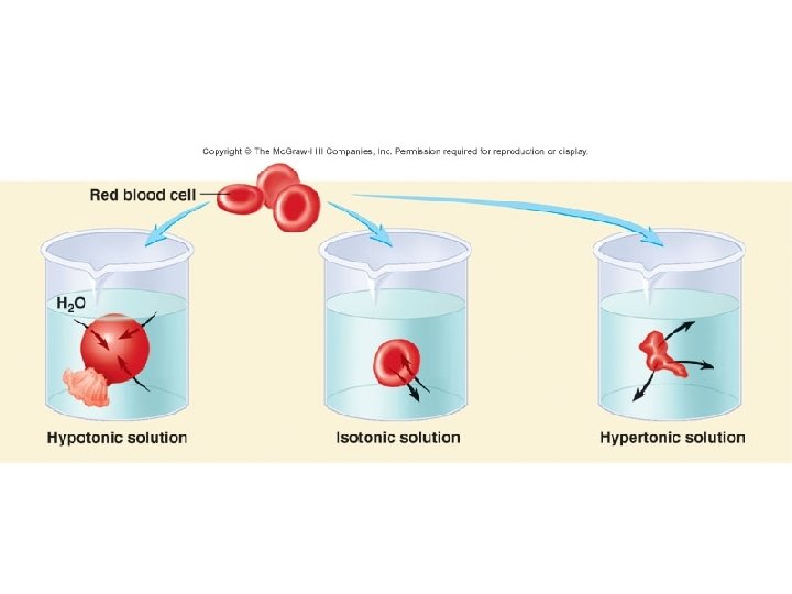
Tonicity § Tonicity –relative term that describes how a cell will behave in a solution. It indicates the conc. of the soln. § Isotonic – body fluids are isotonic to cells; there is an equal concentration of solutes and water on both sides of the cell membrane. § No net movement of water occurs

Tonicity § Tonicity –relative term that describes how a cell will behave in a solution. It indicates the conc. of the soln. § Hypertonic solution – the solution contains a higher concentration of dissolved solutes than the cell. § Net movement of water is out of the cell by osmosis § Cell shrivels/ crenates

Tonicity § Tonicity –relative term that describes how a cell will behave in a solution. It indicates the conc. of the soln. § Hypotonic solution – the solution contains a lower concentration of dissolved solutes than the cell. § Net movement of water is into the cell § Cell swells OR hemolyzes.

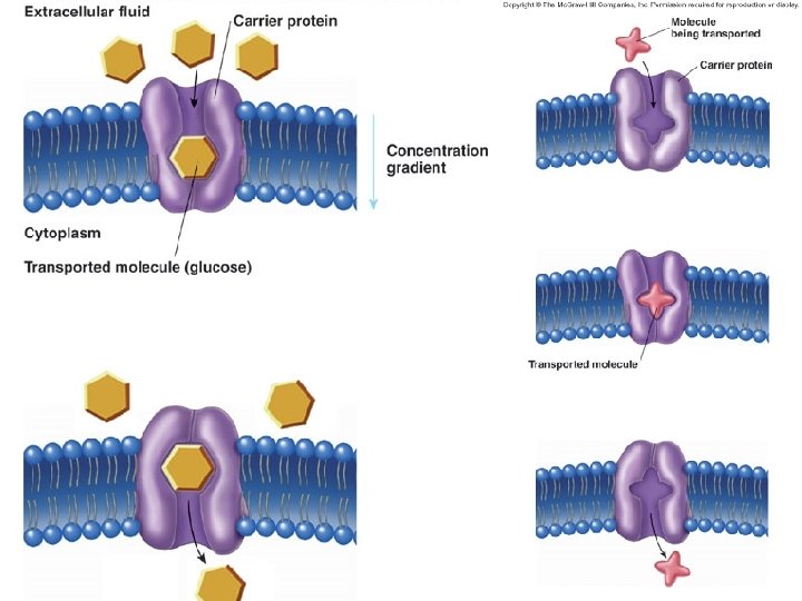
Diffusion/ Osmosis Facilitated diffusion Active transport

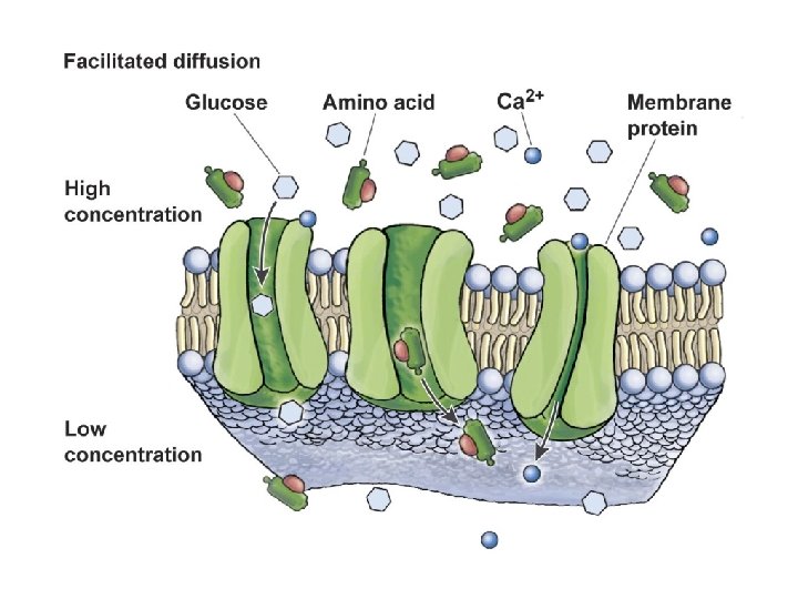
Facilitated Diffusion § Passive transport –facilitated diffusion § Facilitated diffusion involves moving ions and large polar molecules down their concentration gradient. § They cannot diffuse through the membrane due to size or charge so they need transportation. § There are two categories of transport proteins § Channel proteins – Non-gated channel proteins and gated channel proteins (ligand + voltage) § Carrier proteins

Channel Proteins Channel proteins are like pores in the membrane to let small polar molecules OR ions through the membrane. 1. Nongated channels (look like macaroni tubes) Always open in normal cells. Responsible for the permeability of the plasma membrane to ions when the plasma membrane is at rest

Channel Proteins 2. Gated channels= open or close by certain stimuli § Ligand gated channels open in response to small molecules that bind to integral proteins or glycoproteins § Voltage-gated channels open when there is a change in charge across an area of the plasma membrane http: //www. jci. org/articles/view/25505/figure/2

Carrier Proteins § Carrier proteins are integral proteins that carry large nonpolar, or ionic molecules across the plasma membrane down the molc. conc. gradient. § § Tranport amino acids, glucose, and proteins Have specific binding sites Protein changes shape to transport ions or molecules Resumes original shape after transport

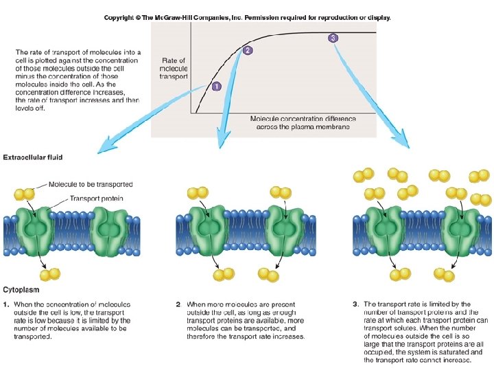
§ Carrier proteins exhibit the following characteristics similar to enzymes: § Specificity for a single type of molecule § Competition among molecules of similar shape § Saturation: rate of transport limited to number of available carrier proteins

Diffusion/ Osmosis Facilitated diffusion Active transport

IVB. Active Transport § Active transport: pumping substances across the membrane against their concentration gradients; this requires ATP. Usually these are referred to as pumps. [low] [high]

Active Transport

Primary Active Transport § 1 o active transport requires ATP allows the cell to accumulate substances against its conc. gradient § Rate of transport depends on [substrate] and [ATP] § Example: Na+/K+ exchange pump creates an electrical potential across membranes

Steps of Na+/ K+ ATPase: • ATP binds to ATP binding site; 3 Na+ ions binds to protein • ATP ADP + Pi • releases energy that pumps Na+ ions to other side • ADP leaves allowing 2 K+ to bind • Pi leaves and protein changes back to original shape and transports 2 K+ to intracellular environment • Cycle repeats

Secondary Active Transport § Use the concentration gradient derived from a primary active transport pump to drive another pump. § Example: Na/K ATPase sets up a Na gradient for Na/Glucose pump to pump glu AGAINST it’s concentration gradient.

Secondary Active Transport

Transport Mechanisms How do we get large molecules into the cell? § Exocytosis and endocytosis – movement of large molecules across the membrane § Exocytosis occurs when a membrane-bound vesicle carrying a substance fuses with the plasma membrane and secretes its contents to the cell’s exterior. § Endocytosis occurs when a substance is brought into the cell and the plasma membrane buds inward.

Exocytosis=accumulated vesicle secretions expelled from cell §Examples § Secretion of digestive enzymes by pancreas § Secretion of mucous by salivary glands § Secretion of milk by mammary glands

Endocytosis § Internalization of substances by formation of a vesicle § Types § Phagocytosis (shown) § Pinocytosis § Receptor-mediated endocytosis 3 -96

Pinocytosis and Receptor-Mediated Endocytosis

Filtration § Works like a sieve § Depends on pressure difference on either side of a partition § Moves from side of greater pressure to lower § Example: blood pressure causes fluid movement out of capillary interstitium § Water and small molecules move through the membrane while large molecules remain in the blood

Other Mechanisms § Secondary messenger systems § c. AMP and G-protein coupled mechanisms

Overall Goal § Move molecules in to and out of cell § Maintain concentrations of particles at a certain level inside and outside cell so H 2 O doesn’t burst cell § Maintain concentration gradients so molecules diffuse where they need to (O 2/CO 2) § Maintain concentration gradients so excitable cells can conduct charges
- Slides: 100