Cell membrane Structure and Functions Dr habil Khidai

Cell membrane – Structure and Functions Dr. habil. Kőhidai László Assoc. Professor Dept. Genetics, Cell- and Immunbiology Semmelweis University - Budapest 2019.

Prokaryotic cell cytopplasm Eukaryotic cell capsule secretory vesicle with cell wall cell membrane cilia microvilli pinocytotic vesicle ribosomes cell membrane microtubuli thylakoid píli flagellum microfilaments chloroplast centroles Golgi complex smooth ER ribosome lysosome nucleolus chromatine mitochondrium nucleoplasm cytoplasm rough ER nuclear pores nuclear envelope nucleus

Prokaryotic cell cytopplasm Eukaryotic cell capsule secretory vesicle with cell wall cell membrane cilia microvilli pinocytotic vesicle ribosomes cell membrane microtubuli thylakoid píli flagellum microfilaments chloroplast centroles Golgi complex smooth ER ribosome lysosome nucleolus chromatine mitochondrium nucleoplasm cytoplasm rough ER nuclear pores nuclear envelope nucleus

Compartmentalization Biochemical pathways are running in isolated, membrane bounded spaces – This provides the optimal environmental conditions to the processes.

Main functions of cytoplasmatic membranes Boundaries – facilitates in- and outward transport of nutrients Anchoring proteins – trasport, bioenergetical processes, sensing signals and chemotaxis Generating and storage of energy – generation of proton gradients

Functions of membranes • 1. Boundaries and selectively permeable – cell membrane/plasma membrane and intracellular membranes – cell organells • 2. Specific functions – they are distinguished upon proteins associated (integrant or periferial proteins). Glucose phosphatase – r. ER-membrane associated enzyme and its marker, too. • 3. Transports are regulated by membranes – a. Simple diffusion (high cc. ->low cc. ): H 2 O, O 2, CO 2, ethanol b. Facilitated diffusion (high cc. ->low cc. ) by the help of transport proteines: saccharides, amino acids c. Proteins of “pump mechanisms”, they are essential in ion-gradients: low cc. -> higy cc. Intake of saccharides, amino acids against concentration gradient. d. Endocytosis and exocytosis, etc.

Functions of membranes • 4. Sensing and transmission of electric and chemical signals – signal transduction: receiving and transmitting external signals E. g. : Estrogen – can cross membranes due to its hydrophobic character; other molecules require receptors which process generates secondary messengers. The signal can reach even the nucleus and can influence levels of gene expression. • 5. Cell-cell communication The communication mediator components are – gap junction (in animal cells) and plasmodesmata (in plant cells) allow direct, intercellular communication.

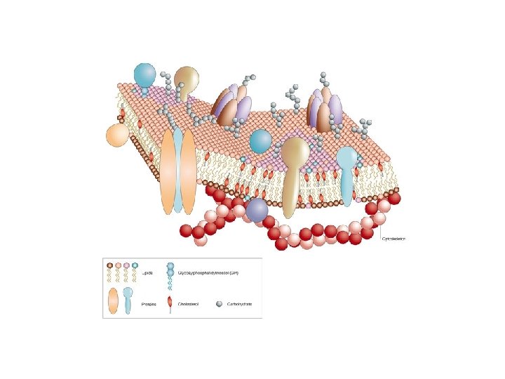
Membrane-models Lipid soluble substances enter the cell rapidly Benzine-lipid mixture, after evaporation of benzine, a molecular lipid film is formed Benzine-soluble lipids form a bilayer film on the surface of water Proteins are integral parts of the cell membrane. The lipid bilayer is covered by a layer of proteins inside and outside. Partially explains the rapid passage of proteins, sugars, ions and other hydrophilic substances. Discovery of electron microscope. The cells are covered with a plasma membrane. "Unit-membrane" model. . Mosaic arrangement of proteins in the membrane. Some proteins cross the membrane, transmembrane proteins

Fluid mosaic membrane model Singer – Nicolson 1972 RBC membrane

Membrane models – Fluid mosaic model: Proteins arranged mosaic-like, scattered and embedded within the liquid lipid bilayer It is the first protein to be described by the transmembrane domain and with its retinol-like and also energy-binding moiety. The energy of sunlight after binding causes a structural change that results in the release of protons from the cell. The resulting proton gradient serves as a source of energy. Lipids, proteins, and carbohydrates are also involved in the construction of most membranes.

General structure of phospholipids

The main lipid components that make up the membrane Phospholipides Glycolipides Other membrane lipids (not phospholipids) Cholesterol Cerebrosides Sphingolipides Ceramide

Membrane lipids: elements responsible for the fluidity of the model Several types of lipids are involved in the formation of membranes • Phospholipids Phosphoglycerides Sphingolipids Phospholipid composition of different membranes

• Glycolipids Cerebrosides and gangliosides Tay-Sachs disease: Lysosomes are deficient in β –N-acetilhexoseaminidase – degradation of gangliyosides is impaired; ganglyoside accumulation in CNS. • Steroids (they are not found in prokaryotes or in the inner membranes of the mitochondria, the color body) Cholesterol Phytosterol The membranes of bacteria and cyanobacteria contain steroid-like molecules - haponoid (rich in petroleum deposits; membrane components of early prokaryotes? )

Thin layer chromatography - an important tool for lipid analysis: Stationary phase - thin silica gel with a hydrophilic layer on a glass or metal plate Mobile phase - a mixture of solvent, chloroform, methanol, and water. Low affinity Cholesterol Phosphatidylethonolamine (PE) Phosphatidylcholine (PC) High affinity Phosphatidylserine (PS) Fatty acids are also important for membrane structure and function Fatty acids are components of all membrane lipids except steroids. Hydrophobic hydrocarbons are generally 16 or 18 -C long and provide stability to the lipid bilayer. . Saturated Fatty Acids: Palmitic (16 C) and Stearic (18 C) Unsaturated Fatty Acids: Oleic and Linoleic Acids (both are 18 C)

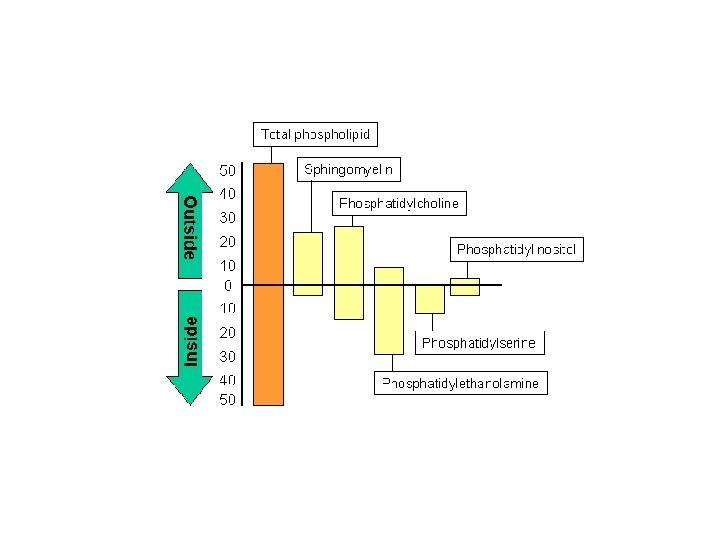
Membrane asymmetry: the majority of lipids are unequally distributed in the two lipid monolayers - differences in quantity and quality Asymmetry is embodied in differences in lipid types and degrees of saturation. Asymmetry occurs during the biogenesis of membranes. The resulting asymmetry, for thermodynamic reasons, prevents the exchange of lipids between the two surfaces. External Membrane: Membrane Glycolipids - Recognition processes and signaling Internal membrane: membrane important for signaling to the cell's interior - phosphatidylethanolamine, phosphatidylinositol, phosphatidylserine Membrane asymmetry: the majority of lipids are unequally distributed in the two lipid monolayers - differences in quantity and quality Movement of lipids in the membrane: - "Flip-flop" or inverse diffusion - rotation - lateral diffusion Flip-flop movement is rare, but is promoted in biological membranes by phospholipid translocators or flippases. Therefore, phospholipid translocation in biological membranes is more common than in artificial lipid bilayers.

Development and maintenance of membrane asymmetry : • Phospholipids are synthesized bound to the ER membrane. • Phospholipids bind to the cytoplasmic surface of the ER • Flippase transfers each lipid to the other side of the bilayer.

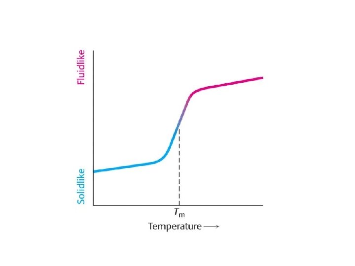
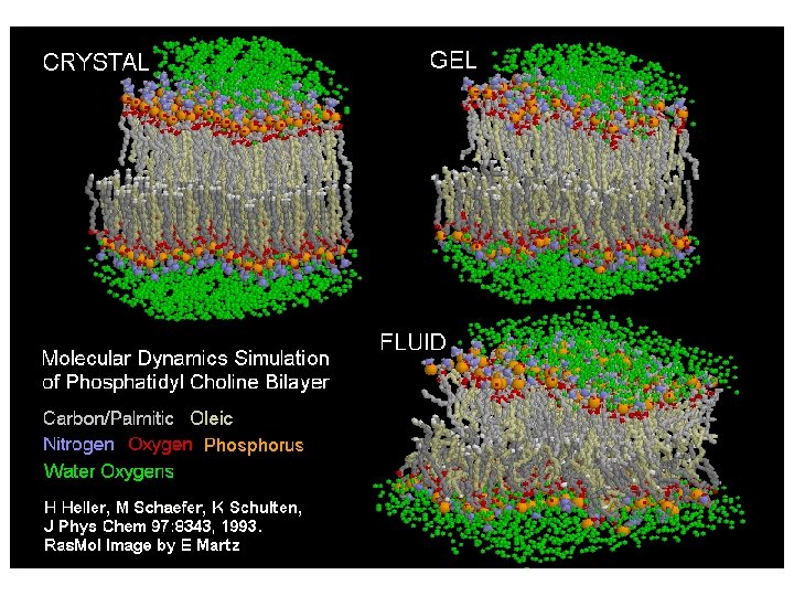
Investigation of the fluidity of the lipid bilayer By lateral diffusion, lipids and proteins, can move a few millimeters (equal to the length of a bacterium) of a lipid of 800 M. S. in 1 second! Due to their large size, proteins move more slowly. Test procedure: fluorescence recovery after photobleaching (FRAP) technique The membrane only functions properly in its "fluid" state Phase transition temperature (Tm): Phase transition: gel-like state -> fluid Differential scanning calorimetry Calorimeter: chamber for measuring heat absorption. The location of the peak of the curve indicates Tm. Unsatuirated: Low Tm Saturated: High Tm

76 o. C Membrane-fluidity and fatty acid composition There are two main aspects: chain length and degree of saturation Membrane-lipids often consist of 1 saturated and 1 unsaturated fatty acid 70 o. C 32 o. C Effect of steroids on membrane fluidity -11 o. C Steroids - cholesterol and phytosterol. Steroids may represent up to 50% of the total membrane lipid content Cholesterol - forms hydrogen bonds with the oxygen atoms of neighboring phospholipids. Paradoxical effect of cholesterol: decreases membrane fluidity at high temperatures (effect of rigid rings) and increases fluidity at low temperatures (inhibiting the natural arrangement of adjacent hydrocarbon chains) HOWEVER, steroids reduce the permeability of the lipid bilayer - filling the space between phospholipids. The organisms can control membrane fluidity Poikelotherms (bacteria, fungi, plants, amphibians, etc. ) vs. homeotherms (warm-blooded animals) Homeoviscopic adaptation - membrane viscosity retained despite changing temperature - shortening or reduction of fatty acid chains by removal of terminal C's. de-saturation (e. g. , E. coli desaturase) 18 -C lipids

The fluidity of the membrane is influenced by: phospholipids: chain length saturation Cholesterol content

Lipid rafts: - High in cholesterol - Micro-domains -Glycolipids + sphingolipids - Rafts are present in the cell surface membrane and on membranes attached to the Golgi -Their importance: to reduce the difference in free energy between each cell membrane layer membrane bound transport processes Other names in the literature: - detergent-insoluble glycolipid-enriched complexes (GEM or DIG) - detergent resistant membranes (DRMs) Detection: fluorescence microscopy - GM 1 gangliosides; AFM

Membrane Proteins: The "mosaic" character of the model Freezing fracturing: rapid freezing (in liquid nitrogen) then diamond blade exploratin after E face (exoplasmatic face) and P face (protoplasmatic face)

Membrane proteins: Integrant membrane proteins: ~20 amino acids, transmembrane segment. Hard to isolate, extraction requires treatment with detergents. Intergrant, monotopic proteins Transmembrane proteins: singlepass multi-subunit - integrant - peripheric - anchored by lipids Glycophorin (vvt. ) Bacteriorhodopsin Hydropathy plot

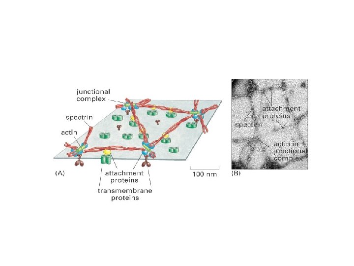
Peripheral membrane proteins are attached to membrane surfaces by weak electrostatic forces or hydrogen bonds. Connection with hydrophilic moieties of integrant proteins or polar head portions of membrane lipids. E. g. : spectrin and ankyrin in RBC plasma membrane. The proteins can be separated by SDS-polyacrylamide gel electrophoresis Isolation of membrane proteins Peripheral membrane proteins: altering p. H or ionic bonds, removing calcium chelating agents (cationic bonds), or urea (breaking down hydrogen bonds). Lipid-bound proteins can be isolated in the same way as peripheral proteins, since covalent bonds must also be cleaved. . Extraction of integral membrane proteins is a more difficult, detergent-intensive task. The detergent breaks down the hydrophobic bonds and interacts with the lipid bilayer. SDS (sodium dodecyl sulfate) treatment SDS-polyacrylamide gel electrophoresis: The use of an electrophoretic electric field to separate electrically charged molecules. The migration speed depends on the charge and size. SDS digestion (proteins coated with negatively charged SDS) -> electrophoresis -> staining with Coomassie brilliant blue. Lipid-bound membrane proteins: covalent bond with lipid. - Fatty acid or prenyl as an anchor - GPI-switching (glycosylphosphatidyinositol, GPI)

Isolation and assay of membrane proteins: The detergents used mimics the phospholipid structure around proteins hydrophilic hydrophobic
 and")
Membrane proteins can perform many functions Enzyme: E. g. : glucose phsphatase (ER-assocated) and glycerolaldehyde-3 phosphate dehydrogenase (GPD), peripheral proteins of RBCs invoolved in the glucose metablism of blood. Transport proteins e. g. transport ATPases use energy of ATP to transfer ions across membrane. Receptors: e. g. hormones and growth factors Other: endocytosis and exocytosis; targeting, sorting, modification of proteins in ER or Golgi; light perception; stabilization of cell membrane structure and determination of shape (ankyrin, spectrin). TRANSPORTER ANCHORING Transmembrane (intergrant) Single-pass Multi-pass ? ? ? RECEPTORS ENZYMES Membrán-associated (periferial) Covalently bound vs non-covalent

Embedding of membrane proteins in bilayer The α-helical structure is very common. This is because the expression of the hydrophobic amino acids facilitates the anchoring of proteins to the hydrophobic layer of the membranes expressed. The a-helical structures hydrophilic components a are important in pore formation

The carbohydrate components of the cell membrane Functions: Surface protection Recognition Cell adhesion - extracellular matrix

Most of the membrane proteins are glycosylated Human RBC membrane: 52% proteins, 40% lipids and 8% carbohydrates. Carbohydrates are found in glycolipid (rare) and glycoprotein (common) in the membrane Glycoproteins: They are N-linked through the amino group of the Asp side chain O-linked – through hydroxyl grup of Ser or. Thr Some O-linked carbohydrates are linked via the hydroxyl group of hydroxy-Lys or hydroxy-Pro

Length of protein-bound carbohydrates: 2 -60 units, straight v. linear Common building blocks: galactose, mannose, N-acetylglucosamine and sialic acid The integral protein (glycophorin) of RBC has a negatively charged sialic acid at its terminal position (15 O-linked and 1 Nlinked), which reduces the tendency of RBCs to adhere and to reduce blood viscosity. Glycoproteins play a primary role in the cell-cell recognition function of cell membranes Experiment: Lectins: plant (animal) proteins with sugarspecific bonds. E. g. : wheat germ agglutinin (wheat) binds oligsaccharides with terminal Nacetylglucoseamine; the concanavalin is isolated from „jack bean” and binds mannoses of oligosaccharides. Ferritin is an iron cntaining protein which is bound to glycoproteins – it is easy to detect its binding due to its high electronedensity.

Mobility of membrane proteins can vary widely Some proteins move freely while others bind. Technique used to detect mobility: cell fusion

Cell membrane protein movement is restricted : E. g. polarized surface of intestinal epithelial cells or glandular epithelium Polarized location, appearance of receptors, transporters, cell coupling structures.

Take home message

Extracellular Intracellular

Reference: Hardin, J. , Kleinsmith, L. , Becker, W. : World of the cell with free solutions, (Benjamin Cummings, 2002) Alberts, B. , Johnson, A. , Lew, J. : Essential Cell Biology, (Garland, 2004)
- Slides: 40