CELL MEMBRANE CHEMICAL COMPOSITION STRUCTURE AND MEMBRANE DYNAMICS

CELL MEMBRANE : CHEMICAL COMPOSITION , STRUCTURE AND MEMBRANE DYNAMICS Presenter: Dr. Dnyanesh Amle Moderator: Dr. Smita Kaushik

• Boundaries of all the cells are defined by biological membrane • Barrier with selective permeability
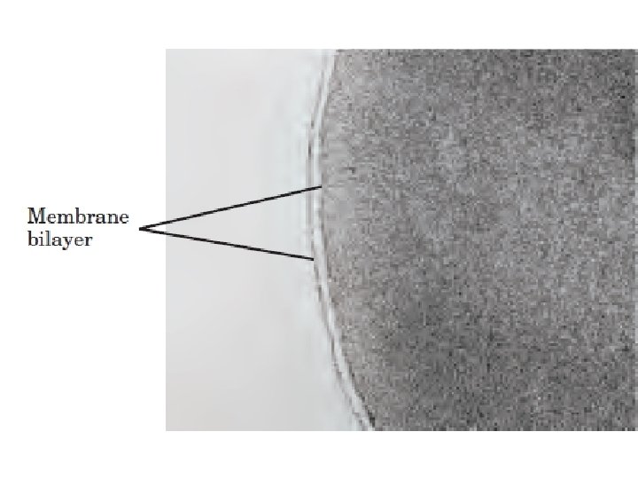

COMMON PROPERTIES: • Sheet like structures • Contains mainly lipids and proteins • Membrane lipids are small amphipathic molecules • Specific proteins mediate distinctive function

COMMON PROPERTIES: • Non covalent assemblies • Asymmetric • Fluid structures • Electrically polarized

• FUNCTIONS: – Maintenance of cell shape – Cellular movements – Controls movement of molecules between inside and outside of the cell – Cell-cell recognition and communication

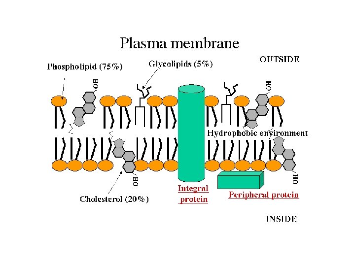
• MAINLY COMPOSED OF : – Lipids – Proteins – Carbohydrates

• LIPIDS : – Phospholipids – Glycolipids – Cholesterol
 PHOSPHOLIPIDS • Based on the platform: – Glycerophospholipids( Phosphoglycerides) – Sphingolipids")
1) PHOSPHOLIPIDS • Based on the platform: – Glycerophospholipids( Phosphoglycerides) – Sphingolipids

PHOSPHOGLYCERIDES HYDROPHOBIC PART FATTY ACID G L Y C E R O L HYDROPHILIIC PART PHOSPHATE ALCOHOL
, the simplest phosphoglyceride • Major phosphoglycerides are derivatives")
PHOSPHOGLYCERIDES • phosphatidate (diacylglycerol 3 -phosphate), the simplest phosphoglyceride • Major phosphoglycerides are derivatives of phosphatidate

• Phosphtidylinositol: – Golgi body – Endosomes – Plasma membrane • Cardiolipin: – Inner mitochondrial membrane • Phosphtidylcholine > Phosphtidylethanolamine • Plasmalogens: – Nervous tissue – Heart

SPHINGOLIPIDS • Derived from sphingosine SS P H I N G O S I N E FATTY ACID PHOSPHATE • Ceramide ALCOHOL
GLYCOLIPIDS: • In animal cells: derived from sphingosine • Sugar unit is attached to")
2)GLYCOLIPIDS: • In animal cells: derived from sphingosine • Sugar unit is attached to primary -OH group • Simple glycolipid : cerebroside – Phrenosine • Complex glycolipid: Ganglioside

• Galactocerebroside: – Brain and nervous tissue • Glucocerebroside: – Non neural tissue • Ganglioside: – 5 -8% lipid in brain
CHOLESTEROL: • Third major membrane lipid")
3)CHOLESTEROL: • Third major membrane lipid

OH

• MEMBRANE LIPIDS : AMPHIPATHIC MOLECULES Sphingo lipids glycero Phospho lipids Cholesterol SHORTHAND DEPICTION

AMPHIPATHIC NATURE: • Micelles • Bilayer: leaflets

PROPERTIES OF LIPID BILAYER: • Formation in aqueous environment is rapid and spontaneous • Hydrophobic interactions: major driving force • Other forces: – Van der waal’s attractive forces – Electrostatic – Hydrogen bonds • Co-operative structures

HYDROPHOBIC INTERACTIONS: • Inherent tendency to be extensive • Tend to close on themselves: forms compartments • Self sealing: As hole in lipid bilayer is energetically unfavorable

• Type of structures formed depends on: – Fatty acyl chains – Degree of saturation – Temperature

• LIPOSOMES: sonicating Gel filtration

Uses – To study the effect of different fatty acids on membranes – Drug delivery – Concentrate in regions of increased blood flow : Cell gating – Selective fusion

MEMBRANE PROTEINS: • Integral • Peripheral • Amphitropic

INTEGRAL MEMBRANE PROTEINS : Inside Outside

PERIFERAL MEMBRANE PROTEINS : Cytoplasmic side Phosphtidylinositol Membrane Anchored protein _____ +++++++

• Membrane proteins structure – Electron microscopy and X-ray crystallography • Membrane spanning α helix BACTERIORHODOPSIN

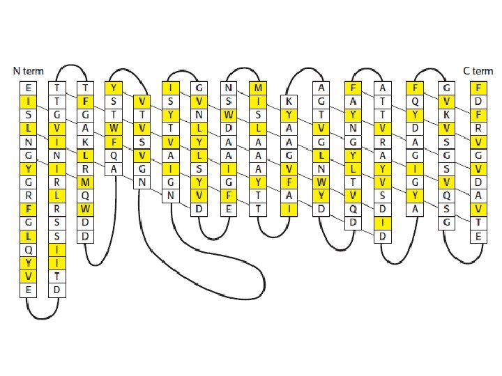
GLYCOPHORIN: • A protein containing single trans-membrane α helical strand • Present in plasma membrane of human erythrocytes • Amino terminus exterior to cell contains various oligosaccharide unit including ABO and MN blood group determinants

74 95 GLYCOPHORINE

• A channel can be formed from β strands PORINE

PROSTAGLANDIN H 2 SYNTHASE: Prostaglandin H 2 synthase

CARBOHYDRATES: • Rarely exists as free component • Present as glycoprotein and glycolipid • Always present on the outer side

BIOLOGICAL MEMBRANES DIFFER IN COMPOSITION: • Myelin: 18% protein , ↑ glycosphingolipids • Plasma membrane : 50% protein • Inner mitochondrial membrane : 75% protein, ↑ cardiolipins

FLUID MOSAIC MODEL: 1972 • S Jonathan Singer & Girth Nicolson • Membranes are two dimensional solution of lipids and globular proteins CARBOHYDRATES INTEGRAL PROTEINS PERIFERAL PROTEINS LIPIDS

MEMBRANE DYNAMICS • ↓ physiological temp. : gel phase • ↑ physiological temp. : liquid-disordered state • Intermediate temp. : liquid-ordered state • Unsaturated fatty acids • Cholesterol

LATERAL DIFFUSION: • Biological membranes are not rigid structures • Lipids > proteins are constantly in a lateral motion • Can be detected by FRAP • S = (4 Dt)1/2 • For lipid : D= 1µm 2/s • S= 2 µm/S

• Proteins differ extremely in mobility – Rhodopsin : D=0. 4 µm/s – Fibronectin : D = 10 -2µm/s • fluidity increases with increase in – No of short chain fatty acids – Unsaturated fatty acids – Temperature • Cholesterol decreases fluidity at high temp • Increases fluidity at low temp

TRANSVERSE DIFFUSION: • Flip flop occurs once in several hours • Flip flop of proteins have not been observed • Thus proteins play important role in preserving the asymmetry of the membrane • But sometimes Flip-Flop is needed

Cytosolic leaflet

MEMBRANE FLUIDITY : CLINICAL CORRELATION • ↑cholesterol : alteration in membrane fluidity • Spur cell anemia • Alcohol intoxication • Abetalipoproteinemia: ↑ sphingomyeline ↓phosphatidylcholine • Lecithin cholesterol acyltransferase deficiency • Hypertension • Alzheimer’s

MEMBRANES : ASYMMETRIC STRUCTURE • Inside-outside asymmetry: – Phospholipids – Proteins – Carbohydrates • Regional asymmetry • Mechanism

MICRO DOMAINS OF LIPID PROTEIN COMPLEX • Micro domain called lipid raft contains distinctly organized bilayer structures • Enriched in sphingolipids and cholesterols • Biological membranes are actually mosaic of Different microdomains

• Outer leaflet : ceramid and glycosphogilipids with long chain fatty acids → thicker • Inner leaflet ↑ saturated fatty acids → closed packing

• Function : to segregate and concentrate specific protein and to facilitate their activity • Proteins are activated when – several rafts fuse together – Ligands binding which favors fusion of rafts

CAVEOLAE: • Caveoline cholesterol binding integral membrane protein • Forces bilayer to curve inwards forming caveolae • Functions : membrane trafficking, signal transduction Caveoline dimer with six fatty acid moeitis

MEMBRANE CURAVATURE : • Central to ability of membrane to undergo fusion with other membrane • Mechanisms – Intrinsically curved protein binding – Many subunits of scaffold protein into proteins assembled into curved supra-molecular complexes – May insert one or more hydrophobic helices into one face of bilayer

FUSION OF SYNAPTIC VESICLE: • v-SNARES • t-SNARES • SNAP-25 • NSF V-SNARE SNAP-25 t-SNARE

TRANSFERRIN RECEPTOR CYCLE: Transferrin receptor cycle

IN A NUTSHELL • Biological membranes define cellular boundaries, divide cells into discrete compartments, organize complex reaction sequences, and act in signal reception and energy transformations. • The lipid bilayer is the basic structural unit explained by Fluid-mosaic model. • Membranes are structarally and functionally asymmetrical.

• Lipid > proteins are continuously in a state of motion in the plane of cell membrane called lateral diffusion • But transverse diffusion or Flip-flop of lipids is very slow except when specifically catalyzed by flippases, floppases and scramblases. • lipid rafts are rich in sphingolipids and cholesterol consist of membrane proteins that are GPI-linked • Specific proteins mediate the fusion of two membranes, which accompanies processes such as viral invasion and endocytosis and exocytosis

THANK YOU

HYDROPATHY PLOT GLYCOPHORINE

STEPS IN FUSION: • Recognition • Close opposing • Local disruption • Fusion proteins

TIGHT JUNCTION: • Present between two cells that lies in close a approximation • Forms narrow hydrophilic channels • Prevents the diffusion of macromolecules • Only three layers of plasma membrane are present DESMOSOMES: provide attachment of cells to the basal tissue • Mostly seen in epithelial cells
- Slides: 57