Cell Membrane Cell Transport Cell Division Chapter 7

Cell Membrane, Cell Transport & Cell Division Chapter 7: Membrane Structure and Function Chapter 12: The Cell Cycle

Plasma Membrane

Plasma Membrane Structure Boundary that separates the living cell from its surroundings Ø Phospholipids are the most abundant lipid l Arranged in a bilayer Ø l • hydrophobic region (tails) • hydrophilic region (heads) Ø Exhibits , allowing some substances to cross it more easily than others Ø The states that a membrane is a fluid structure with a “mosaic” of various proteins embedded in it

Phospholipid Structure

Selective Permeability A cell must exchange materials with its surroundings, a process controlled by the plasma membrane Ø Regulates what enters and leaves the cell Ø Hydrophobic (nonpolar) molecules, such as hydrocarbons, can dissolve in the lipid bilayer and pass through the membrane rapidly Ø Polar molecules, such as sugars, Ø Maintains Ø Allows for Ø inside the cell of cells in same organism

Fluid Mosaic Model Ø Phospholipid molecules can Ø Makes the membrane act like a Ø Fluid Mosaic Model Animation

Membrane Cholesterol is a Ø Adds to plasma membrane Ø Helps keep fatty acid tails of phospholipids separated Ø

Membrane Proteins Different proteins are embedded in the fluid matrix of the lipid bilayer Ø They determine most of the Ø Spread throughout the membrane like raisins in raisin bread Ø Allow membrane to “ ” with its environment Ø

Membrane proteins are bound to the surface of the membrane Ø penetrate the hydrophobic core Ø l l Integral proteins that span the membrane are called The hydrophobic regions of an integral protein consist of one or more stretches of nonpolar amino acids, often coiled into

Membrane proteins Ø Six major functions of membrane proteins: l l l Signal transduction l Intercellular joining l Attachment to the cytoskeleton and extracellular matrix (ECM) l l *Includes a carbohydrate chain

Carbohydrate chains Carbohydrates on the external side of the plasma membrane vary among species, individuals, and even cell types in an individual Ø Attached to phospholipids ( ) or proteins ( ) l These a cell l Individual (Mr. Fusco cell) l Species (Human cell) l Type (kidney cell) Ø l • Cells recognize each other by binding to surface molecules, often carbohydrate chains, on the plasma membrane

LABEL THESE PARTS Cholesterol Glycolipid Phospholipid Glycoprotein Integral protein Carbohydrate chain Peripheral protein Inside/Outside of cell

Cell Transport Because of the numerous amount of activities associated with the cell, substances must constantly move in and out of the cell Ø There are 2 types of cell transport: Ø l l

Passive versus Active Passive Active Ø Ø Ø the concentration gradient ( Ø From random molecular motion the Ø ) concentration gradient ( )

Concentration Gradient Ø From High Low Ø NO energy needed ØSubstances diffuse down their concentration gradient easily ØNo work must be done to move substances the concentration gradient ØWork is required to move substances the concentration gradient From Low High Ø Energy Needed

Transport Types Passive Ø Diffusion Ø Osmosis Ø Facilitated Diffusion Active Ø Protein pumps Ø Endocytosis Ø Exocytosis

Diffusion Ø Ø Ø Diffusion is the tendency for molecules to into the available space Although each molecule moves randomly, diffusion of a population of molecules may exhibit a net movement in one direction the concentration gradient: l HIGH LOW require energy Results in (as many molecules cross one way as cross in the other direction) Diffusion Animation
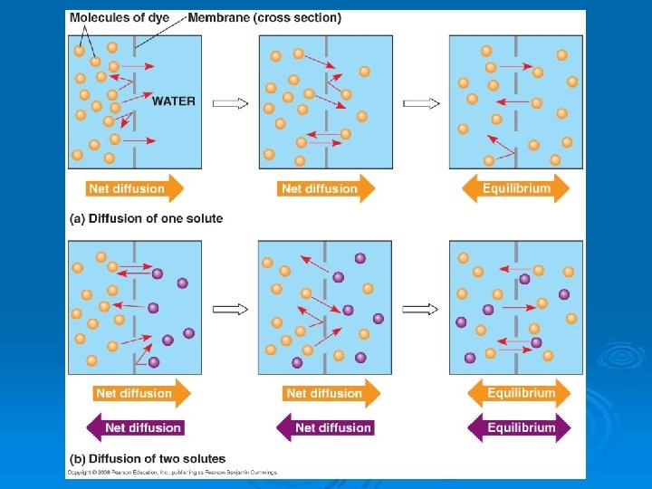

Osmosis Ø Osmosis is the across a selectively permeable membrane Ø Water diffuses across a membrane from the region of lower solute concentration to the region of higher solute concentration

Simple Rule for Osmosis ØSalt is a ØWhen it is concentrated inside or outside the cell, it will draw the water in its direction

Tonicity is the ability of a solution to cause a cell to gain or lose water Ø Three types of solutions: Ø l l l solution Hypertonic or hypotonic environments create osmotic problems for organisms Ø , the control of water balance, is a necessary adaptation for life in such environments Ø
 concentrations inside and outside the cell Ø")
Isotonic Solutions Ø water and solute (salt) concentrations inside and outside the cell Ø Dynamic Equilibrium l Water in = water out Cells keep normal shape Solute concentration is the same as that inside the cell Ø across the plasma membrane Ø Ø
 concentration outside than inside Since salt sucks, water will")
Hypotonic Solutions Ø solute (salt) concentration outside than inside Since salt sucks, water will move into the cell Ø Solute concentration is less than that inside the cell Ø Ø (swells) and may burst

Plant Cells in Hypotonic Solution is the pressure inside plant cells Ø Cell walls help maintain water balance Ø A plant cell in a hypotonic solution swells until the wall opposes uptake Ø l Ø Cell is now If a plant cell and its surroundings are isotonic, there is no net movement of water into the cell l Cell becomes , and the plant may wilt
 concentration outside than inside Ø Water sucked out of")
Hypertonic Solutions Ø solute (salt) concentration outside than inside Ø Water sucked out of the cell Ø Solute concentration is greater than that inside the cell Ø

Cells in Hypertonic Solutions Animal Cells Ø Plant Cells Ø Turgor pressure drops Ø In a hypertonic environment, plant cells lose water Ø Eventually, the membrane pulls away from the wall, a usually lethal effect called

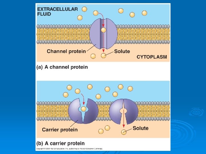
Facilitated Diffusion Ø Some molecules are so small that they pass through the membrane with little resistance Ø Oxygen & Carbon Dioxide Ø Lipid molecules (even though very large) also pass easily Ø In speed the passive movement of molecules across the plasma membrane Ø (transmembrane) provide corridors that allow a specific molecule or ion to cross the membrane Ø Ø , for facilitated diffusion of water that open or close in response to a stimulus (gated channels)
 ØRequires energy in the form")
Active Transport ØMoves substances their concentration gradient: (Low High) ØRequires energy in the form of ØActive transport is performed by specific proteins embedded in the membranes
 to function Ø Allows cells to maintain")
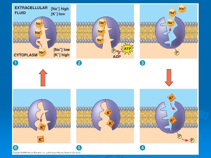
Protein Pumps Protein 'pump' requires energy (ATP) to function Ø Allows cells to maintain concentration gradients that differ from their surroundings Ø The is one type of active transport system l Exchanges in animal cells l Transported molecules enter the in the membrane l The energy causes a shape change in the protein that allows it to move the molecule to the other side of the membrane Ø

Ø Endocytosis Ability of a cell to Ex. large molecules, groups of molecules, or whole cells Requires Energy Cell takes in macromolecules by forming vesicles from the plasma membrane There are three types of endocytosis: l Phagocytosis (“ ”) l Pinocytosis (“ ”) l Receptor-mediated endocytosis l Ø Ø Ø

Types of Endocytosis In a cell engulfs a particle in a vacuole l The vacuole fuses with a lysosome to digest the particle Ø In , molecules are taken up when extracellular fluid is “gulped” into tiny vesicles Ø In , binding of ligands to receptors triggers vesicle formation l A is any molecule that binds specifically to a receptor site of another molecule Ø

Phagocytosis

Pinocytosis

Receptor-Mediated Endocytosis

Exocytosis Ø Opposite of endocytosis l To expel wastes or secrete hormones Ø Requires energy Ø Transport vesicles Ø Many secretory cells use exocytosis to Ø Endocytosis and Exocytosis Animation

Cell Division ØLife is based on the ability of cells to ØRudolf Virchow (German physician) stated “omnis cellula e cellula” meaning Øcell division = one cell divides into two cells
")
Why Cells Divide Ø Unicellular organisms l cell division = asexual reproduction (new organism) Ø Multicellular organisms l (single cell to trillions) l Ø Cell division is an integral part of the cell cycle, the life of a cell from formation to its own division

Cell Cycle - Eukaryotes Ø Ø Ø Defined nucleus houses DNA as chromosomes, a condensed form of chromatin Most cell division results in daughter cells with identical genetic information (DNA) A special type of division produces nonidentical daughter cells (gametes, or sperm and egg cells) Goal: l Chromosomes are from 1 parent cell (before division) to each daughter cell (after division) Includes: l l

Cell Cycle - Eukaryotes

Cell Cycle Ø Ø Ø Interphase ( can be divided into 3 subphases: l (“first gap”) l (“synthesis”) l (“second gap”) The cell grows during all three phases l Makes proteins l Copies organelles Chromosomes are duplicated only during the S phase Mitosis is the division of the Cytokinesis is the division of the )

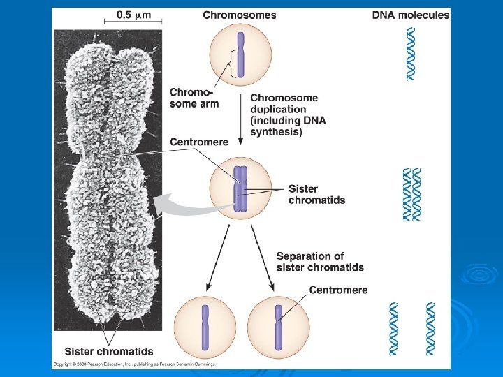
Eukaryotic Chromosomes Ø Chromatin l Long strands of DNA wrapped around proteins l In preparation for cell division, DNA is replicated and condenses to form chromosomes l Each duplicated chromosome has two , which separate during cell division l The is the narrow “waist” of the duplicated chromosome, where the two chromatids are most closely attached Ø Chromosomes must be copied before a cell divides l Each new cell must have a complete set l Contain thousands of genes l Vital for organisms to properly function

Human Example Ø 23 pairs of chromosomes l l 1 originally from mom, 1 from dad Ø Every time our body cells ( ) reproduce l Each NEW cell must also end up with 23 pairs of chromosomes

Chromosome Duplication Ø One chromosome = Ø 2 sister chromatids = l l Exact copies Attached by a centromere Ø Cell division separates sister chromatids Ø Each new cell gets one copy of each chromosome

Mitotic Phase Mitosis is conventionally divided into five phases: l Prophase l Prometaphase* l Metaphase l Anaphase l Telophase Ø Cytokinesis is well underway by late telophase Ø *Some texts do not recognize this as a separate phase

Mitosis

Prophase Pro = “before” Chromosomes Ø Nucleolus and nuclear envelope Ø Ø l l l Microtubules that controls chromosome movement during mitosis Includes the centrosomes, the spindle microtubules, and the asters • An aster is a radial array of short microtubules During prophase, assembly of spindle microtubules begins in the centrosome, the microtubule organizing center

Prophase

Prometaphase Ø Nuclear envelope completely gone Ø Microtubules Ø Some spindle microtubules attach to the of chromosomes and begin to move the chromosomes • Kinetochores are specialized protein structures at the centromere

Prometaphase
 Ø Chromosomes line up at Also called the metaphase")
Metaphase Meta = “with” (middle) Ø Chromosomes line up at Also called the metaphase plate (midway point between the spindle’s two poles) Ø Microtubules attach l
 Ø Sister chromatids Move along microtubules connected to")
Anaphase Ana= “upward” or “back” (apart) Ø Sister chromatids Move along microtubules connected to kinetochore towards opposite ends of cell l Chromatids now chromosomes Ø Other microtubules elongate cell l

Telophase Telos = “end” Ø Reverse of prophase l Begins when chromosomes reach poles l l l Chromosomes “ (less condensed) ” l Ø Genetically identical daughter nuclei form at opposite ends of the cell

Cytokinesis Ø Division of cellular contents l Cytoplasm l Organelles Ø In animal cells, cytokinesis occurs by a process known as

Cytokinesis Ø In plant cells, a Ø Formed from vesicles that pinch off of the Golgi body, move along microtubules, and join in the middle of the cell

Some Animations Ø Mitosis Animation Ø Mitosis and Cytokinesis Animation Ø Mitosis Animation 2

Mitosis Ho-Down MITOSIS is a process that helps one cell become two It happens when a cell dies or makes some brand new This is how a cut heals and how a baby grows It works all o’er the body from your head down to your toes PROPHASE is the first phase where chromosomes you'll see Then comes METAPHASE where they line up perfectly After that is ANAPHASE where they are pulled apart Finally is TELOPHASE, cells split then go back to start

Cell Cycle Controls The sequential events of the cell cycle are directed by a distinct , which is similar to a clock Ø The cell cycle control system is regulated by both internal and external controls Ø The clock has specific where the cell cycle stops until a goahead signal is received Ø l G 1, G 2, M checkpoints

Cell Cycle Controls Ø G 1 checkpoint seems to be l A go-ahead signal at the G 1 checkpoint allows cell to complete the S, G 2, and M phases and divide l No go-ahead signal and the cell will exit the cycle, switching into a G 2 checkpoint l Assesses success of l Triggers start of mitosis Ø Mitosis checkpoint l Assesses accuracy of mitosis l Occurs during Ø

Cell Cycle Controls Ø Two types of regulatory proteins are involved in cell cycle control: Activity of cyclins and Cdks fluctuates during the cell cycle Ø MPF (maturation-promoting factor) l A cyclin-Cdk complex l Triggers a cell’s passage past the G 2 checkpoint into the M phase l Also called “mitosis-promoting factor” Ø Cell Cycle Control Animation l

Cancer Ø l l Disease of cell cycle DNA mutation changes genes that normally control growth l Ø Cancer cells do not respond normally to the body’s control mechanisms

Features of Cancer Cells l Normal cells divide about 50 times before dying Ø l Aging, toxins (smoking), mutagens (UV light), DNA replication errors Ø l Instead of “sticking” to neighbors, cancer cells become “round”, allowing for metastasis (spread) Ø l Keep growing after touching neighbor

Tumors Ø Cancer cells form l Masses of abnormal cells within otherwise normal tissue Ø l l Abnormal mass of essentially normal cells Remain at original site Ø l l l Invade surrounding tissues Can • Move to other sites and create a new tumor (secondary) Cells send out signals for blood vessel production • Gives them food, oxygen, escape route

Review Questions 1. Identify and describe the parts of the fluid mosaic model of the plasma membrane. 2. Describe the various regions of a phospholipid molecule as they apply to the arrangement of the plasma membrane. 3. Explain the role of cholesterol in the membrane. 4. Describe the 2 main types and 6 various functions of membrane proteins. 5. Differentiate between passive and active transport. 6. Explain the idea of a concentration gradient, along with moving down and against it. 7. Define tonicity and explain hypertonic, isotonic, and hypotonic solutions. 8. Define osmoregulation and turgor pressure. 9. Differentiate between diffusion, osmosis, and facilitated diffusion, naming the parts of the membrane that help these transports. 10. Differentiate between protein pumps, endocytosis, and exocytosis, naming that parts of the membrane that help these transports. 11. Name and describe 3 types of endocytosis. 12. Explain the importance of cell division. 13. Name the parts of the cell cycle and state the events that occur in each stage. 14. Differentiate between chromatin, chromosome, and chromatid.

Review Questions 15. Define somatic cells. 16. Name the 5 steps to the cell cycle. 17. Define cytokinesis and explain how it differs in plant and animal cells. 18. Describe the 3 events that occur in prophase. 19. Name 2 events that occur in prometaphase. 20. Name 2 events that occur in metaphase. 21. Name the main event of anaphase. 22. Name 3 events that occur in telophase. 23. Explain how the cell plate forms in plant cell cytokinesis. 24. Name the 3 checkpoints of the cell cycle control system and explain what occurs in each step. 25. Differentiate between the roles of cyclins and cyclin-dependent kinases. 26. Define the importance of MPF. 27. Relate the formation of cancer to the cell cycle. 28. Name 4 major features of cancer cells. 29. Define tumor and differentiate between benign and malignant. 30. Define metastasis.
- Slides: 67