Cell Growth and Division Cell Growth When a


, the rest of the process of")


 – *HOMOLOGOUS")


- Slides: 89
Cell Growth and Division
Cell Growth • When a living thing grows, what happens to its cells? • Does an animal get larger because each cell increases in size or because it produces more of them? – In most cases, living things grow by producing more cells • On average, the cells of an adult animal are no larger than those of a young animal—there are just more of them
Limits to Cell Growth • There are two main reasons why cells divide rather than continuing to grow indefinitely: – The larger a cell becomes, the more demands the cell places on its DNA – The cell has more trouble moving enough nutrients and wastes across the cell membrane
DNA Overload • As you may recall, the information that controls a cell's function is stored in a molecule known as DNA • In eukaryotic cells, DNA is found in the nucleus of the cell • When a cell is small, the information stored in that DNA is able to meet all of the cell's needs • But as a cell increases in size, it usually does not make extra copies of DNA • If a cell were to grow without limit, an “information crisis” would occur
DNA Overload • To help understand why a larger cell has a more difficult time functioning efficiently than a smaller cell, compare the cell to a growing town • Suppose a small town has a library with a few thousand books • If more people move into the town, the town will get larger • There will be more people borrowing books, and sometimes people may have to wait to borrow popular titles – Similarly, a larger cell would have to make greater demands on its available genetic “library” • In time, the cell's DNA would no longer be able to serve the increasing needs of the growing cell
Exchanging Materials • There is another reason why the size of cells is limited • You may recall that food, oxygen, and water enter a cell through its cell membrane • Waste products leave in the same way • The rate at which this exchange takes place depends on the surface area of the cell, which is the total area of its cell membrane – However, the rate at which food and oxygen are used up and waste products are produced depends on the cell's volume • Understanding the relationship between a cell's volume and its surface area is the key to understanding why cells must divide as they grow
Ratio of Surface Area to Volume • • Imagine a cell that is shaped like a cube, like those in the table below If this cell has a length of 1 cm, its surface area would be equal to length × width × number of sides, or 1 cm × 6 = 6 cm 2 The volume of the cell would be equal to length × width × height, or 1 cm × 1 cm = 1 cm 3 To obtain the ratio of surface area to volume, divide the surface area by the volume – In this case, the ratio of surface area to volume would be 6 / 1, or 6 : 1
Ratio of Surface Area to Volume
Ratio of Surface Area to Volume • If the length of the cell doubled, what would happen to the cell's surface area compared to its volume? • The cell's surface area would be equal to 2 cm × 6 = 24 cm 3 • The volume would be equal to 2 cm × 2 cm = 8 cm 3 • The cell's ratio of surface area to volume would be 24 / 8, or 3 : 1
Ratio of Surface Area to Volume • What if the length of the cell triples? • The cell's surface area now would be 3 cm × 6 = 54 cm 2 • The volume would be 3 cm × 3 cm = 27 cm 3 • The ratio of surface area to volume would be 54 / 27, or 2 : 1
Ratio of Surface Area to Volume • Note that the volume increases much more rapidly than the surface area, causing the ratio of surface area to volume to decrease • This decrease creates serious problems for the cell
Ratio of Surface Area to Volume • To use the town analogy again, suppose that the small town has a two-lane main street • As the town grows, more people will begin to use this street • The main street leading through town, however, has not increased in size • As a result, people will encounter more traffic as they enter and leave the town • A cell that continues to grow larger would experience similar problems • If a cell got too large, it would be more difficult to get sufficient amounts of oxygen and nutrients in and waste products out • This is one reason why cells do not grow much larger even if the organism of which they are a part does
Division of the Cell • Before it becomes too large, a growing cell divides forming two “daughter” cells • The process by which a cell divides into two new daughter cells is called cell division
Division of the Cell • Before cell division occurs, the cell replicates, or copies, all of its DNA • This replication of DNA solves the problem of information storage because each daughter cell gets one complete set of genetic information – Thus, each daughter cell receives its own genetic “library” • Cell division also solves the problem of increasing size by reducing cell volume – Each daughter cell has an increased ratio of surface area to volume • This allows efficient exchange of materials with the environment
Cell Division • What do you think would happen if a cell were simply to split into two, without any advance preparation? – Would each daughter cell have everything it needed to survive? – Because each cell has only one set of genetic information, the answer is no • Every cell must first copy its genetic information before cell division begins • Each daughter cell then gets a complete copy of that information
Cell Division • In most prokaryotes (NO NUCLEUS), the rest of the process of cell division is a simple matter of separating the contents of the cell into two parts • In eukaryotes, cell division is more complex and occurs in two main stages: – The first stage, division of the cell nucleus, is called mitosis – The second stage, division of the cytoplasm, is called cytokinesis
Cell Division • Many organisms, especially unicellular ones, reproduce by means of mitosis and cytokinesis • Reproduction by mitosis is classified as asexual, since the cells produced by mitosis are genetically identical to the parent cell • Mitosis is also the source of new cells when a multicellular organism grows and develops • In humans, for example, mitosis begins shortly after the egg is fertilized, producing the vast numbers of cells needed for the embryo to take form
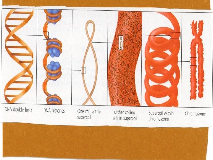
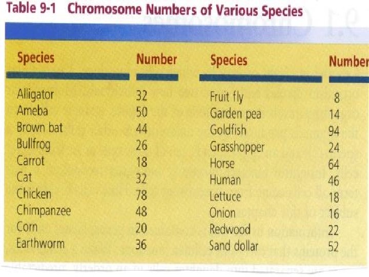
Chromosomes • In eukaryotic cells, the genetic information that is passed on from one generation of cells to the next is carried by chromosomes • Chromosomes are made up of DNA—which carries the cell's coded genetic information— and proteins • The cells of every organism have a specific number of chromosomes • The cells of: – Fruit flies have 8 chromosomes – Human cells have 46 chromosomes – Carrot cells have 18 chromosomes
Chromosomes • Chromosomes are not visible in most cells except during cell division • This is because the DNA and protein molecules that make up the chromosomes are spread throughout the nucleus • At the beginning of cell division, however, the chromosomes condense into compact, visible structures that can be seen through a light microscope
CHROMOSOME STRUCTURE • When replicated each chromosome has two identical parts: – Each called a chromatid (often called sister chromatids) • Point at which each pair of chromatids is attached is called the centromere
CHROMOSOME NUMBER • Every species has a characteristic number of chromosomes in each cell • In all sexually reproducing organisms chromosomes occur in pairs – The two members of each pair are called homologous chromosomes or homologues • Each chromosome of a pair has the same size and shape as its homologue but the genetic information can vary – One from each biological parent • Structurally different from all other homologous pairs in the cell
CHROMOSOME NUMBER • A cell that contains both chromosomes of a homologous pair is termed diploid – In a human the diploid number is: 2 N = 46 • N represents the number of homologous pairs • A cell that has only one chromosome of each homologous pair is termed haploid (monoploid) – In a human the haploid (monoploid) number of the human egg/sperm cell is: N = 23 – There are no homologous chromosomes in either cell
• CHROMOSOME NUMBER: chromosomes are in pairs (one from each parent) – *HOMOLOGOUS CHROMOSOMES: the pairs (one from each parent ) – DIPLOID NUMBER (2 n): both members of each pair – *HAPLOID (MONOPLOID) NUMBER (1 n): one member of each pair
• MITOSIS/MEIOSIS • CHROMATIN: the less tightly coiled DNA-protein complex in the nucleus of a non-dividing cell • CHROMOSOME: DNA and protein in a coiled, rod-shaped form that occurs during cell division • CHROMATID: one of two identical parts of a chromosome that has replicated. • *CENTROMERE(Kinetochore): constricted area of each chromatid / holds the two chromatids together
Chromosomes • Well before cell division, each chromosome is replicated, or copied • Because of this, each chromosome consists of two identical “sister” chromatids, as shown to the chromatids right • When the cell divides, the “sister” chromatids separate from each other • One chromatid goes to each of the two new cells
Chromosomes
Chromosomes • This is a human chromosome shown as it appears through an electron microscope • Each chromosome has two sister chromatids attached chromatids at the centromere
Chromosomes • Each pair of chromatids is attached at an area called the centromere • Centromeres are usually located near the middle of the chromatids, although some lie near the ends • A human body cell entering cell division contains 46 chromosomes, each of which consists of two chromatids
Cell Cycle • • At one time, biologists described the life of a cell as one cell division after another separated by an “in -between” period of growth called interphase We now appreciate that a great deal happens in the time between cell divisions, and use a concept known as the cell cycle to represent recurring events in the life of the cell The cell cycle is the series of cell cycle events that cells go through as they grow and divide During the cell cycle, a cell grows, prepares for division, and divides to form two daughter cells, each of which then begins the cycle again
Cell Cycle
Cell Cycle • • • The cell cycle consists of four phases Mitosis and cytokinesis take place during the M phase Chromosome replication, or synthesis, takes place during the S phase – When the cell copies the chromosomes, it makes a duplicate set of DNA • Between the M and S phases are G 1 and G 2 – The G in the names of these phases stands for “gap, ” but the G 1 and G 2 are definitely not periods when nothing takes place • They are actually periods of intense growth and activity
Events of the Cell Cycle • During the normal cell cycle, interphase can be quite long, whereas the process of cell division takes place quickly • Interphase is divided into three phases: – G 1 –S – G 2
Events of the Cell Cycle Interphase • The G 1 phase is a period of activity in which cells do most of their growing • During this phase, cells increase in size and synthesize new proteins and organelles
Events of the Cell Cycle Interphase • G 1 is followed by the S phase, in which chromosomes are replicated and the synthesis of DNA molecules takes place • Key proteins associated with the chromosomes are also synthesized during the S phase • Usually, once a cell enters the S phase and begins the replication of its chromosomes, it completes the rest of the cell cycle
INTERPHASE • Chromosomes not visible • DNA replicates • Chromosomes are long thin strands • Nucleus enclosed by nuclear membrane • Nucleolus visible • Centrioles in animals
INTERPHASE
Events of the Cell Cycle • When the DNA replication is completed, the cell enters the G 2 phase • G 2 is usually the shortest of the three phases of interphase • During the G 2 phase, many of the organelles and molecules required for cell division are produced • When the events of the G 2 phase are completed, the cell is ready to enter the M phase and begin the process of cell division
Mitosis • Biologists divide the events of mitosis into four phases: – – Prophase Metaphase Anaphase Telophase • Depending on the type of cell, the four phases of mitosis may last anywhere from a few minutes to several days
Prophase • The first and longest phase of mitosis, prophase, can take as much as 50 to 60 percent of the total time required to complete mitosis • During prophase, the chromosomes become visible • The centrioles, two tiny structures located in the cytoplasm near the nuclear envelope, separate and take up positions on opposite sides of the nucleus
Prophase
Prophase • The centrioles lie in a region called the centrosome that helps to organize the spindle, a fanlike microtubule structure that spindle helps separate the chromosomes • During prophase, the condensed chromosomes become attached to fibers in the spindle at a point near the centromere of each chromatid • Interestingly, plant cells do not have centrioles, but still organize their mitotic spindles from similar regions
Prophase
Prophase • Near the end of prophase, the chromosomes coil more tightly • In addition, the nucleolus disappears, and the nuclear envelope breaks down
Prophase
PROPHASE • Centrioles form poles in animals • Spindle fibers form • Chromosomes become shorter and thicker • Chromatids (replicated chromosomes) held together by kinetochore (centromere) • Chromatids attach to the spindle fibers
Metaphase • The second phase of mitosis, metaphase, often lasts only a few minutes • During metaphase, the chromosomes line up across the center of the cell • Microtubules (spindle fibers) connect the centromere of each chromosome to the two poles of the spindle
Metaphase
METAPHASE • Chromatid pairs line up at equator
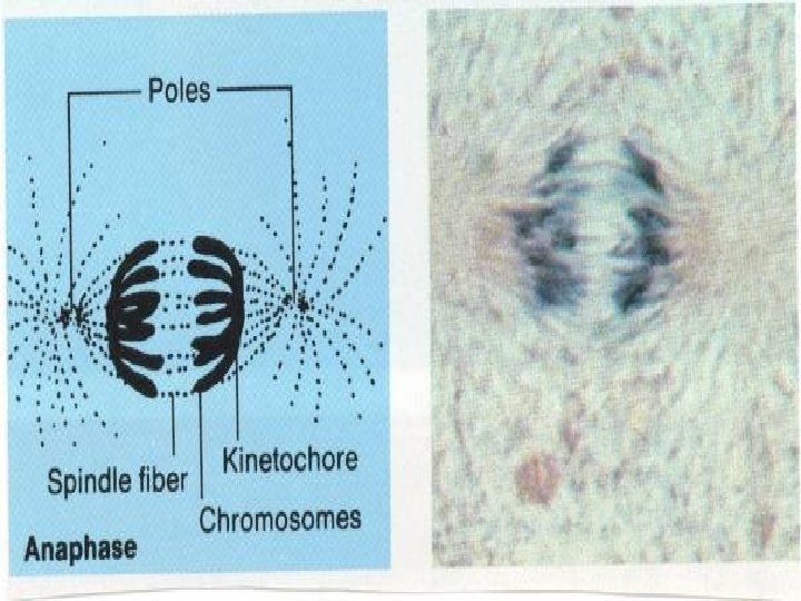
Anaphase • Anaphase is the third phase of mitosis. • During anaphase, the centromeres that join the sister chromatids split, allowing the sister chromatids to separate and become individual chromosomes • The chromosomes continue to move until they have separated into two groups near the poles of the spindle • Anaphase ends when the chromosomes stop moving
Anaphase
ANAPHASE • Separated chromosomes move to opposite poles along the spindle fibers
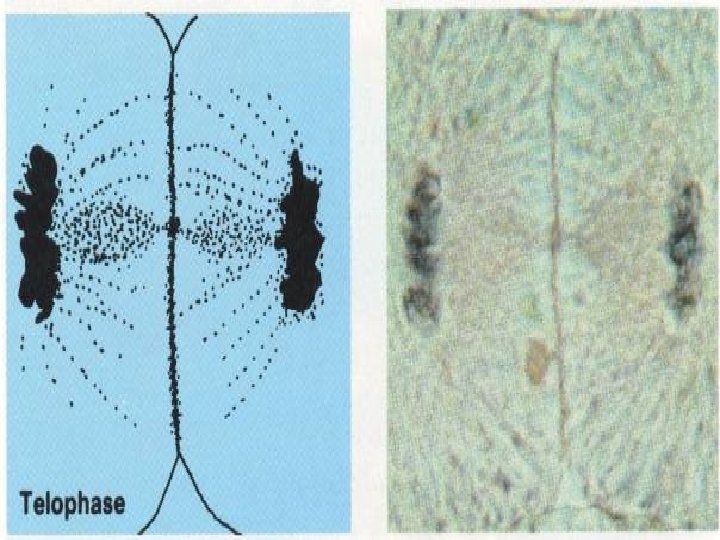
Telophase • • • Following anaphase is telophase, the fourth and final phase of mitosis. In telophase, the chromosomes, which were distinct and condensed, begin to disperse into a tangle of dense material A nuclear envelope re-forms around each cluster of chromosomes The spindle begins to break apart, and a nucleolus becomes visible in each daughter nucleus Mitosis is complete However, the process of cell division is not complete
Telophase
TELOPHASE • Chromosomes reach opposite poles • Chromosomes thin and become invisible • Spindle fibers disappear • Nucleolus reappears • New nuclear membranes form around chromosomes • Daughter cells formed are exact copies
Cytokinesis • As a result of mitosis, two nuclei—each with a duplicate set of chromosomes—are formed, usually within the cytoplasm of a single cell • All that remains to complete the M phase of the cycle is cytokinesis, the division of the cytoplasm itself • Cytokinesis usually occurs at the same time as telophase
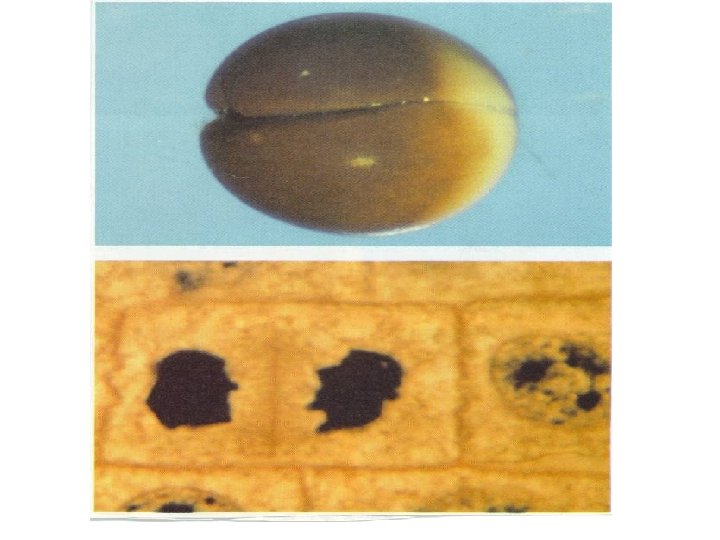
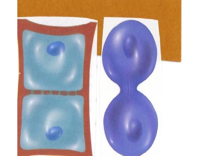
Cytokinesis • • • Cytokinesis can take place in a number of ways In most animal cells, the cell membrane is drawn inward until the cytoplasm is pinched into two nearly equal parts (cleavage furrow) – Each part contains its own nucleus and cytoplasmic organelles In plants, a structure known as the cell plate forms midway between the cell plate divided nuclei, as shown below – The cell plate gradually develops into a separating membrane – A cell wall then begins to appear in the cell plate – During cytokinesis in plant cells, the cytoplasm is divided by a cell plate – The thin line you can see between the two dark nuclei in the drawing of onion cells dividing is the cell plate forming
CYTOKINESIS • The division of the cytoplasm of a parent cell and its contents (organelles) into two daughter cells • Each newly formed cell has one of the two nuclei formed during mitosis • Animal Cell: – Cleavage furrow separates the daughter cells • pinching in of the cell membrane • Plant Cell: – Cell Plate separates the daughter cells • Vesicles formed by the Golgi bodies fuse at the equator and form the cell plate (membrane across the middle of the cell) • New cell wall forms on both sides of the cell plate
Cytokinesis
Cytokinesis
Life Spans of Human Cells
Regulating the Cell Cycle • One of the most striking aspects of cell behavior in a multicellular organism is how carefully cell growth and cell division are controlled • Not all cells move through the cell cycle at the same rate • In the human body, most muscle cells and nerve cells do not divide at all once they have developed • In contrast, the cells of the skin and digestive tract, and cells in the bone marrow that make blood cells, grow and divide rapidly throughout life – Such cells may pass through a complete cycle every few hours – This process provides new cells to replace those that wear out or break down
Controls on Cell Division • Scientists can observe the effects of controlled cell growth in the laboratory by placing some cells in a petri dish containing nutrient broth • The nutrient broth provides food for the cells • Most cells will grow until they form a thin layer covering the bottom of the dish, as shown in the figure at right • Then, the cells stop growing • When cells come into contact with other cells, they respond by not growing
Controls on Cell Division
Controls on Cell Division • If cells are removed from the center of the dish, however, the cells bordering the open space will begin dividing until they have filled the empty space • These experiments show that the controls on cell growth and cell division can be turned on and off
Controls on Cell Division • Something similar happens within the body • When an injury such as a cut in the skin or a break in a bone occurs, cells at the edges of the injury are stimulated to divide rapidly – This action produces new cells, starting the process of healing • When the healing process nears completion, the rate of cell division slows down, controls on growth are restored, and everything returns to normal
Cell Cycle Regulators • For many years, biologists searched for a substance that might regulate the cell cycle—something that would “tell” cells when it was time to divide, duplicate their chromosomes, or enter another phase of the cycle • In the early 1980 s, biologists found the substance
Cell Cycle Regulators
Cell Cycle Regulators • Several scientists, including Tim Hunt of Great Britain and Mark Kirschner of the United States, discovered that cells in mitosis contained a protein that when injected into a nondividing cell, would cause a mitotic spindle to form • Such an experiment is shown in the figure at right • To their surprise, they discovered that the amount of this protein in the cell rose and fell in time with the cell cycle • They decided to call this protein cyclin because it seemed to regulate the cell cycle • Investigators have since discovered a family of closely related proteins, known as cyclins, that are involved in cell cycle regulation • Cyclins regulate the timing of the cell cycle in eukaryotic cells
Cell Cycle Regulators • The discovery of cyclins was just the beginning • More recently, dozens of other proteins have been discovered that also help to regulate the cell cycle • There are two types of regulatory proteins: – Those that occur inside the cell – Those that occur outside the cell
Internal Regulators • Proteins that respond to events inside the cell are called internal regulators • Internal regulators allow the cell cycle to proceed only when certain processes have happened inside the cell – Example: • Several regulatory proteins make sure that a cell does not enter mitosis until all its chromosomes have been replicated • Another regulatory protein prevents a cell from entering anaphase until all its chromosomes are attached to the mitotic spindle
External Regulators • Proteins that respond to events outside the cell are called external regulators • External regulators direct cells to speed up or slow down the cell cycle • Growth factors are among the most important Growth factors external regulators – They stimulate the growth and division of cells • Growth regulators are especially important during embryonic development and wound healing • Molecules found on the surfaces of neighboring cells often have an opposite effect, causing cells to slow down or stop their cell cycles – These signals prevent excessive cell growth and keep the tissues of the body from disrupting each other
Uncontrolled Cell Growth • Why is cell growth regulated so carefully? – The principal reason may be that the consequences of uncontrolled cell growth in a multicellular organism are very severe • Cancer, a disorder in which some of the body's own cells lose the ability to control growth, is one such example – Cancer cells do not respond to the signals that regulate the growth of most cells – As a result, they divide uncontrollably and form masses of cells called tumors that can damage the surrounding tissues – Cancer cells may break loose from tumors and spread throughout the body, disrupting normal activities and causing serious medical problems or even death
CANCER • Tumor: an abnormal mass of cells that results from ungoverned cell division – Benign: cells remain in the mass • Generally no threat to life – Malignant: undergo metastasis (break away) causing new tumors to form in other locations • Disease cause by malignant tumors are collectively referred to as cancer • Categorized according to the types of tissue they infect – Carcinomas: grow in skin and nerves – Sarcomas: grow in bone and muscle – Lymphomas: solid tumors that grow in the tissues that form blood cells – Leukemia: an abnormal growth of immature white blood cells
CANCER • Causes: – Carcinogen: any substance that causes cancer • Whether a person actually develops cancer depends on many factors, including genetic predisposition, the number of exposures, and the amount of carcinogen in each exposure • Tobacco, asbestos, UV light, viruses – Oncogenes: genes that when expressed cause normal cells to become cancerous
Uncontrolled Cell Growth • What causes the loss of growth control that characterizes cancer? – The various forms of cancer have many causes, including smoking tobacco, radiation exposure, and even viral infection • All cancers, however, have one thing in common: The control over the cell cycle has broken down – Some cancer cells will no longer respond to external growth regulators, while others fail to produce the internal regulators that ensure orderly growth
Uncontrolled Cell Growth • An astonishing number of cancer cells have a defect in a gene called p 53, which normally halts the cell cycle until all chromosomes have been properly replicated • Damaged or defective p 53 genes cause the cells to lose the information needed to respond to signals that would normally control their growth
Uncontrolled Cell Growth • Cancer is a serious disease • Understanding and combating cancer remains a major scientific challenge, but scientists at least know where to start • Cancer is a disease of the cell cycle, and conquering cancer will require a much deeper understanding of the processes that control cell division
Stem Cells: Promises and Problems • Where do the different cells and tissues in your body come from? • Incredible as it seems, every cell was produced by mitosis from a small number of cells called stem cells • Stem cells are unspecialized cells that have the potential to Stem cells are unspecialized cells differentiate—to become specialized in structure and function—into a wide variety of cell types • In early embryonic development, stem cells produce every tissue in the body • Evidence indicates that stem cells also are found in adults • Stem cells in the bone marrow, for example, produce more than a dozen types of blood cells, replacing those lost due to normal wear and tear
Stem Cells in Medicine • Although your body produces billions of new cells every day, it is not always able to produce the right kind of cell to replace those damaged by injury or disease – For example: • The body is not able to produce new neurons to repair serious spinal cord injuries, such as those that cause paralysis • Because of this, at present, there is no way for doctors to restore movement and feeling to people who are paralyzed
Stem Cells in Medicine • Stem cells may be the perfect solution to this problem – Recently, researchers have found that implants of stem cells can reverse the effects of brain injuries in mice – There is hope that the same will hold true for humans and that stem cells might be used to reverse brain and spinal cord injuries – It also may be possible to use stem cells to grow new liver tissue, to replace heart valves, and to reverse the effects of diabetes
Sources of Stem Cells • Human embryonic stem cells were first isolated in 1998 by scientists in Wisconsin • In 2004, Korean scientists produced such cells by transferring adult cell nuclei into the cytoplasms of egg cells – However, since such cells are taken from human embryos, these techniques raise serious moral and ethical questions – Because of such issues, embryonic stem cell research is highly controversial
Sources of Stem Cells • Researchers have also found that nerve, muscle, and liver cells sometimes can be grown from adult stem cells isolated from the bone marrow and other tissues in the body • Experiments such as these, although still in the early stages of development, may usher in a new era of therapy in which replacement tissue is grown from a person's own stem cells