Cell Growth and Division 10 1 Cell growth


 and haploid (n) If a cell has 2 sets of chromosomes")


 phase, new DNA is synthesized")


- Slides: 67
Cell Growth and Division 10. 1: Cell growth, Division, and Reproduction 10. 2: The Process of Cell Division 10. 3: Regulating the Cell Cycle 10. 4: Cell Differentiation
Cell Division About 2 trillion cells- about 25 million cells per second- are produced by an adult human body every day. All cells come from the division of preexisting cells. (cell theory) Cell division or reproduction is the process by which cells produce offspring cells. Cell division differs in prokaryotes and eukaryotes. Cell reproduction in both prok and euk produce the same result – two cells from one!! 2
Review What are three parts to the cell theory? 1. All living organisms are composed of one or more cells. 2. Cells are the basic units of structure and function in an organism. 3. Cells come only from the reproduction of existing cells. Four things common in all cells: cell membrane, cytoplasm, DNA, Ribosomes 3
Information “Overload” Living cells store critical information in DNA. As a cell grows, that information is used to build the molecules needed for cell growth. As size increases, the demands on that information grow as well. If a cell were to grow without limit, an “information crisis” would occur.
Exchanging Materials Food, oxygen, and water enter a cell through the cell membrane. Waste products leave in the same way. The rate at which this exchange takes place depends on the surface area of a cell. The rate at which food and oxygen are used up and waste products are produced depends on the cell’s volume. The ratio of surface area to volume is key to understanding why cells must divide as they grow.
Ratio of Surface Area to Volume Imagine a cell shaped like a cube. As the length of the sides of a cube increases, its volume increases faster than its surface area, decreasing the ratio of surface area to volume. If a cell gets too large, the surface area of the cell is not large enough to get enough oxygen and nutrients in and waste out.
Division of the Cell Before a cell grows too large, it divides into two new “daughter” cells in a process called cell division. Before cell division, the cell copies all of its DNA. It then divides into two “daughter” cells. Each daughter cell receives a complete set of DNA. Cell division reduces cell volume. It also results in an increased ratio of surface area to volume, for each daughter cell.
Cell Division and Reproduction How do asexual and sexual reproduction compare? The production of genetically identical offspring from a single parent is known as asexual reproduction. Offspring produced by sexual reproduction inherit some of their genetic information from each parent.
Cell Division -Eukaryotes Two main types of division: 1. Mitosis- results in new cells with genetic material that is identical to the genetic material of original cell During: growth, development, repair, asexual reproduction (production of offspring from one parent) 2. Meiosis- occurs during the formation of gametes (reproductive cells, 1 n), reduces the chromosome number by ½ in new cells, often combine to make 2 n cells 9
Asexual Reproduction In multicellular organisms, cell division leads to growth. It also enables an organism to repair and maintain its body. In single-celled organisms, cell division is a form of reproduction.
Asexual Reproduction Asexual reproduction is reproduction that involves a single parent producing an offspring. The offspring produced are, in most cases, genetically identical to the single cell that produced them. Asexual reproduction is a simple, efficient, and effective way for an organism to produce a large number of offspring. Both prokaryotic and eukaryotic single-celled organisms and many multicellular organisms can reproduce asexually. Bacteria cells reproduce by binary fission
Sexual Reproduction - In sexual reproduction, offspring are produced by the fusion of two sex cells – one from each of two parents. These fuse into a single cell before the offspring can grow. The offspring produced inherit some genetic information from both parents. Most animals and plants, and many single-celled organisms, reproduce sexually.
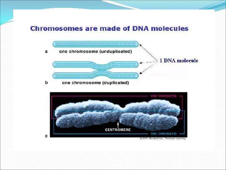
Chromosomes The genetic information that is passed on from one generation of cells to the next is carried by chromosomes. Every cell must copy its genetic information before cell division begins. Each daughter cell gets its own copy of that genetic information. Cells of every organism have a specific number of chromosomes.
Prokaryotic Chromosomes Prokaryotic cells lack nuclei. Instead, their DNA molecules are found in the cytoplasm. Most prokaryotes contain a single, circular DNA molecule, or chromosome, that contains most of the cell’s genetic information.
Eukaryotic Chromosomes In eukaryotic cells, chromosomes are located in the nucleus, and are made up of chromatin.
Chromatin is composed of DNA and histone proteins. DNA coils around histone proteins to form nucleosomes. The nucleosomes interact with one another to form coils and supercoils that make up chromosomes.
Chromosomes structure cont: Chromosomes contain 2 identical halves each half of the chromosome is called a chromatid Chromatids form when the DNA makes a copy of itself before cell division When the cell divides each ½ gets a copy Chromatids are attached by the centromere When DNA is not tightly packed it is call chromatin Prokaryotes DNA is simple, circular, one chromosome, very compact to fit in the cell
Chromosomes Numbers Each species has a set number of chromosomes Must know terms: 1. Sex chromosomes: are chromosomes that determine the sex of an organism, they may also carry genes for other characteristics (X, Y) (2 humans) 2. Autosomes- all non sex chromosomes (44 of humans )
Chromosomes Numbers cont: 3. Homologous chromosomes- they are chromosomes that are the same sex and shape and carry genes from the same traits 4. Karyotype- photomicrograph of chromosomes in a normal dividing cell found in human cells (46 chromosomes) 22 homologous pairs of autosomes 2 sex chromosomes
Karyotype Male VS Female Chromosomal abnormalities and karyotypes
Diploid (2 n) and haploid (n) If a cell has 2 sets of chromosomes it is diploid Reproductive cells like sperm and eggs (called gametes) have 1 set of chromosomes, they are haploid. Ex: For humans the 2 n or diploid number of chromosomes is 46 (23 pairs) All cells of the body except for the sex cells are autosomes
The Cell Cycle During the cell cycle, a cell grows, prepares for division, and divides to form two daughter cells.
The Prokaryotic Cell Cycle The prokaryotic cell cycle is a regular pattern of growth, DNA replication, and cell division. Most prokaryotic cells begin to replicate, or copy, their DNA once they have grown to a certain size. When DNA replication is complete, the cells divide through a process known as binary fission.
Cell division in prokaryotes Have no true nuclei or membrane-bound organelles DNA is not coiled around proteins- it is circular attached to the inner surface of the plasma membrane like a rope attached to the inner wall of a tent Process of cell division is Binary fission
Binary fission Is the division of a prok into two offspring cells 1. DNA is copied get two identical chromosomes 2. A new cell membrane then begins to develop between the two DNA copies 3. Cell grows till its 2 x the original size 4. Cells split containing identical chromosomes
The Eukaryotic Cell Cycle The eukaryotic cell cycle consists of four phases: G 1, S, G 2, and M. Interphase is the time between cell divisions. It is a period of growth that consists of the G 1, S, and G 2 phases. The M phase is the period of cell division.
G 1 Phase: Cell Growth In the G 1 phase, cells increase in size and synthesize new proteins and organelles.
S Phase: DNA Replication In the S (or synthesis) phase, new DNA is synthesized when the chromosomes are replicated.
G 2 Phase: Preparing for Cell Division In the G 2 phase, many of the organelles and molecules required for cell division are produced.
M Phase: Cell Division In eukaryotes, cell division occurs in two stages: mitosis and cytokinesis. Mitosis is the division of the cell nucleus. Cytokinesis is the division of the cytoplasm. Or a G 0: stage in which the cell is mature and no longer dividing
Important Cell Structures Involved in Mitosis Chromatid – each strand of a duplicated chromosome Centromere – the area where each pair of chromatids is joined Centrioles – tiny structures located in the cytoplasm of animal cells that help organize the spindle Spindle (Mitotic Spindle) – a fanlike microtubule structure that helps separate the chromatids
Prophase During prophase, the first phase of mitosis, the duplicated chromosome condenses and becomes visible.
Prophase During prophase, the first phase of mitosis, the duplicated chromosome condenses and becomes visible. The centrioles move to opposite sides of nucleus and help organize the spindle.
Prophase During prophase, the first phase of mitosis, the duplicated chromosome condenses and becomes visible. The centrioles move to opposite sides of nucleus and help organize the spindle. The spindle forms and DNA strands attach at a point called their centromere.
Prophase During prophase, the first phase of mitosis, the duplicated chromosome condenses and becomes visible. The centrioles move to opposite sides of nucleus and help organize the spindle. The spindle forms and DNA strands attach at a point called their centromere. The nucleolus disappears and nuclear envelope breaks down.
Metaphase During metaphase, the second phase of mitosis, the centromeres of the duplicated chromosomes line up across the center of the cell.
Metaphase During metaphase, the second phase of mitosis, the centromeres of the duplicated chromosomes line up across the center of the cell. The spindle fibers connect the centromere of each chromosome to the two poles of the spindle.
Anaphase During anaphase, the third phase of mitosis, the centromeres are pulled apart and the chromatids separate to become individual chromosomes.
Anaphase During anaphase, the third phase of mitosis, the centromeres are pulled apart and the chromatids separate to become individual chromosomes. The chromosomes separate into two groups near the poles of the spindle.
Telophase During telophase, the fourth and final phase of mitosis, the chromosomes spread out into a tangle of chromatin.
Telophase During telophase, the fourth and final phase of mitosis, the chromosomes spread out into a tangle of chromatin. A nuclear envelope re- forms around each cluster of chromosomes.
Telophase During telophase, the fourth and final phase of mitosis, the chromosomes spread out into a tangle of chromatin. A nuclear envelope re- forms around each cluster of chromosomes. The spindle breaks apart, and a nucleolus becomes visible in each daughter nucleus.
Cytokinesis completes the process of cell division – it splits one cell into two. Cytokinesis is the division of the cytoplasm. The process of cytokinesis is different in animal and plant cells.
Cytokinesis in Animal Cells The cell membrane is drawn in until the cytoplasm is pinched into two equal parts at the Cleavage furrow Each part contains its own nucleus and organelles.
Cytokinesis in Plant Cells In plants, the cell membrane is not flexible enough to draw inward because of the rigid cell wall. Instead, a cell plate forms between the divided nuclei that develops into cell membranes. A cell wall then forms in between the two new membranes.
Biology notes-Chapter 8 47
The Stages of the Cell Cycle
Cell Cycle Rate Do all cells move through the cell cycle at the same rate? No! In the human body, most muscle and nerve cells do not divide at all once they have developed. The cells of the skin, bone marrow and digestive tract cells grow and divide rapidly throughout life.
Controls on Cell Growth The controls on cell growth and division can be turned on and off. For example, when an injury such as a broken bone occurs, cells are stimulated to divide rapidly and start the healing process. The rate of cell division slows when the healing process nears completion.
The Discovery of Cyclins are a family of proteins that regulate the timing of the cell cycle in eukaryotic cells. This graph shows how cyclin levels change throughout the cell cycle in fertilized clam eggs.
Regulatory Proteins – The cell cycle is controlled by regulatory proteins both inside and outside the cell. – Internal regulators are proteins that respond to events inside a cell. – – They allow the cell cycle to proceed only once certain processes have happened inside the cell. Ex: several proteins make sure that a cell does not enter mitosis until all its chromosomes have been replicated. – External regulators are proteins that respond to events outside the cell. – – They direct cells to speed up or slow down the cell cycle. Imp ortant during embryonic development and wound healing. – Growth factors are external regulators that stimulate the growth and division of cells. – Important during embryonic development and wound healing.
Apoptosis is a process of programmed cell death. Apoptosis plays a role in development by shaping the structure of tissues and organs in plants and animals. For example, the foot of a mouse is shaped the way it is partly because the toes undergo apoptosis during tissue development.
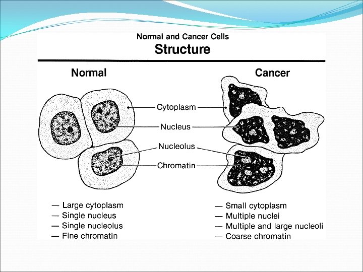
Uncontrolled Cell Growth Why is cell growth regulated so carefully? Consequences of uncontrolled cell growth can be severe. Cancer is one of these consequences! They divide uncontrollably and form masses of cells called tumors. Cancer cells may break loose and spread throughout the body.
Cancer: Uncontrolled Cell Growth – Cancer cells do not respond to the signals that regulate the growth of most cells. – Cancer is a disorder in which cells lose the ability to control cell growth. – Cancer cells divide uncontrollably to form a mass of cells called a tumor.
Cancer • A benign tumor is noncancerous. • Does not spread to surrounding healthy tissue. • A malignant tumor is cancerous. • Invades and destroys surrounding healthy tissue • Can spread to other parts of the body. The spread of cancer cells is called metastasis. • Cancer cells absorb nutrients needed by other cells, block nerve connections, and prevent organs from functioning.
What Causes Cancer? Cancers are caused by defects in genes that regulate cell growth and division. – Some sources of gene defects are smoking tobacco, radiation exposure, defective genes, and viral infection. – A damaged or defective p 53 gene is common in cancer cells. – – – P 53 normally halts the cell cycle until all chromosomes have been properly replicated Damage causes cells to lose the information needed to respond to growth signals. – Cancer is a disease of the cell cycle!
Treatments for Cancer Some localized tumors can be removed by surgery. Many tumors can be treated with targeted radiation. Chemotherapy is the use of compounds that kill or slow the growth of cancer cells.
Cell Differentiation All organisms start life as just one cell Embryo Early stage of development most multicellular organisms pass through Cells become more differentiated and specialized.
Differentiation Process of cells becoming specialized. Cell’s role can be determined at a specific point in development.
Differentiation in Mammals Controlled by a number of interacting factors in the embryo Chemical signals Regulatory factors Interaction between cells Adult cells generally can no longer differentiate.
Stem Cells Unspecialized cells from which differentiated cells develop Totipotent Can form all the tissues of the body Only the fertilized egg and first few divisions
Embryonic Stem Cells Pluripotent Capable of developing into many, but not all, cell types Located in inner mass of cells of blastocyst Blastocyst Early embryo stage with a hollow ball of cells with a cluster of cells inside
Adult Stem Cells Multipotent Can produce many types of differentiated cells Typically produce only the types of cells that are unique to that tissue where they are located.
Potential Benefits Research may lead to new ways to repair the cellular damage that results from heart attack, stroke, and spinal cord injuries.
Ethical Issues Most techniques for harvesting, or gathering, embryonic stem cells cause destruction of the embryo Now you have the option of saving your babies umbilical cord Government funding vs. Private funding.