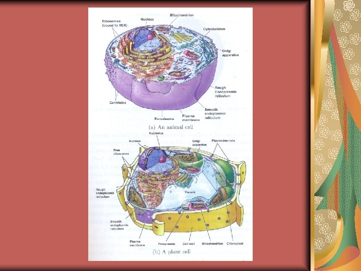
cell Eukaryotes meaning true nucleus are organisms with
 are organisms with cells within which the genetic material")

, polyploid (>4 n) as a result of. DNA replication without cell")


; starts when synapsis is")
: the synaptonemal")


- Slides: 62
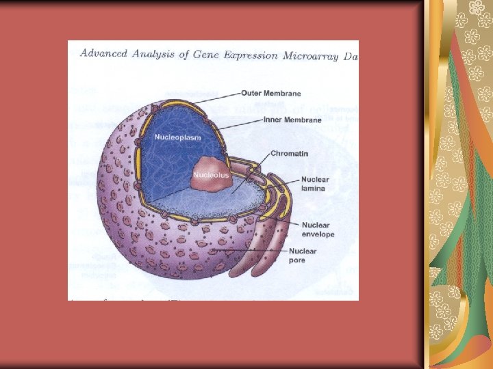
cell Eukaryotes (meaning true nucleus) are organisms with cells within which the genetic material (DNA) is located in the nucleus (a discrete structure bounded by a nuclear envelope). In contrast to eukaryotes, prokaryotes (meaning prenuclear) do not have a nucleare envelope surrounding their DNA.
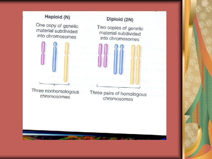
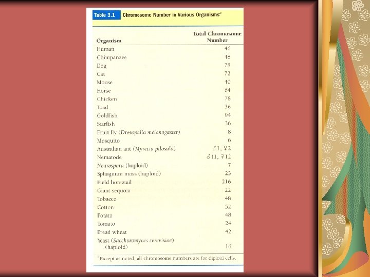
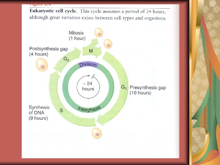
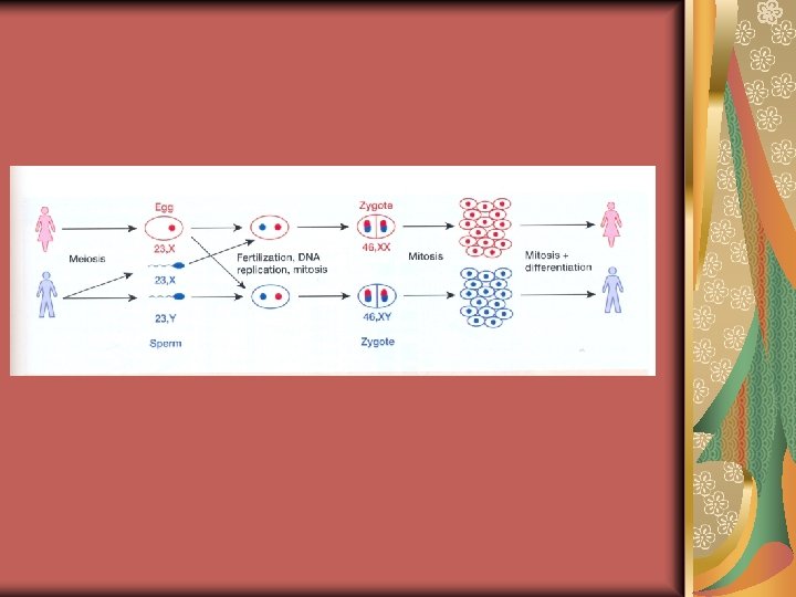
Ploidy and the cell cycle The number of different chromosomes in any nucleated cell, the chromosome set, and the associated DNA content are designated n and C respectively. For human, n=23 and C=approximately 3. 5 pg. Sperm and egg cells carry a single chromosome set and are said to be haploid (n chromosomes and DNA content=C). Most human cells, however, carry two copies of the chromosome set and are diploid (2 n chromosomes and DNA content=2 C).
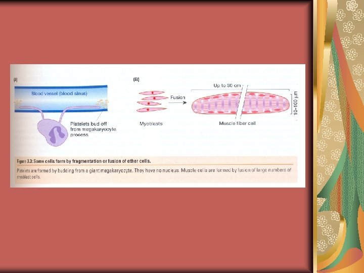
Tetraploid (4 n), polyploid (>4 n) as a result of. DNA replication without cell devision (endomitosis) or as a result of cell fusion the ploidy of hepatocytes ranges from 2 n to 8 n or cardiomyocytes from 4 n to 8 n or giant megakaryocytes of the bone marrow from 16 n to 64 n which give rise to thousands of nulliploid platelet cells, triploidy (3 n)=less common because triploids have problems with meiosis. Nulliploid in some terminally differentiated cells, such as red blood cells, keratinocytes and platelets.
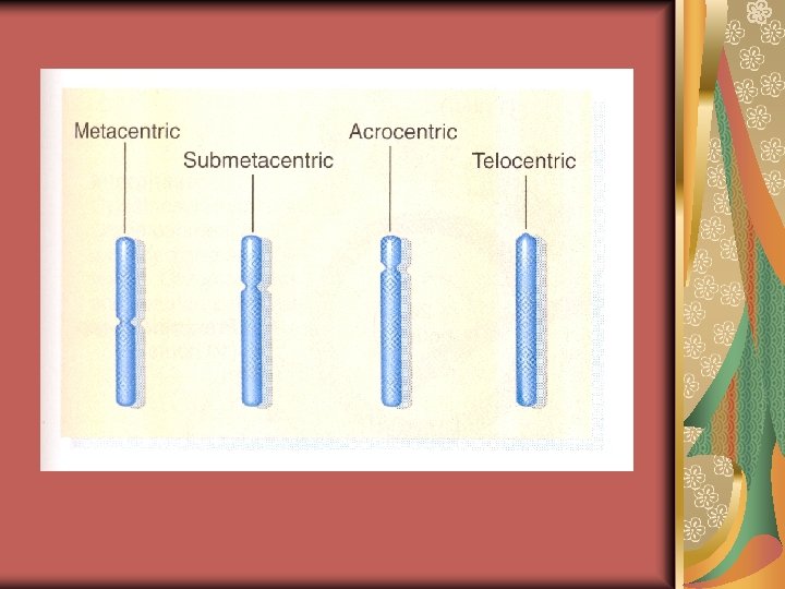
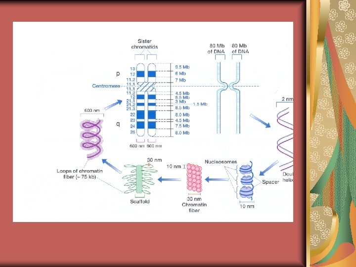
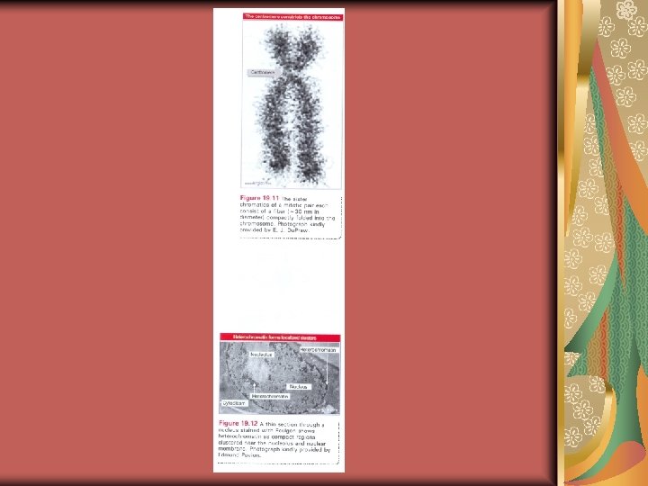
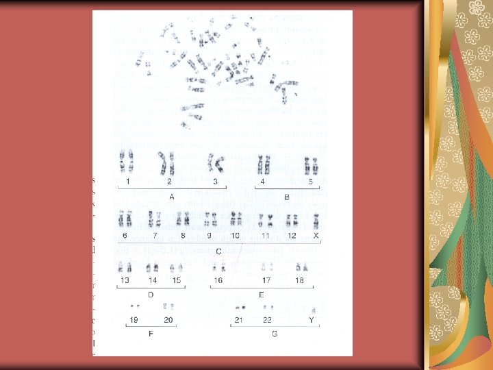
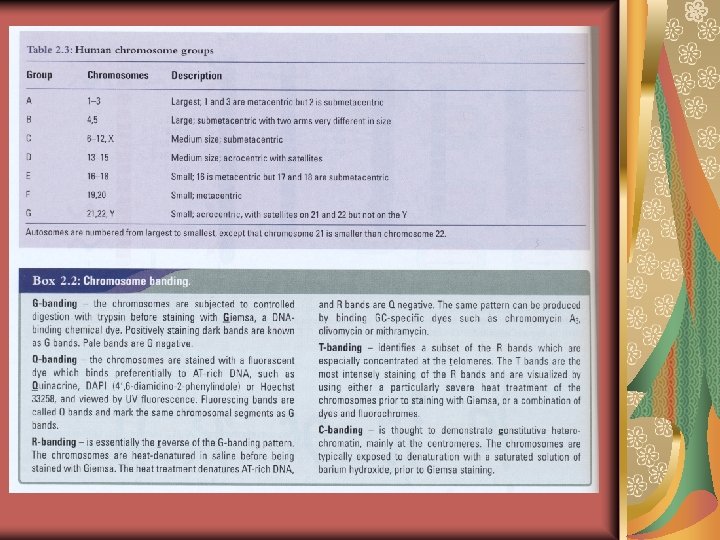
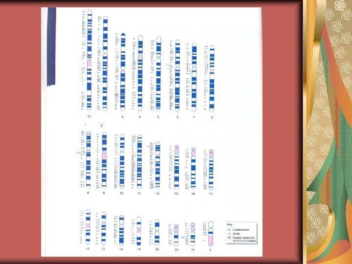
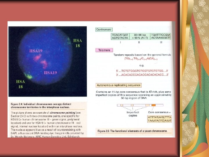
Chromosome The chromosomes are distinguishable on the basis of their size, centromere position, and banding pattern. The centromere may be in the middle, off-center, or close to one end- metacentric (1, 3, 19, 20), submetacentric, and acrocentric, respectively. Acrocentric chromosomes have one arm with a stalk and often with a ‘’bulb’’ (called a satellite) on it ; 13, 14, 15, 21, 22 (repeated bp of r. RNA genes). Telocentric chromosomes have only one arm, because the centromere is at the end.
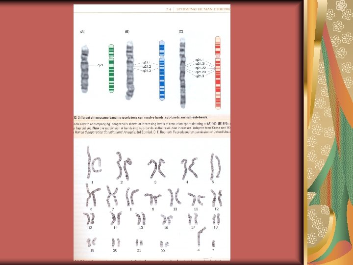
Ø Originally the chromosomes were assigned to groups A through G according to their general size and the position of the centromere. With banding, each chromosome is individually distinguishable. Ø The diagrammatic representation of the banding pattern is the idiogram.
chromosome Chromosomes have a linear appearance: two arms are continuous at the centromere. The shorter arm is designated p (for petit, french) and the longer is q (next letter in the alphabet). In the early part of the cell cycle, each chromosome is present as a single structure, a chromatid, a single DNA molecule. During the cell cycle the chromosomes replicate, and two sister chromatids form.
ØThe autosomes are numbered from largest to smallest, no. 1 through 22 (to split hairs, this order is not exact: for example, chromosome 10 and 11 are shorter than chromosome 12, and chromosome 21 is smaller than 22). ØThe 46 chromosomes come in 23 matching pairs, and constitute the genome.
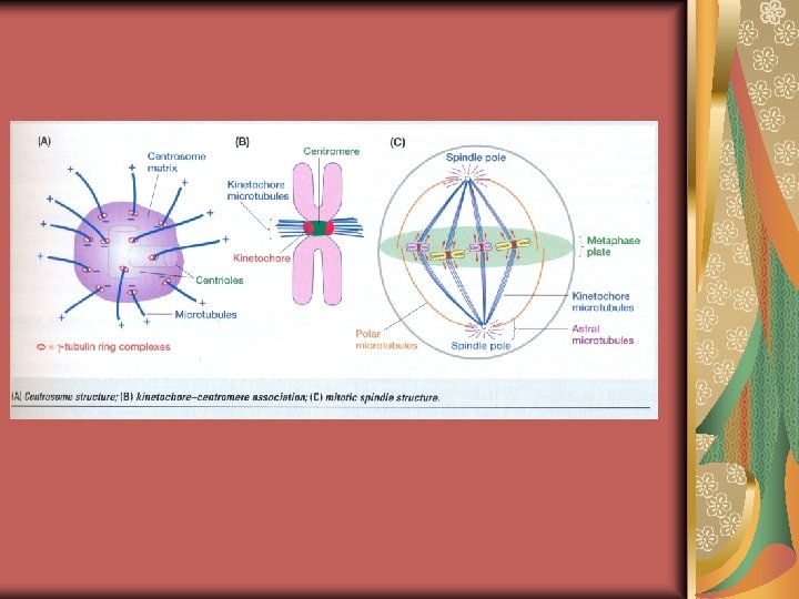
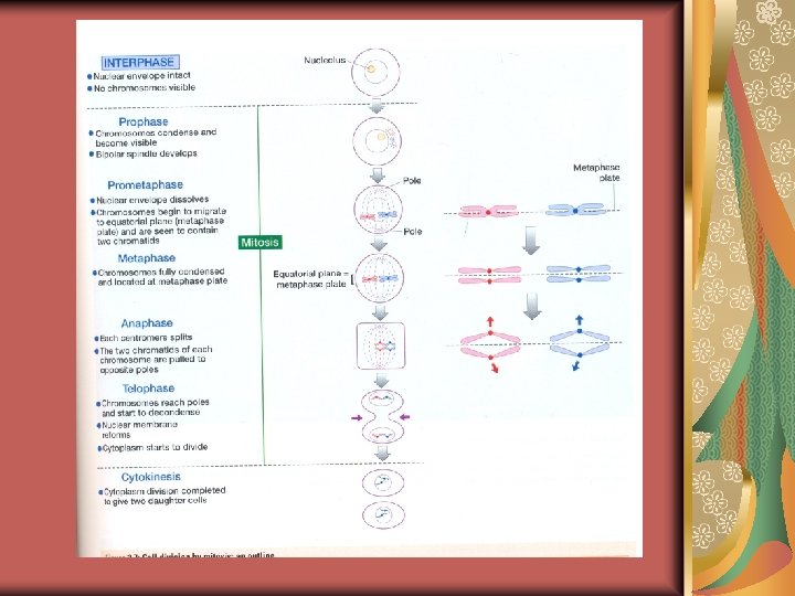
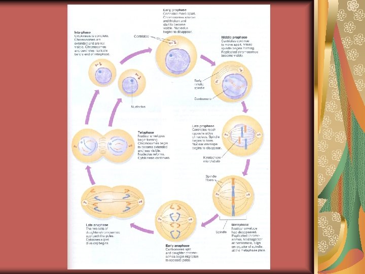
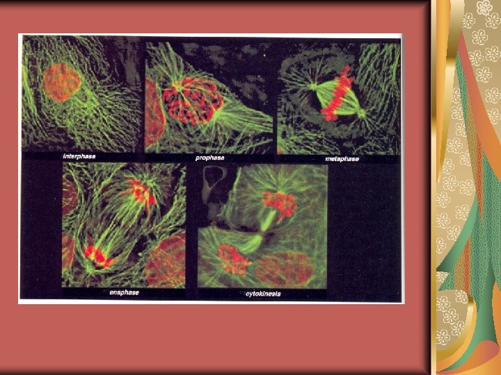
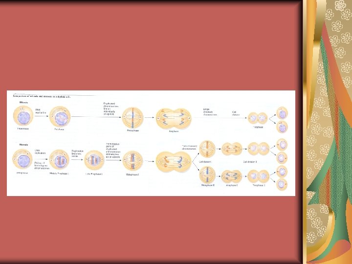
Mitosis The M phase of the cell cycle consists of the various stages of nuclear division; prophase, prometaphase, anaphase and telophase of mitosis, and cell division (cytokinesis) which overlaps the final stages of mitosis. The chromosomes contract during late prophase to become thicker and shorter (chromosomal condensation). During late prophase of mitosis, a pair of large multiprotein complexes known as kinetochores, forms at each centromere, one attached to each sister chromatid. Microtubules attach to each kinetochore, linking the centromere of a chromosome and the two spindle poles.
Ø At anaphase, the kinetochore microtubules pull the two sister chromatids toward opposite poles of the spindle. Ø Kinetochores play a central role in this process, by controlling assembly and disassembly of the attached microtubules.
Mitosis In late prophase, the nuclear membrane disappears and metaphase begins. At this point, the mitotic spindle becomes visible as thin threads. It begins at two polelike structures (centrioles). The interaction between the different spindle fibres pulls the chromosomes toward the center, and by metaphase each chromosome is aligned on the equatorial plane (metaphase plate). During mitosis each chromosome in the diploid set behaves independently and paternal and maternal homologs do not associate at all.
ØAt anaphase the centromeres divide leading to physical separation of what were previously sister chromatids, and the pull by the spindle fibers ensure that the separated sister chromatids go to oppsite poles. ØThe DNA of the two sister chromatids is identical, barring any errors in DNA replication.
The stages of Mitosis Prophase: the chromatides are very elongated and they begin to coil tightly. So they appear shorter and fatter under the microscope. Metaphase: the nuclear envelope has completely disappeared. The microtubules attached to the kinetochores orient the chromosomes so that their centromeres become aligned in one plane halfway between the two spindle poles. Anaphase: It begins when the joined centromeres of sister chromatids separate, giving rise to two daughter chromosomes. Telophase: During this stage the migration of daughter chromosomes to the two poles is completed.
Result of Mitosis Thus the effect of mitosis is to generate daughter cells that contain precisely the same set of DNA sequences.
Interphase and the stages of mitosis in whitefish early embryo cells
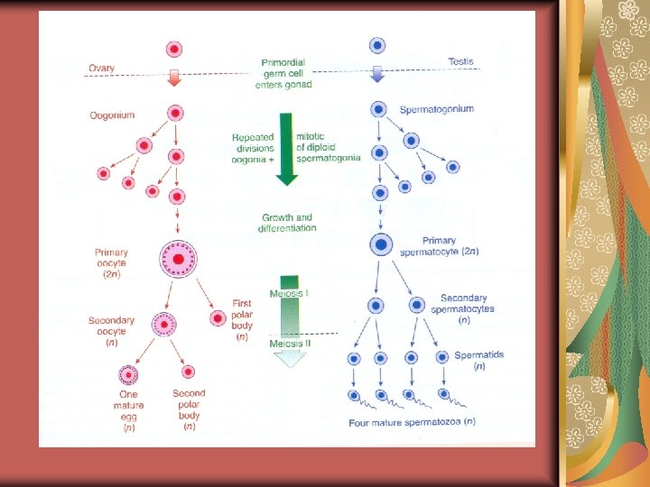
Meiosis is the two successive division of a diploid nucleus after only one DNA replication (chromosome duplication) cycle. Primordial germ cells migrate into the embryonic gonad and engage in repeated rounds of mitosis to form oogonia in females and spermatogonia in males (this involves many more mitosis in males than females which may be a significant factor in explaining sex differences in mutation rate). Further growth and differentiation produces primary oocytes in the ovary and primary spermatocytes in the testis.
Ø In males, meiosis’ product is four spermatozoa; in females, however, there is asymmetric cell division because the cytoplasm divides unequally at each stage: the products of meiosis I (the first meiotic division) are a large secondary oocyte and a small cell (polar body). Ø During meiosis II (the second meiotic division) the secondary oocyte then give rise to the large mature egg cell and a second polar body.
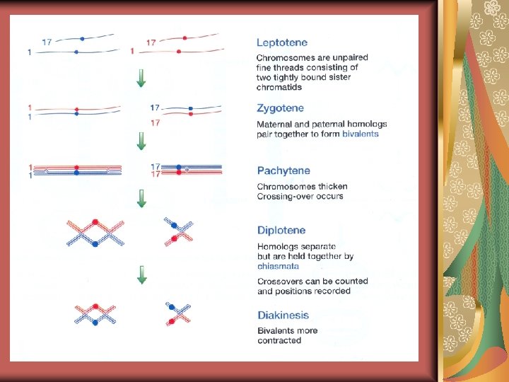
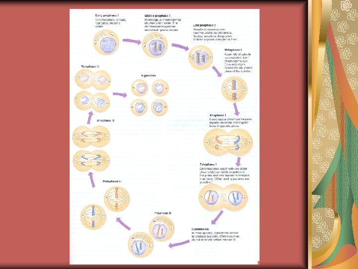
Meiosis’ stages 1 Meiosis I: Prophase I; 1. Leptonema (early prophase I, the leptotene stage): the extended chromosomes begin to coil and become visible as long, thin threads. 2. Zygonema (early to midprophase I, the zygotene stage): the chromosomes continue to shorten. § The homologous pairs of chromosomes actively find each other and align roughly along their length. § Each pair of homologs then undergoes synapsis –the formation along the length of chromatids of a zipperlike structure called the synaptonemal complex, which aligns the two homologs precisely; base pair for base pair.
§ The telomeres of chromosomes play an important role in the initiation of synapsis. § They clustered on the nuclear envelope to produce an arrangement called a bouquet, because of its resemblance to the stems from a bouquet of cut flowers. § In some way, the telomeres move the chromosomes around so that homologous chromosomes align and undergo synapsis.
Meiosis’ stages 2 3. Pachynema: (midprophase I, the pachytene stage); starts when synapsis is completed and each synapsed set of homologous chromosomes consists of 4 chromatids which called a bivalent or a tetrad. § Crossing-over is a significant event for genetic which the reciprocal physical exchange of chromosome segments at corresponding positions along pairs of homologous chromosomes occurs. § This exchange is facilitated by the alignment of the homologous chromosomes brought about by the synaptonemal complex. § If there are genetic differences between the homologs, crossing-over can produce new gene combinations in a chromatid (The new chromosomes called recombinant chromosome).
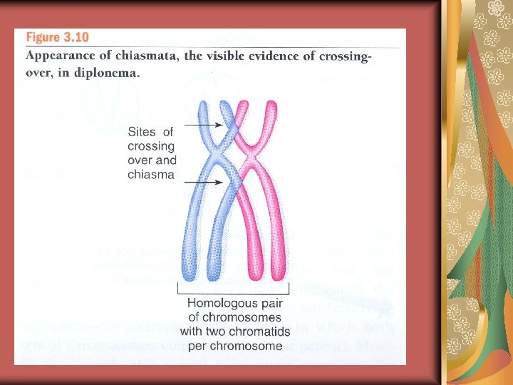
Meiosis’ stages 3 4. Diplonema: (mid-to late prophase I, the diplotene stage): the synaptonemal complex disassembles and the homologous chromosomes begin to move apart. Ø The result of crossing-over becomes visible during diplonema as a cross-shaped structure called a chiasma (plural, chiasmata). Ø Even though the sex chromosomes are not homologous, the Y chromosome of eutherian (placental) mammals has small regions at each end that are homologous to regions on the X chromosome. Ø These pseudoautosomal regions (PARs) pair in male meiosis, and crossing-over occurs between them. Ø When the PAR is deleted from the short arm of the Y chromosome, pairing between the X and Y chromosomes does not occur, and male is sterile.
• In most organisms, diplonema is followed rapidly by the remaining stages of meiosis, However, in many animals, the oocytes (egg cells) can remain in diplonema for very long periods. • In human females, for example, oocytes go through meiosis I up to diplonemaby the seventh month of fetal development and then remain arrested in this stage for many years. • At the onset of puberty and until menopause, one oocyte per menstrual cycle completes meiosis I and is ovulated. 5. Diakinesis (late prophase), the nucleoulus and nuclear envelope break down. Simultaneously, the spindle is assembled. • The chromosomes can be counted most easily at this stage of meiosis.
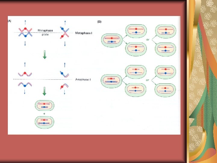
Meiosis’ stages 4 Metaphase I: The nuclear envelope has completely broken down and the bivalents become aligned on the equatorial plane of the cell. The spindle is completely form now, and the microtubules are attached to the kinetochores of the homologs. Anaphase I: The chromosomes in each bivalent separate, so the chromosomes of each pair disjoin and migrate toward opposite poles (in this stage, each of separated chromosomes called a dyad). Telophase I: The dyads complete their migration to opposite poles of the cell, and (in most cases) new nuclear envelopes form around each grouping.
Meiosis’ stages 5 Meiosis II: The second meiotic division is similar to a meiotic division. 1. Prophase II: The chromosomes condense. 2. Metaphase II: Each of the two daughter cells organizes a spindle apparatus which attaches to the centromeres that still connect the sister chromatids. The centromers line up on the equator of the second-division spindles.
1. Anaphase II: The centromeres split, and the chromatids are pulled to the opposite poles of the spindle. • One sister chromatid of each pair goes to one pole, and the other goes to the opposite pole. 1. Telophase II: A nuclear envelope forms around each set of chromosomes, and cytokinesis takes place.
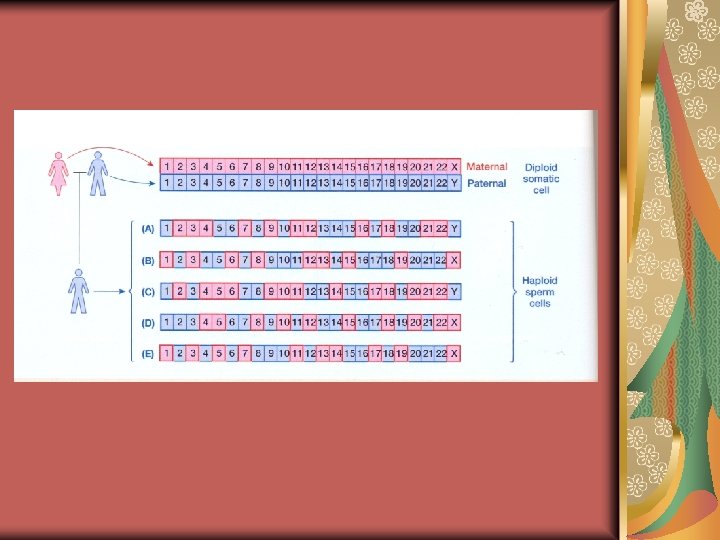
Gene segregation in Meiosis has three significant results: 1. Meiosis generates haploid cells which fusion of haploid nuclei restores the diploid number. 2. In metaphase I, each maternally derived chromosome and each paternally derived chromosome has an equal chance of aligning on one or the other side of the equatorial metaphase plate. The number of possible chromosome arrangements at the metaphase n-1 plate in meiosis is 2 which is more than 4 million combination for human.
Meiosis results 3. The crossing-over between maternal and paternal chromatid pairs during meiosis I generates still more variation in the final combinations.
Mitosis and meiosis differences There are two crucial differences between mitosis and meiosis 1. The products of mitosis are diploid; the products of meiosis are haploid. 2. The products of mitosis are genetically identical; the products of meiosis are genetically different. Another key difference between mitosis and meiosis I is that sister chromatids remain joined after metaphase in meiosis I, whereas they separated in mitosis.
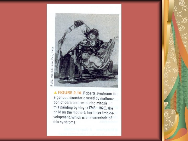
Roberts syndrome Autosomal recessive disorder characterised by: craniofacial anomalies, limb reduction defects and loss of cohesion at heterochromatin regions of centromeres (chromosome puffing)
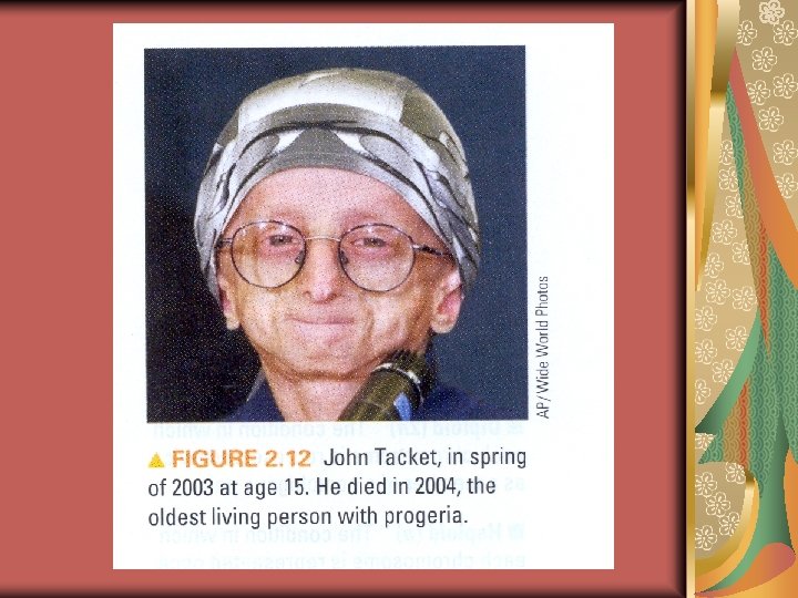
Cancer is a disease of the cell cycle Cells grow in vitro undergo a characteristic number of divisions. Once this number, known as the Hayflick limit, is reached, the cells stop dividing. Cells from human embryos have a limit of about 50 divisions. Cells from adults can divide only about 10 to 30 times. Several genetic disorders that affect cell division are associated with accelerated aging (Progeria).
Hutchinson-Gilford Progeria syndrome The word progeria is derived from the Greek for "prematurely old". The condition is distinguished by limited growth, alopecia and a characteristic appearance with small face and jaw and pinched nose. Later the condition causes wrinkled skin, atherosclerosis and cardiovascular problems. Mental development is not affected. Individuals with the condition rarely live more than 16 years; the longest recorded life-span was 29 years. The development of symptoms is comparable to aging at a rate six to eight times faster than normal, although certain age-related conditions do not occur. Specifically, victims show no neurodegeneration or cancer predisposition.
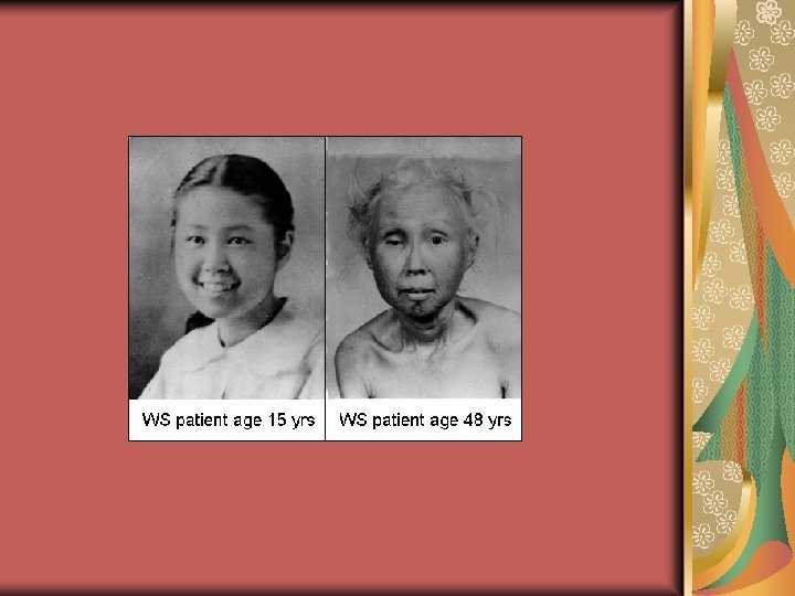
Cancer is a disease of the cell cycle Werner Syndrome is an other genetic disorder associated with premature aging. In this case, the disease process begins between the ages of 15 and 20 years, and affected individuals die of age-related problems by 45 t 0 50 years. It is characterised by: premature greying an thinning of the hair, short stature, prematurely aged appearance, osteoporosis, diabetes mellitus type 2, hypogonadism, premature atherosclerosis, a weak or hoarse voice, and cataracts.
Questions 1. 2. 3. 4. 5. A cell from a human female has just undergone mitosis. For unknown reasons, the centromere of chromosome 7 failed to divide. Describe the chromosomal contents of the daughter cells? How can errors in the cell cycle lead to cancer in humans? Mitosis and Meiosis always differ in regard to the presence of: Chromatids Homologs Bivalents Centromeres spindles