CELL DIVISION Subtopic 1 6 Cell division is

CELL DIVISION Subtopic 1. 6

Cell division is essential, but must be controlled • Cells need to divide for: • growth • tissue repair • reproduction • to prevent the inefficiency of exchange of substances as cell size increases. • However, this process must be controlled, otherwise, it can lead to uncontrolled growth, such as tumors.

Cell cycle • Defines the cycle of events that occurs in the life of a cell from the point when it is created until it divides to give rise to new cells. • These events are grouped into defined stages of the cell cycle.

1. 6. 1 Cell cycle and cyclins Cell cycle is divided into three stages: 1. Interphase 2. Mitosis 3. Cytokinesis

Chromosomes • Eukaryotic chromosomes consist of DNA which is tightly wound around Histones. • Histones are basic (alkaline) proteins that form part of nucleosomes. • Many nucleosomes are coiled together in a specific pattern to form a superstructure called a chromosome. • During interphase, chromosomes are unwound, so that protein synthesis and replication can take place. • Chromatin is considered as a lower order of DNA organization while chromosomes are the higher order of DNA organization


Chromatin, Chromosome or sisterchromatid? • Most of the time, DNA-histone complex material is diffused, or spread out, in the nucleus of a cell, and it is called chromatin. • When getting ready to divide, the DNA condenses into denser bodies, called chromosomes. • When a single chromosome has been replicated in copies, it is called a sisterchromatid. Centromeres, hold the two sister chromatids together. • Sister chromatids are used in cell division, while homologous chromosomes are used in reproductive division.

Sister Chromatids • During interphase, DNA is visible as chromatin. • Following prophase, the phase when DNA supercoiling takes place, each chromosome is visible as a pair of sister chromatids that are identical to each other.

Interphase – G 1, S & G 2 • Most active • Longest phase of the cell cycle • Involves many processes that are occurring both in the nucleus and cytoplasm. • It has three important stages G 1 (Gap 1) • S (synthesis) • G 2 (Gap 2) •

G 1 – Gap 1 • Takes place in the cytoplasm. • Rapid protein synthesis takes place allowing the cell to grow in size. • Proteins required for DNA synthesis (the next phase) are made.

S phase - synthesis • Takes place in the nucleus. • DNA replication takes place and each chromosome is converted to a pair of sister chromatids connected by the centromere. • Amount of DNA doubles.

G 2 – Gap 2 • Takes place in the cytoplasm. • Protein synthesis occurs to produce a variety of proteins of particular significance to the cell cycle such as microtubule proteins. The cell is actively preparing for cell division. • Replication of mitochondria and chloroplast (in the case of plant cells).

Mitosis • Division of the nucleus • Produces two identical daughter nuclei.

Cytokinesis • Cytoplasmic division. • Divides the cytoplasm of a parental cell among the two daughter cells.

Cyclins – regulate the cell cycle • Family of proteins that control the progression of cells through the cell cycle. • Cells cannot progress to the next stage of the cell cycle unless the specific cyclin reaches its threshold. • Cyclins bind to enzymes called cyclin-dependent kinases (CDKs) and activate them. The activated CDKs then attach phosphate groups to other proteins in the cell. The attachment of phosphate triggers the other proteins to become active and carry out tasks (specific to one of the phases of the cell cycle). The diagram below shows how the concentration of four types of cyclins fluctuate during the cell cycle. • Thus, the main regulatory elements of cell cycle control are cyclins and CDKs. • Depending on the presence and action of these proteins, the cell cycle can be fast or slow, and it may even stop altogether.

Cyclin Concentration Throughout the Cell Cycle G 1 phase S phase G 2 phase During this phase G 1 cyclin (red line) levels gradually fluctuate. The G 1/S cyclins (purple line) S cyclins (blue line) induce are instrumental for DNA replication and also promote centromere duplication. Mitosis M cyclins (yellow line) are essential for the formation of mitotic spindles and the alignment of chromatids.

Nature of Science Luck has a role in scientific discovery. • Tim Hunt was investigating the control of protein manufacture in developing sea urchin eggs. During his experiments, he accidentally discovered a set of proteins whose concentration increased and then decreased in coinciding with progression through the stages of the cell cycle. He named the proteins cyclins. • He was awarded the 2001 Nobel Prize for Physiology for this discovery and in his acceptance speech made many allusions to serendipity – the role of luck in science.

TOK What is the role of intuition and serendipity in scientific discovery? • • Intuition can mean the ability to develop an idea prior to deriving it from rational thought. As such, an intuitive thought can be thought of as a “hunch” and the subsequent explanation of a phenomenon as serendipitous. To quote Albert Szent-Gyorgyi, a 1937 Nobel Laureate in Physiology or Medicine: “A discovery is said to be an accident meeting a prepared mind”. Intuitive thoughts are regarded as a function of the mind that is not dependent upon logic. The intuitive thoughts that eventually lead to a new idea are often known as the “eureka moment” from the ancient Greek meaning “found it”. Karl Popper, an eminent philosopher wrote in 1959 “The initial stage, the act of conceiving or inventing a theory seems to me neither to call for logical analysis nor to be susceptible of it”. A scientist is an observer, an intuitive thought comes as a projection to an initial observation in a trained mind. Serendipity also plays a role in scientific discovery, for example, if the contamination of an agar plate by Penicillium had not occurred, Fleming could not have made the observation that led to the discovery of the antibiotic penicillin. A scientific discovery must be evidentially verified experimentally but without the original “flash of inspiration” the investigation leading to a discovery would never have been pursued.

1. 6. 2 Mitosis occurs after G 2 phase Involves 4 phases namely: • Prophase • Metaphase • Anaphase • Telophase

Prophase • DNA supercoils causing the chromatin to condense to form chromosomes that appear as sister chromatids held together by the centromere. • Nucleolus disappears. • Nuclear membrane disintegrates. • Spindle fibers (made of microtubules) start to form (and are completely formed by the end of prophase). • Centrioles (absent from plant cells) move to opposite poles.

Metaphase • Spindle fibers bind to centromere of chromosome/sister chromatids and cause their movement towards the equatorial plate. • Chromosomes are aligned at the equatorial plate at the end of metaphase.

Anaphase • Starts with the splitting of the centromere due to shortening of the microtubule of the spindle fibers. • Sister chromatids are separated (now known as chromosomes) and pulled to opposite poles by the spindle fibers. • Because chromosomes are pulled by their centromeres, the latter is the first to reach the poles.

Telophase • The chromosomes have reached the poles. • The nuclear membrane starts to reform at each pole. • The nucleolus reappear in each new nucleus. • The spindle fibers disintegrate. • The cell elongate in preparation for cytokinesis. • In some cases the invagination of the membrane is also visible (marking the beginning of cytokinesis).

1. 6. 3 Cytokinesis • Though mitosis is similar for animal and plant cells, cytokinesis is very different due to the presence of a cell wall in plant cells. It is the division of the parental cytoplasm among the two daughter cells after mitosis (though it is often started in telophase).

Animal cells • Microfilaments, a ring of contractile protein, located immediately beneath the plasma membrane at the equator pulls the plasma membrane inward. • The inward pull on the plasma membrane produces the characteristic cleavage furrow. • When the cleavage furrow reaches the center of the cells it is pinched apart to form two daughter cells.

Plant cells • During telophase, vesicles derived from the Golgi apparatus move to the equator of the cell. This give rise to a row of vesicles. • Vesicles fuse to form tubular structures. • The tubular structures merge to form two layers of plasma membrane (i. e. the cell plate). • The cell plate grows from the center towards the lateral walls until it connects with the existing cell’s plasma membrane. • This completes the division of the cytoplasm and the formation of two daughter cells. • Vesicles deposit, by exocytosis, pectins and other substances in the lumen between the daughter cells to form the middle lamella (‘gluing’ the cells together). • Both daughter cells secrete cellulose to form their new adjoining cell walls.

Mitotic Index •

Cell division phases
,")
1. 6. 4 Tumorigenesis • The formation of a tumor (or several of them), which is itself defined as a mass of cells. • 2 types: • Benign – non-cancer causing usually localized • does not spread to other parts of the body. • respond well to treatment • • Malignant – cancer causing cancerous growth that is often resistant to treatment. • may spread to other parts of the body • sometimes recur after it has been removed. •

What causes tumors? • Events of the cell cycle are disrupted because of a mutation in one of the cyclins or CDKs. • A single cell can trigger the beginning of a tumor. The cell may have lost its ability to enter interphase, but instead continues to divide rapidly. • The mutation is passed on to the daughter cells, and a clump of cells start to form. • When this uncontrolled growth continues, a tumor is formed, as can be seen in the diagram below

How cancer happens

Mutation • A mutation is a change in an organism’s genetic code. • A change in the base sequence of a certain gene can result in tumor formation. • Due to non-coding sequences in genes, not all gene mutations lead to uncontrolled cell division.

Mutagens • These are agents that cause gene mutations. Although not all mutations result in cancers, anything that causes a mutation has the potential to cause a cancer. Mutagens can be: • Chemicals that cause mutations that are referred to as carcinogens. • High energy radiation such as X-rays. • Short-wave ultraviolet light. • Some viruses.

Oncogenes

Oncogene • An oncogene is a mutated gene that contributes to the development of a tumor. • In their normal, un-mutated state, oncogenes are called proto-oncogenes, and they help in the regulation of cell division. • Metastasis is the movement of cells from a primary tumor to set up secondary tumors in other parts of the body.

Primary and secondary tumors • Once abnormal growth has started at a particular place in the body and a malignant tumor has been initiated, a primary tumor is formed. • Secondary tumors develop from primary. The steps involved are: • Cancerous cells detach from the primary tumor. • Some cancerous cells gain the ability to penetrate the walls of lymph or blood vessels and hence circulate around the body. • The circulating cancerous cells invade tissues at different locations and develop, by uncontrolled cell division, into secondary tumors.

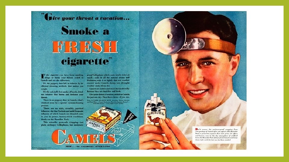
1. 6. 5 Smoking and cancer • Smoking not only causes lung cancer, but also a range of other types of cancer. • The link between smoking and the incidence of cancer was once aggressively denied by the tobacco industry. Initially, research was able to establish a strong correlation, but over many years of study, causal relationships between some of the compounds in cigarette smoke and tumorigenesis were established. Only then did the tobacco industry grudgingly admit that the link existed.

• https: //www. youtube. com/watch? v=ysk. YG-EVl. BY

Link between smoking and the incidence of lung cancer With increasing consumption of cigarettes, as well as the number of years a person smokes, the risk of developing lung cancer increases.

Link between smoking and the incidence of lung cancer There is an age-related connection too. Below the age of 29, the risks of developing lung cancer are relatively small, and are not so different for those who smoke less than 10 cigarettes per day, compared to those who smoke more than 20 per day.
- Slides: 42