Cell Division in Eukaryotes Mitosis Eukaryotic Chromosomes Composed


 primary growth phase, usually")
 resting state after G 1, may remain")

 M (metaphase)")


- Slides: 15
Cell Division in Eukaryotes: Mitosis
Eukaryotic Chromosomes � Composed of chromatin: complex of DNA and proteins � Chromosome number varies in species ◦ Humans have 46 (23 identical pairs) ◦ Drosophila has 8 (4 identical pairs)
Eukaryotic Chromosomes � Telomere: specialized structure that caps the end of each chromosome � Centromere: contains repeated DNA sequences that bind specific proteins; make up the kinetochore where the microtubules bind during cell division � Microtubules: composed of the protein tubulin; moves the chromosomes into position for cell Kinetochore division
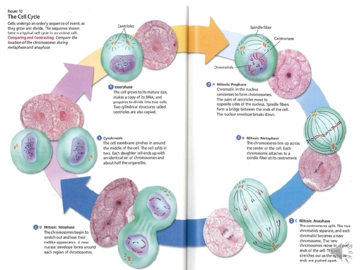
Eukaryotic Cell Cycle � Requires the duplication of the genome, segregation, and division of cellular contents � Divided into 5 phases, which can be grouped into two based on functions
Interphase: Preparation for Mitosis � Gap Phase 1: (G 1) primary growth phase, usually the longest phase ◦ “Gap phase” refers to gap between cytokinesis and DNA synthesis � Synthesis: (S) cell synthesizes a replica of the genome � Gap Phase 2: (G 2) secondary growth phase ◦ Fills in gap between DNA synthesis and start of mitosis ◦ Mitochondria and other organelles replicate while microtubules begin to assemble at a spindle
� Gap Interphase Phase 0: (G 0) resting state after G 1, may remain in this state for days to years ◦ Muscle and nerve cells remain in G 0 permanently ◦ Most of the cells in an animal’s body are in G 0 at any given time
Interphase
Mitosis: “M Phase” Chromosome Segregation and Division of Cytoplasmic Contents P (prophase) M (metaphase) A (anaphase) T (telophase) C (cytokinesis)
� Prophase: chromosomes condense and become visible while the nuclear envelope breaks down; the spindle begins to assemble P
� Metaphase: microtubules become organized into a spindle, attach to chromosomes from opposite poles, and move them to the equatorial plane M
� Anaphase: initiated by sister chromatids separating – daughter chromosomes move to opposite poles of the cell and spindle poles move apart A
� Telophase: chromosomes cluster at opposite poles and decondense while nuclear envelopes re-form around them T
� Cytokinesis: not technically mitosis, but part of the “M phase” ◦ Involves cleavage of cell at cleavage furrow into roughly equal halves C (cleavage furrow)
Recap �Interphase �M Phase G 1 G 0 S G 2 Prophase Metaphase Anaphase Telophase Cytokinesis Mitosis