Cell Division Genetics Molecular Biology 20 1 a

Cell Division, Genetics, Molecular Biology 20. 1 a History of DNA and Structure
 �Found in nucleus of all organisms (within chromosomes) �DNA only")
DNA �Deoxyribonucleic acid (DNA) �Found in nucleus of all organisms (within chromosomes) �DNA only molecule capable of replicating itself �Contains instructions that ensure continuity of life - coded within chemical messages of DNA - regulates the production of proteins �Ability to change due to mutations and new combinations of genes

Frederich Miescher � 1869 – extracted viscous white substance from bandages of wounded soldiers - slightly acidic, phosphorus & nitrogen rich - called it nuclein �Nuclein composed of acidic portion (nucleic acid) and alkaline portion (protein) �Single nucleic acid was later shown to be 2 nucleic acids - deoxyribonucleic acid (DNA) - ribonucleic acid (RNA) �DNA material of heredity: early focus was on proteins
 �Nucleus in foot:")
Joachim Hammerling �Acetabularia: green algae, 3 distinct regions (cap, stalk, foot) �Nucleus in foot: cut off cap and new cap regenerated, cut off foot, no new foot regeneration �Suggested hereditary material located in nucleus

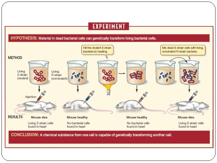
Frederick Griffith �Lab Exercise pg 644 �Streptococcus pneumoniae – 2 forms - virulent: S-form (coated) - harmless: R-form �S-form cells heated and killed, injected into mice and they lived �Heated cells mixed with R-form cells, killed mice �Concluded there must be something chemical altering the living cells: transformation - transformed into virulent cells

Avery, Mc. Carty, Mac. Leod �Lab Exercise pg 645 � 1944 – experiments with Streptococcus pneumoniae in test tubes �Treated heat-killed virulent bacteria with a proteindestroying enzyme: transformation still occurred �Treated heat-killed virulent bacteria with DNAdestroying enzyme: transformation DID NOT occur �Concluded DNA was “transforming principle” - likely source of hereditary information
 that infect bacterial")
Alfred Hershey & Martha Chase � 1952 – used bacteriophages (virus) that infect bacterial host (2 components: DNA and protein coat) �Infects by injecting DNA into it, virus multiplies within and then bursts out, killing the cell �Hershey & Chase concluded that only the DNA, not protein coat, enters bacteria - tagged viral proteins with isotope of sulfur (not component of DNA) - tagged viral DNA with isotope of phosphorus (component of DNA) �Allowed tagged bacteriophage to infect bacterial cell �Cells blended to remove protein coats and centrifuged to

�Bacterial cells found to contain isotope of phosphorus, not isotope of sulfur �Isotope of sulfur found in culture medium �Conclusion! DNA was hereditary material

James Watson & Francis Crick �Known that DNA comprised of chains of nucleotides - consist of 5 -carbon cyclic ring: deoxyribose sugar - one of 4 nitrogenous bases attached to 1’ carbon - phosphate group attached to 5’ carbon � 4 bases: adenine (A), guanine (G), thymine (T), cytosine (C) - A & G: purines (double ring) - C & T: pyrimidines (single ring) �Evidence from Edwin Chargaff: calculated that amount of adenine always equal to amount of thymine (same for guanine and cytosine). Observed for almost all species �Evidence from Rosalind Franklin: x-ray diffraction, photograph taken

James Watson & Francis Crick �All the evidence compiled, Watson & Crick created a 3 D model �Portrayed relationship between bases as well as bond angles and spacing of atoms - consistent with observations from other researchers to that point �Won Nobel Prize in 1962 along with Maurice Wilkins (researcher in charge of

DNA Structure � 2 strands of nucleotides �Each nucleotide contains: - deoxyribose sugar - phosphate group - nitrogenous base �Covalently bonded into double helix like a twister ladder - hydrogen bonds keep helix together �Base pairs are rungs, sugar/phosphate backbones are struts �Complementary base pairing to form rungs - A pairs with T

DNA Structure

DNA Structure �Opposite strand always have the complementary sequence of bases 5’ – ATGCCGTTA – 3’ 3’ – TACGGCAAT – 5’ �Antiparallel: run parallel but in opposite directions - one strand has 5’ carbon & phosphate group at one end and 3’ carbon & hydroxyl group of deoxyribose sugar at other end - other strand runs opposite 3’ to 5’ �Direction important to enzymes interacting with DNA - only read or copy DNA in one direction DNA Structure

Nobel Prize - DNA
- Slides: 15