Cell Division Chapter 9 Evolution of Cell Division

 � � � A. No")


 �One double helix �Two chromatids �Two double")

 Tubulin subunits in centrosome begin to assemble into �")


 G 1 checkpoint – cycle initiation")


 Density-dependent Inhibition � cells that")

 �Transformed cell")


- Slides: 48
Cell Division Chapter 9
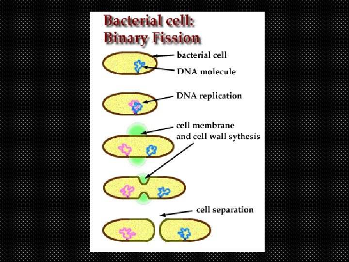
Evolution of Cell Division �I. Prokaryote Cell Division (Bacteria/Archaea) � � � A. No nucleus so no mitosis B. No microtubules or motor proteins to move chromosome. C. Divide by Prokaryotic fission 1. single circular chromosome binds to cell membrane 2. DNA replication in both directions around circle 3. Cell divides by adding to cell membrane
�II. � � � � Eukaryotic Cell Division A. DNA contained in nuclear membrane B. DNA replicated prior to nuclear division ( in interphase) C. Cell division divided into two parts 1. mitosis = division of nucleus 2. cytokinesis = division of cytoplasm D. Microtubules and microfilaments needed E. Motor proteins and ATP required
Two Types of Eukaryotic Nuclear Division �I. Mitosis � � � � A. produces clones (daughter cells) B. unicellular organisms : reproduction C. Multicellular organisms : 1. asexual reproduction (budding) 2. growth 3. replacement 4. repair
�II. Meiosis � � � A. produces haploid cells 1. chromosome number cut in ½ 2. non-identical cells 3. gametes B. only done for sexual reproduction
Vocab. �Somatic cell – �normal diploid body cell �Diploid cell – �has 2 copies of each chromosome �Haploid cell – �has 1 copy of each chromosome �Chromosome – �naturally occurring segment of DNA and associated proteins
Chromosome forms �I. Chromatin - A. DNA wrapped around histones � B. no supercoiling � C. Most DNA available for transcription � D. not visible under microscope �II. Chromatid � A. nucleosomes supercoiled into compact ‘arms’ � B. DNA packaged for transport not use � C. condensed chromosomes visible �
Condensed Chromatids �Constriction in center = � centromere �= a region of DNA that binds to cohesin proteins that function to hold sister chromatids together �Other cohesins hold sister chromatids together more loosely along their lengths
Sister Chromatids �Identical �Formed by semi-conservative replication �While joined at centromere = 1 chromosome
Semi-conservative Replication
unduplicated �One chromosome �One chromatid �(one centromere) �One double helix �Two chromatids �Two double helixes
Genome �Genome = all of a cells DNA �All eukaryotes have set # Chromosome in their genome �Humans have 46 � Two of each type… 23 different types
Mitotic Spindle formation � 1) Tubulin subunits in centrosome begin to assemble into � � microtubules. http: //www. youtube. com/watch? v=Rbbtbt 2 i 8 x. A&list=PLCF 9 FC 302 EC 1 CA 125 � 2) microtubules grow toward the center to form spindle fibers � 3) short microtubules form a radial array called an aster � 4) centrioles present in animals but not needed �
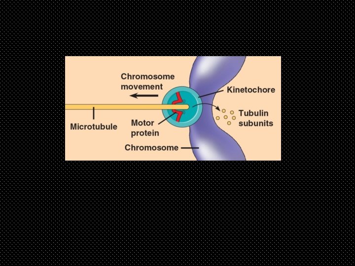
Kinetochore �Proteins located at centromere �Attachment site for some microtubules of spindle �Polar microtubules overlap with microtubules from opposite pole at center of cell
Mitotic Spindle includes: centrosomes, spindle microtubules, & aster �Polar microtubules centrosome
Prophase �Centrosomes begin producing microtubules & moving toward opposite poles �Chromosomes condense into… � �Nucleoli disappear. chromatids �(pro-metaphase) �Nuclear envelope breaks down �Microtubules attach to … � kinetochores �Polar microtubules overlap at equator
Metaphase �Chromosomes lined up at equator �Pulled by kinetochore microtubules �C line up single file, �One sister chromatid on each side �Centrosomes reach poles
Anaphase �Cohesin proteins cleaved by Separase enzymes �Separated sister chromatids move toward opposite poles �Kinetochore microtubules shrink as they depolymerize at centrosome �Motor proteins drag chromatids along shrinking microtubules toward poles �Cell elongates as motor proteins push polar microtubules past each other �
Telophase �Begins when chromatids reach poles �Microtubules disassemble �Nuclear envelope reforms �Chromosomes �de-condense into � chromatin
Cytokinesis �Cytokinesis begins before mitosis is complete �Different in plants and animals �Does not always take place
Animal cell cytokinesis http: //cc. scu. edu. cn/G 2 S/Template/View. aspx? course. Type=1&course. Id=17&top. Menu. Id=113305&menu. Type=1&action=view&type=&name=&linkpage. ID=113700 �Contractile Ring Mechanism � 1) a band of microfilaments of the cell cortex contracts � 2) indentation forms : cleavage furrow � 3) ring contracts until cell membrane is pinched in 2
�Myosin motor proteins � Move actin filaments Past each other to tighten ring
Plant Cytokinesis �Cell Plate Formation �Vesicles containing cell wall components move �from golgi to equator �Merging vesicle membranes form new cell membrane �Cell wall components assembled in center of merging vesicles form new primary cell wall
Primary & Secondary Cell Walls �Primary cell wall : flexible stretchy allows growth �Secondary cell wall: deposited inside primary wall � solid � inflexible � support wall
Cell Cycle – Pattern of stages in cell life �Interphase – time spent between cell divisions (90% of cell cycle) �Mitosis – nuclear division �Cytokinesis – cytoplasmic division �
Interphase : 3 sub-phases �G 1 – gap 1 - cell grows ( max size based on. . � SA: V ratio) � cell performs its function for the body � cell may never leave G 1 (ex nerve cells = G 0) �S – synthesis : entire genome is synthesized �by semi-conservative replication �Growth and cell function continue �G 2 - gap 2 – cell grows & prepares to divide duplicates centrosomes & centrioles (not required: present in animals)
�Usually, cells will take � interphase 18 -20 hours. �G 1 – highly variable � - some cells are in G 1 only long enough to grow � - some almost never divide and are said to enter G 0 �S phase 5 -6 hours �G 2 3 -4 hours in most cells. �Mitosis, & cytokinesis only takes about 2 hours
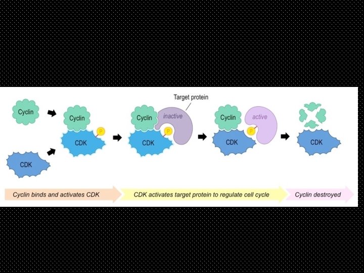
Cell cycle controlled by Checkpoints �Check points = places where the cell cycle stops �Cell cycle only resumes if certain criteria are met. �Cells signal that the criteria have been met by making checkpoint proteins called cyclins. �Cyclins activate Cyclin Dependent Kinases (CDKs) �CDKs act to signal the cell to move on to the next step in the cell cycle.
Cyclin production �When all criteria of a checkpoint are met the specific checkpoint gene for that checkpoint is activated. �The Checkpoint gene contains the instructions for making one specific checkpoint protein (cyclin). �Production of that particular cyclin activates a specific CDK which sets of a chain of reactions that lets the cell cycle pass that checkpoint and continue the cell cycle. �Cyclin breaks down after one use and levels of that
CDK levels remain constant Cell cycle is controlled by changing levels of Cyclins
CDK s : Cyclin Dependent Kinase function �Kinase = group of enzymes that phosphorylate proteins (activate them) �CDKs are a type of Kinase that only functions when bound to cyclin. �Different versions of cyclin activate different CDKs. �Each activated CDK phosphorylates a different protein. �Activating (phosphorylating) that proteins sets off a transduction cascade that transduces the signal to proceed to the next checkpoint
Animal cells stay in G 1 or G 0 unless signaled by growth factors
Check points – regulated by CDKs � 1) G 1 checkpoint – cycle initiation � a)controlled by cell size � b) growth factors � c) environment � 2) G 2 checkpoint – transition to M � a) DNA replication complete � b) DNA damage/mutations � 3) M-spindle checkpoint � a) spindle attachment
�Growth Factors … � 1. are signal molecules � 2. released by cells to signal nearby cells to divide � 3. diffuse through intracellular fluids � 4. bind to membrane receptors on target cell � 5. transduction of signal causes cyclin production � 6. cyclins activate CDK � 7. CDK phosphorylates first protein of cascade that moves cell past G 1 checkpoint � 8. are an example of cell-to- cell communication
Example growth factor = PDGF Platelet-derived Growth Factor �PDGF released by platelets cause Fibroblast(wound repair) cells to divide � 1) PDGF binds to recepter on Fibroblast � � 2) signal transduction pathway initiated � 3) cell passes G 1 checkpoint and starts to divide
Animal cells stay in G 1 or G 0 unless signaled by growth factors
Cyclin-CKD example : MPF M-phase Promoting Factor = � CDK-cyclin complex �STEPS � checkpoint gene for MPF cyclin is activated �MPF cyclin levels build up & build MPF levels �High enough concentration of MPF allows �Cell to move from G 2 into M phase �MPF concentration reduced in Anaphase by breakdown of � cyclin causing MPF to revert to � inactive CDK
CKI = Cyclin Dependent Kinase Inhibitors �Inhibitors stop things �CKIs stop the CDK enzymes from working �Example: �CKI p 21 stops CDK 2 from working…thus �Stopping the transition from G 1 – S phase �The CKI inhibitor molecule p 21 is only active when tumor suppressor gene p 53 is transcribed (copied) *** know p 53 ***
Normal Cell Division �Cell Division limited by: � 1) Density-dependent Inhibition � cells that are crowded stop dividing � 2) Anchorage dependency – cell must be anchored to extra-cellular matrix of a tissue to divide. �
Cell Division in Cancer Cells �Cancer Cells NOT inhibited by density or anchorage �CC do NOT stop dividing when out of Growth Factor �CC do not follow signals of check point genes �CC do not self-destruct by apoptosis
Origin of Cancer Cells � 1 cell undergoes transformation (damage to DNA) �Transformed cell avoids immune system � avoids apoptosis � ignores regular cell cycle signals � uncontrolled cell division �Benign tumor : cells stay anchored �Malignant tumor cells spread = cancer �Metastasis = spread of cancer cells �
Uncontrolled cell division �Results from the failure of more than one checkpoint gene �Which causes non-functional checkpoint proteins �Causes tumor development �May cause cancer
Cancer Cell Changes �Mutation of Check Point Genes �Change in chromosome number/structure �Abnormal/irregular cell membrane lacks attachment proteins � damaged signal/receptor proteins �Secrete signal molecules that encourage blood vessel growth �
Cancer Treatment �Radiation for localized tumor �Chemotherapy – poisons most damaging to dividing cells