CELL DIVISION AND REPRODUCTION 2015 Pearson Education Inc


 reproduce")


 •")


 90 80")


 n Egg cell n Sperm")


- Slides: 67
CELL DIVISION AND REPRODUCTION © 2015 Pearson Education, Inc.
8. 1 Cell division plays many important roles in the lives of organisms • Cell division • is reproduction at the cellular level, • produces two “daughter” cells that are genetically identical to each other and the original “parent” cell, • requires the duplication of chromosomes, the structures that contain most of the cell’s DNA © 2015 Pearson Education, Inc.
8. 1 Cell division plays many important roles in the lives of organisms • Living organisms reproduce by two methods. • Asexual reproduction • produces offspring that are identical to the original cell or organism and • involves inheritance of all genes from one parent. • Sexual reproduction • produces offspring that are similar to the parents but show variations in traits and • involves inheritance of unique sets of genes from two parents. © 2015 Pearson Education, Inc.
8. 1 Cell division plays many important roles in the lives of organisms • Cell division is used for • reproduction of single-celled organisms, • growth of multicellular organisms from a fertilized egg into an adult, • repair and replacement of cells, and • production of sperm and eggs. © 2015 Pearson Education, Inc.
8. 2 Prokaryotes reproduce by binary fission • Prokaryotes (single-celled bacteria and archaea) reproduce by binary fission (“dividing in half”). • Binary fission of a prokaryote occurs in three stages: 1. duplicate chromosome and separate copies, 2. elongation of the cell 3. division into two daughter cells. © 2015 Pearson Education, Inc.
THE EUKARYOTIC CELL CYCLE AND MITOSIS © 2015 Pearson Education, Inc.
8. 3 The large, complex chromosomes of eukaryotes duplicate with each cell division • Eukaryotic cells • store most of their genes on multiple chromosomes within the nucleus. • Each eukaryotic species has a characteristic number of chromosomes in each cell nucleus. • Humans have 46 chromosomes • Fruit Flies have 8 chromosomes © 2015 Pearson Education, Inc.
8. 3 The large, complex chromosomes of eukaryotes duplicate with each cell division • To prepare for division, the chromatin (DNA and proteins) becomes • highly compact and • visible with a microscope. © 2015 Pearson Education, Inc.
8. 3 The large, complex chromosomes of eukaryotes duplicate with each cell division • Before a eukaryotic cell begins to divide, it duplicates all of its chromosomes, resulting in two copies called sister chromatids. • The sister chromatids are joined together and cinched at a “waist” called the centromere. • When a cell divides, the sister chromatids • separate from each other and are then called chromosomes, and • sort into separate daughter cells. © 2015 Pearson Education, Inc.
8. 4 The cell cycle includes growing and division phases • The cell cycle is an ordered sequence of two stages that starts from the time a cell is formed until its own division. Interphase: duplication of cell contents • G 1—growth, increase in cytoplasm • S—duplication of chromosomes • G 2—growth, preparation for division Mitotic phase: division • Mitosis—division of the nucleus • Cytokinesis—division of cytoplasm © 2015 Pearson Education, Inc.
8. 5 Cell division is a continuum of dynamic changes • A mitotic spindle • is required to divide the chromosomes, • guides the separation of the two sets of daughter chromosomes • Spindle microtubules emerge from two centrosomes, organizing regions in the cytoplasm of eukaryotic cells. © 2015 Pearson Education, Inc.
Figure 8. 5 -1 INTERPHASE Centrosomes Chromatin Nuclear envelope © 2015 Pearson Education, Inc. Plasma membrane MITOSIS Prometaphase Prophase Early mitotic spindle Centrosome Fragments of the nuclear envelope Kinetochore Centromere Chromosome, consisting of two sister chromatids Spindle microtubules
8. 5 Cell division is a continuum of dynamic changes • Interphase • The cytoplasmic contents double. • Two centrosomes form. • Chromosomes duplicate in the nucleus during the S phase.
8. 5 Cell division is a continuum of dynamic changes • Prophase (chromosomes become present) • In the nucleus, chromosomes become more tightly coiled and folded.
8. 5 Cell division is a continuum of dynamic changes • Prometaphase • The nuclear envelope breaks into fragments and disappears. • Spindle microtubules form and extend from the centrosomes into the nuclear region and attach to kinetochores.
Figure 8. 5 -6 MITOSIS Metaphase Anaphase Telophase and Cytokinesis Metaphase plate Cleavage furrow Mitotic spindle © 2015 Pearson Education, Inc. Separated chromosomes Nuclear envelope forming
8. 5 Cell division is a continuum of dynamic changes • Metaphase • The mitotic spindle is fully formed. • Chromosomes align at the cell equator (middle). • Kinetochores of sister chromatids are facing the opposite poles of the spindle.
8. 5 Cell division is a continuum of dynamic changes • Anaphase (apart) • Sister chromatids separate at the centromeres. • Daughter chromosomes are moved to opposite poles of the cell as motor proteins move the chromosomes along the spindle microtubules • At the end of anaphase, the two ends of the cell have equal collections of chromosomes. © 2015 Pearson Education, Inc.
8. 5 Cell division is a continuum of dynamic changes • Telophase • The cell continues to elongate. • The nuclear envelope forms around chromosomes at each pole, establishing daughter nuclei. • Chromatin uncoils. • The mitotic spindle disappears. © 2015 Pearson Education, Inc.
8. 6 Cytokinesis differs for plant and animal cells • During cytokinesis, the cytoplasm is divided into separate cells. In animal cells, cytokinesis occurs as a cleavage furrow forms and deepens to separate the contents into two cells. In plant cells, cytokinesis occurs as a cell plate forms in the middle and extends to form new cell walls © 2015 Pearson Education, Inc.
8. 7 Anchorage, cell density, and chemical growth factors affect cell division • The cells within an organism’s body divide and develop at different rates. • Cell division is controlled by • anchorage dependence, the need for cells to be in contact with a solid surface to divide, • density-dependent inhibition, in which crowded cells stop dividing, • the presence of essential nutrients, and • growth factors, proteins that stimulate division. © 2015 Pearson Education, Inc.
8. 8 Growth factors signal the cell cycle control system • The cell cycle control system is a cycling set of molecules that trigger and coordinate key events in the cell cycle. • Checkpoints in the cell cycle can • stop an event or • signal an event to proceed. © 2015 Pearson Education, Inc.
8. 9 CONNECTION: Growing out of control, cancer cells produce malignant tumors • Cancer currently claims the lives of 20% of the people in the United States. • Cancer cells escape controls on the cell cycle. • Cancer cells divide excessively and invade other tissues of the body. © 2015 Pearson Education, Inc.
8. 9 CONNECTION: Growing out of control, cancer cells produce malignant tumors • A tumor is a mass of abnormally growing cells within otherwise normal tissue. • Benign tumors remain at the original site but may disrupt certain organs if they grow in size. • Malignant tumors can spread to other locations in a process called metastasis. • An individual with a malignant tumor is said to have cancer. © 2015 Pearson Education, Inc.
8. 9 CONNECTION: Growing out of control, cancer cells produce malignant tumors • Localized tumors can be • removed surgically and/or • treated with concentrated beams of high-energy radiation. • Metastatic tumors are treated with chemotherapy. • It is increasingly possible to personalize cancer treatment by • sequencing the genome of tumor cells and • tailoring treatment based upon the tumor’s specific genetic profile. © 2015 Pearson Education, Inc.
MEIOSIS AND CROSSING OVER © 2015 Pearson Education, Inc.
8. 11 Chromosomes are matched in homologous pairs • Homologous chromosomes are matched in • length, • centromere position, and • staining pattern. • A locus (plural, loci) is the position of a gene. • Different versions of a gene may be found at the same locus on the homologous pair.
8. 11 Chromosomes are matched in homologous pairs • In humans, somatic cells (body cells) have 46 chromosomes forming 23 pairs of homologous chromosomes. • The human sex chromosomes X and Y differ in size and genetic composition. • The other 22 pairs of chromosomes are autosomes with the same size and genetic composition. © 2015 Pearson Education, Inc.
8. 12 Gametes have a single set of chromosomes • Humans and many animals and plants are diploid (2 n), because all somatic cells contain pairs of homologous chromosomes. • Gametes • are eggs and sperm and • are said to be haploid (n) because each cell has a single set of chromosomes. • n = chromosomes sets © 2015 Pearson Education, Inc.
8. 12 Gametes have a single set of chromosomes • The human life cycle begins when a haploid sperm fuses with a haploid egg in fertilization. • The zygote, formed by fertilization, is now diploid. • Mitosis of the zygote generates all the somatic cells of an adult © 2015 Pearson Education, Inc.
8. 12 Gametes have a single set of chromosomes • Meiosis is a type of cell division that produces haploid gametes in diploid organisms in the ovaries and testes. • Meiosis reduces the chromosome number by half.
8. 13 Meiosis reduces the chromosome number from diploid to haploid • Interphase: Like mitosis, meiosis is preceded by an interphase, during which the chromosomes duplicate. © 2015 Pearson Education, Inc.
8. 13 Meiosis reduces the chromosome number from diploid to haploid • Meiosis I – key events • Homologous chromosomes separate • Chromatids of homologous chromosomes exchange segments in a process called crossing over.
8. 13 Meiosis reduces the chromosome number from diploid to haploid • Each of the two haploid products enters meiosis II. • Meiosis II – key event • Sister chromatids separate
8. 14 VISUALIZING THE CONCEPT: Mitosis and meiosis have important similarities and differences • Mitosis and meiosis both begin with diploid parent cells that have chromosomes duplicated during the previous interphase. • However, the end products differ. • Mitosis produces two genetically identical diploid somatic daughter cells. • Meiosis produces four genetically unique haploid gametes. © 2015 Pearson Education, Inc.
Figure 8. 14 -6 MEIOSIS I MITOSIS MEIOSIS II One division of the nucleus and cytoplasm Result: Two genetically identical diploid cells Used for: Growth, tissue repair, asexual reproduction © 2015 Pearson Education, Inc. Two divisions of the nucleus and cytoplasm Result: Four genetically unique haploid cells Used for: Sexual reproduction
8. 15 Independent orientation of chromosomes in meiosis and random fertilization lead to varied offspring • Genetic variation in gametes results from • crossing over • independent orientation at metaphase I • random fertilization.
Figure 8. 15 -3 Possibility A Possibility B Two equally probable arrangements of chromosomes at metaphase I Metaphase II Gametes Combination 1 Combination 2 © 2015 Pearson Education, Inc. Combination 3 Combination 4
8. 17 Crossing over further increases genetic variability • Genetic recombination is the production of new combinations of genes due to crossing over. • Crossing over is an exchange of corresponding segments between nonsister chromatids of homologous chromosomes. • Nonsister chromatids join at a chiasma (plural, chiasmata), the site of attachment and crossing over. • Corresponding amounts of genetic material are exchanged between maternal and paternal (nonsister) chromatids.
Animation: Crossing Over © 2015 Pearson Education, Inc.
Figure 8. 17 a-0 Chiasma Tetrad © 2015 Pearson Education, Inc. Sister chromatids
Figure 8. 17 b-0 C E c e 1 Breakage of nonsister chromatids C E c e 2 C Tetrad (pair of homologous chromosomes in synapsis) Joining of nonsister chromatids E Chiasma c e 3 Separation of homologous chromosomes at anaphase I C E C c e E c e 4 Separation of chromatids at anaphase II and completion of meiosis C E C e c E c e Parental type of chromosome Recombinant chromosome Parental type of chromosome Gametes of four genetic types © 2015 Pearson Education, Inc.
ALTERATIONS OF CHROMOSOME NUMBER AND STRUCTURE © 2015 Pearson Education, Inc.
8. 18 Accidents during meiosis can alter chromosome number • Nondisjunction is the failure of chromosomes or chromatids to separate normally during meiosis. • Fertilization after nondisjunction yields zygotes with altered numbers of chromosomes. © 2015 Pearson Education, Inc.
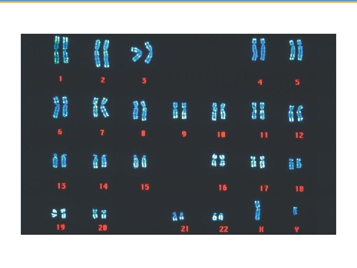
8. 19 A karyotype is a photographic inventory of an individual’s chromosomes • A karyotype is an ordered display of magnified images of an individual’s chromosomes arranged in pairs. • Karyotypes • are often produced from dividing cells arrested at metaphase of mitosis and • allow for the observation of • homologous chromosome pairs, • chromosome number, and • chromosome structure. © 2015 Pearson Education, Inc.
Figure 8. 19 -1 -3 Packed red and white blood cells Blood culture Centrifuge Fluid © 2015 Pearson Education, Inc. Hypotonic solution Fixative White blood cells Stain
Figure 8. 19 -2 © 2015 Pearson Education, Inc.
Figure 8. 19 -3 Centromere Sister chromatids Pair of homologous chromosomes Sex chromosomes © 2015 Pearson Education, Inc.
8. 20 CONNECTION: An extra copy of chromosome 21 causes Down syndrome • Trisomy 21 • involves the inheritance of three copies of chromosome 21 and • is the most common human chromosome abnormality. © 2015 Pearson Education, Inc.
8. 20 CONNECTION: An extra copy of chromosome 21 causes Down syndrome • A person with trisomy 21 has a condition called Down syndrome, which produces a characteristic set of symptoms, including • • characteristic facial features, short stature, heart defects, susceptibility to respiratory infections, leukemia, and Alzheimer’s disease, and • varying degrees of developmental disabilities. • The incidence increases with the age of the mother. © 2015 Pearson Education, Inc.
Figure 8. 20 b Infants with Down syndrome (per 1, 000 births) 90 80 70 60 50 40 30 20 10 0 © 2015 Pearson Education, Inc. 20 25 30 35 Age of mother 40 45
8. 21 CONNECTION: Abnormal numbers of sex chromosomes do not usually affect survival • Sex chromosome abnormalities seem to upset the genetic balance less than an unusual number of autosomes. This may be because of • the small size of the Y chromosome or • X chromosome inactivation. © 2015 Pearson Education, Inc.
8. 22 EVOLUTION CONNECTION: New species can arise from errors in cell division • Errors in mitosis or meiosis may produce polyploid species, with more than two chromosome sets. • The formation of polyploid species is • widely observed in many plant species but • less frequently found in animals. Tetraploid Grey Tree Frog © 2015 Pearson Education, Inc.
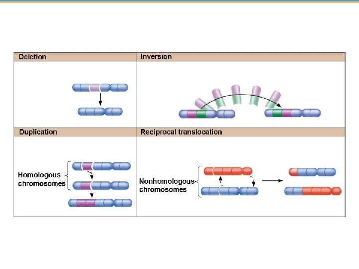
8. 23 CONNECTION: Alterations of chromosome structure can cause birth defects and cancer • Chromosome breakage can lead to four types of changes in chromosome structure. 1. A deletion is the loss of a chromosome segment. 2. A duplication is the repeat of a chromosome segment. 3. An inversion is the reversal of a chromosome segment. 4. A translocation is the attachment of a segment to a nonhomologous chromosome. A translocation may be reciprocal. © 2015 Pearson Education, Inc.
8. 23 CONNECTION: Alterations of chromosome structure can cause birth defects and cancer • Inversions are less likely than deletions or duplications to produce harmful effects, because in inversions all genes are still present in their normal number. • Many deletions cause serious physical or mental problems. • Translocations may or may not be harmful. © 2015 Pearson Education, Inc.
You should now be able to 1. Compare the parent-offspring relationship in asexual and sexual reproduction. 2. Explain why cell division is essential for prokaryotic and eukaryotic life. 3. Explain how daughter prokaryotic chromosomes are separated from each other during binary fission. 4. Compare the structure of prokaryotic and eukaryotic chromosomes. 5. Describe the stages of the cell cycle. © 2015 Pearson Education, Inc.
You should now be able to 6. List the phases of mitosis and describe the events characteristic of each phase. 7. Compare cytokinesis in animal and plant cells. 8. Explain how anchorage, cell density, and chemical growth factors control cell division. 9. Explain how cancerous cells are different from healthy cells. 10. Describe the functions of mitosis. 11. Explain how chromosomes are paired. 12. Distinguish between somatic cells and gametes and between diploid cells and haploid cells. © 2015 Pearson Education, Inc.
You should now be able to 13. Explain why sexual reproduction requires meiosis. 14. List the phases of meiosis I and meiosis II and describe the events characteristic of each phase. 15. Compare mitosis and meiosis, noting similarities and differences. 16. Explain how genetic variation is produced in sexually reproducing organisms. 17. Explain how and why karyotyping is performed. 18. Describe the causes and symptoms of Down syndrome. © 2015 Pearson Education, Inc.
You should now be able to 19. Describe the consequences of abnormal numbers of sex chromosomes. 20. Define nondisjunction, explain how it can occur, and describe what can result. 21. Explain how new species form from errors in cell division. 22. Describe the main types of chromosomal changes. Explain why cancer is not usually inherited. © 2015 Pearson Education, Inc.
Figure 8. 0 -0 © 2015 Pearson Education, Inc.
Figure 8. UN 01 G 1 Genetically identical daughter cells es n i k o Cytokinesis (division of the cytoplasm) Mitosis (division of the nucleus) © 2015 Pearson Education, Inc. S (DNA synthesis) M is s M it i s o G 2
Figure 8. UN 02 Haploid gametes (n = 23) n Egg cell n Sperm cell Fertilization Meiosis Human life cycle 2 n 2 n Multicellular diploid adults (2 n = 46) Mitosis © 2015 Pearson Education, Inc. Diploid zygote (2 n = 46)
Figure 8. UN 03 © 2015 Pearson Education, Inc.
Figure 8. UN 04 © 2015 Pearson Education, Inc.