CELL CYCLE Cell Cycle 3 main parts Interphase


")


- Slides: 31
CELL CYCLE
Cell Cycle • 3 main parts – Interphase – Mitosis – Cytokinesis
Cell Cycle: Interphase n 3 parts of interphase: Pre-DNA synthesis 2. DNA synthesis > Each chromosome is copied (Each strand called a chromatid) 3. Post DNA synthesis 1.
Interphase n Sometimes called the “resting stage” between divisions n BUT cells are metabolically very active n The amount of DNA in the nucleus doubles n New organelles such as mitochondria are made.
Interphase n No chromosomes are visible n Chromosomes appear as threadlike coils (Chromatin)
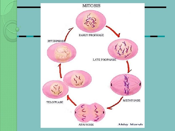
MITOSIS P rophase M etaphase A naphase T elophase
MITOSIS Primary purpose is to increase the number of cells n Daughter cells are genetically identical to the parents n Occurs during growth and asexual reproduction. n Mitosis is a continuous process which is divided for convenience into a number of stages. n
MITOSIS Prophase The longest stage n Chromosomes become visible n In animal cells, the centrioles divide and move to opposite ends of the nucleus
MITOSIS Prophase n Protein microtubules develop from each centriole forming spindle fibres. n (In plant cells there are no centrioles and the spindle forms independently)
MITOSIS Prophase n Towards the end of prophase each chromosome can be seen to consist of two chromatids held together by a centromere.
MITOSIS Prophase n At the end of prophase the nucleolus disappears and the nuclear envelope breaks down.
MITOSIS Metaphase n Chromosomes line up on the equator of the spindle. n They attach themselves to the spindle by their centromere
MITOSIS Anaphase n The centromeres divide n The free chromatids move to the poles
MITOSIS Anaphase n This movement results from the contraction of the spindle fibres. n As they shorten they pull the chromatids apart.
MITOSIS Telophase n The chromatids have reached the poles and are now regarded as distinct chromosomes again. n A nuclear envelope forms around each group of chromosomes
MITOSIS Telophase n The chromosomes uncoil returning to chromatin n The cytoplasm divides by cytokinesis.
MITOSIS n P - production of spindle fibres n M - middle alignment of chromosomes n A - "alone" chromosomes (chromosomes have moved to opposite poles) n T - two separate cells form (leading to cytokinesis)
CYTOKINESIS Animal cells n The centre of the cell ‘pinches in’ to form a division furrow. n As the division deepens, the cell surface membrane on each side joins up. n Two separate cells result.
CYTOKINESIS Plant cells n Vesicles produced by the Golgi body collect on equator of cell n These vesicles fuse to form a cell plate. n The cell plate eventually stretches right across the cell forming the middle lamella. n Cellulose builds up on lamella to form cell walls.
CYTOKINESIS
1. What phase is this?
2. How about this one?
3. This one?
4. This one too?
5. Last one!
TASKS: • Complete Mitosis Worksheet • Complete Cell Cycle Glossary
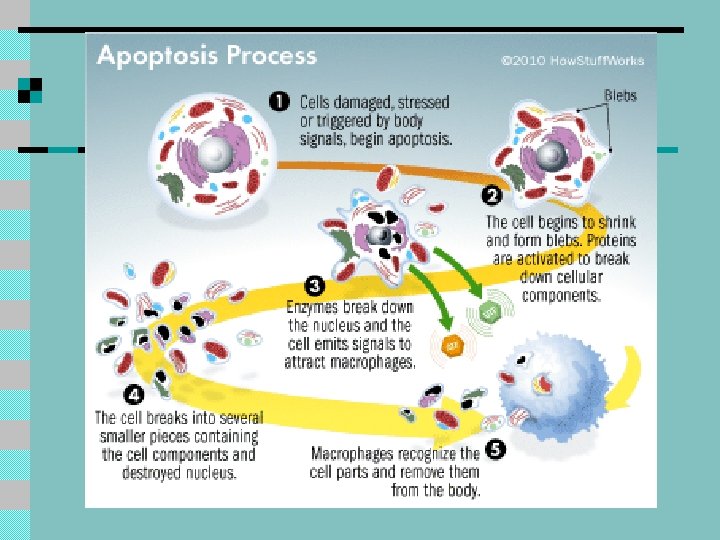
APOPTOSIS – ‘Programed Cell Death’ 2 important types – Good and Bad Good – Scheduled or programmed cell death – Apoptosis - internal enzymes are released to break down cell. – Dying cell is eaten or phagocytosed by special cells – This system removes excess tissue – Helps to shape organisms Bad – Explodes cellular material all over environment – triggering inflammation, infection and damaging surrounding cells – Hard for body to control
Apoptosis & Disease Many diseases and disorders are linked with the life and death of cells -- increased apoptosis is a characteristic of AIDS, Alzheimer's and Parkinson's disease, while decreased apoptosis can signal lupus or cancer. n Anti-cancer drugs and radiation, for example, work by triggering apoptosis in diseased cells. n