Cell Biology Lecture 16 Cytoskeleton Microtubules I Introduction

Cell Biology: Lecture 16 Cytoskeleton
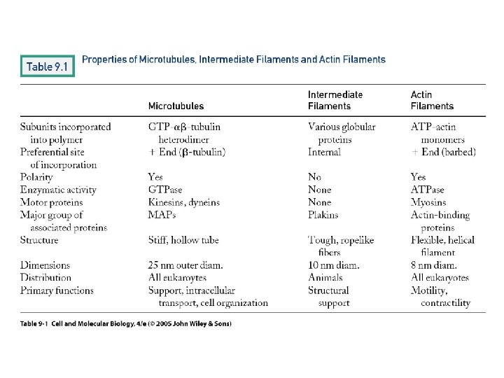

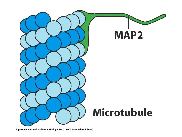
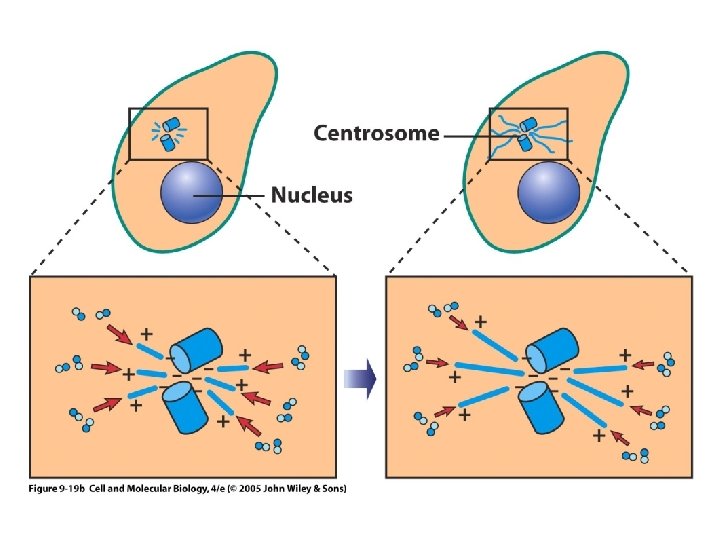
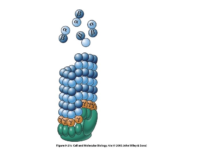
Microtubules • I. Introduction: Long, hollow cylinders, 25 nm in diameter, made of tubulin. The basic subunit is a heterodimer of α and β tubulin (9. 8); 13 protofilaments in a typical cylinder. See below about GTP binding, treadmilling, growth and dynamic instability (9. 26). There is a + end, fast growing, w/β tubulin at its end, and a – end, slow growing, w/α tubulin at its end. The GTP’s are important in assembly • A. They have MAPs, that influence their use- linking them together, stabilizing them, or destabilizing them. • B. They form a network, coming from the microtubule organizing center, which is usually the centrosome or centriole, w/ the – end anchored there. • C. Also form cilia and flagella, and spindle fibers in mitosis.


13 protofilaments
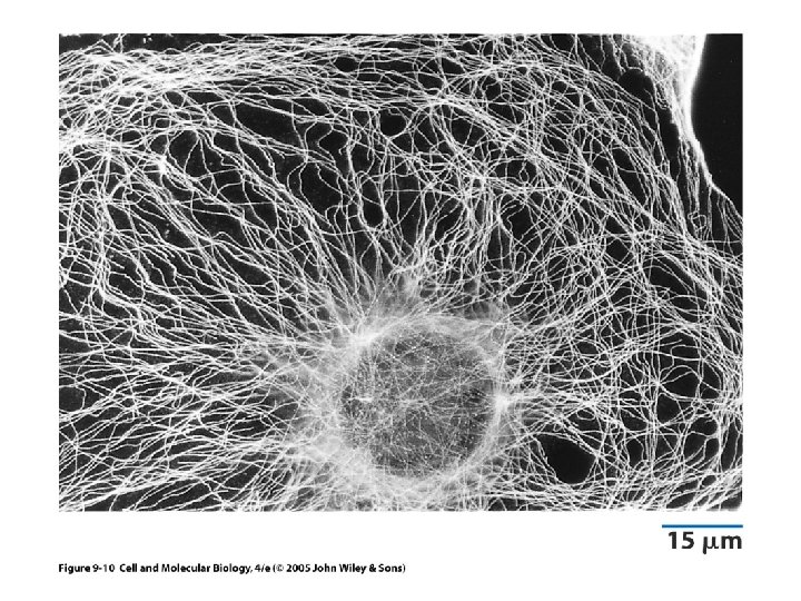

Nucleation • Gamma tubulin in MTOC/centriole- MT’s grow from there

• F. MICROTUBULE DYNAMICS: Key points- the cap means that subunits are added easily- loss of GTP = harder to add subunits, need higher subunit conc. to add. • Produces microtubule catastrophes!

W/ cap- slowly growing; w/o- rapidly shrinking- MT catastrophe!

MT drugs • Colchicine- prevents MT formation- arrests cells at metaphase • Taxol-Stabilizes MT’s – cancer drug • Useful in determining role of MT’s in a process

Intermediate filaments • • 10 nm in diameter Only in animals! (? ? plant/fungal nucleus? ? ) Variety of types- 60 genes! Seem to be involved in providing strength to cells. • Able to interact with both MT's and microfilaments (actin filaments).

Some types to remember • Keratin- epithelial cells, hair, nails • Neurofilaments- in, well, nerves • Lamins- lines the nucleus
 • • • Where we’re going: Basic structure, polarity, treadmilling It’s good")
Microfilaments (Actin) • • • Where we’re going: Basic structure, polarity, treadmilling It’s good buddy, myosin, w/ all its types Muscle contraction Amoeboid movement

Minus end Domains 1 -4 Subunits= G actin-bound w/ATP; F-actin= microfilaments ATP binding cleft Looks like a double helix!

S 1 is a myosin fragment that binds to actin- the points point to the minus end

Poisons! • Cytochalasin. B-depolymerizes • Phalloidin- stabilizes. The Amanita mushroom has TWO nasty toxins that cell biologists like. We’ll meet alpha amanitin later.

Muscle Contraction • Three types of muscle fibers: • Skeletal, striated, voluntary • Heart- more like skeletal, but not multinucleated. Its structure allows the propagation of an action potential (the heart beats by itself, w/o outside signals) • Involuntary, smooth muscle- gut, uterus, etc.

Things to know • Three major cytoskeletal components • Major stories- cilia movement, muscle contraction, amoeboid movement • Major motor proteins for both MT and actin, major poisons • Structure of the ends of MT’s and actin • Accessory proteins that we’ve mentioned.
- Slides: 21