Cell Biology and Physiology Signal Transduction and G

Cell Biology and Physiology Signal Transduction and G Protein. Coupled Receptors Chapter 15 Dr. Capers Molecular and Cell Biology, Lodish, 8 th edition

• No cell lives in isolation • Many types of cell signaling molecules are released by one cell and induce a response in a different cell • Even single celled organisms communicate through extracellular signals

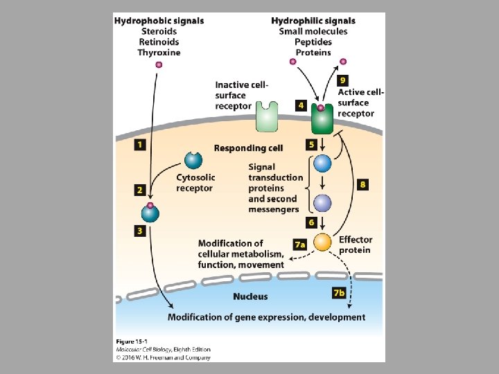
• In cells, a signal produces a specific response only in target cells with receptor proteins that bind that signal • In this chapter and the next, we will focus on extracellular signaling molecules that are too large and hydrophilic to diffuse through the plasma membrane • Need cell surface receptors


• Cell-surface receptors • Integral membrane proteins • 3 discrete domains: » Extracellular domain » Plasma-membrane spanning domain » Intracellular (cystolic) domain • Signaling molecule • Acts as the ligand for the cell-surface receptor • Bind to the extracellular or plasma-membrane spanning domain • Binding of this ligand to the receptor results in conformational change that is transmitted to cystolic domain • Can result in: – Activation or inhibition of other proteins attached to membrane or in the cytosol – Change intracellular ion concentration (such as Ca 2+) – Catalyze synthesis of small molecules
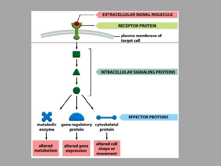

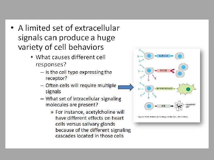
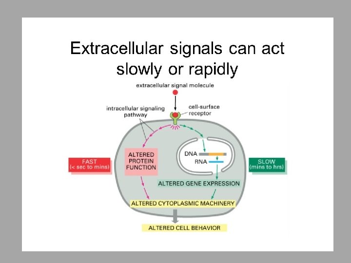
• Signal transduction • Process of converting extracellular signals into intracellular responses • Chain of intermediates is the signal transduction pathway – Results in a variety of outcomes depending on cell type and signal: survival, mitosis, differentiation, production of effector proteins, etc. • Highly conserved through evolution

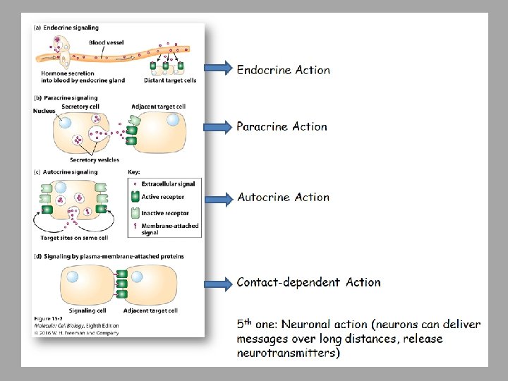
• Signaling molecules can act locally or at a distance
 binds the")
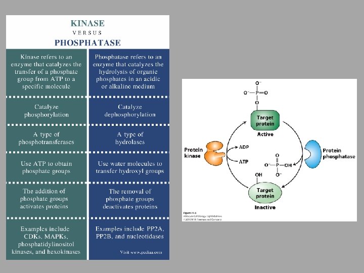
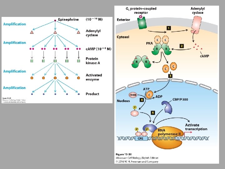
• Cell surface receptors bind specific signals • Signal molecule (ligand) binds the receptor • “First Messengers” • Conformational change of the receptor • Leads to activation of protein kinases – Enzymes that add phosphate groups to specific residues of target proteins • OR leads to activation of phosphatases – Enzymes that remove phosphate groups to specific residues of target proteins • These are “Second Messengers” • There can be an amplification process • Activation of effector protein (trigger movements, metabolism, gene expression)

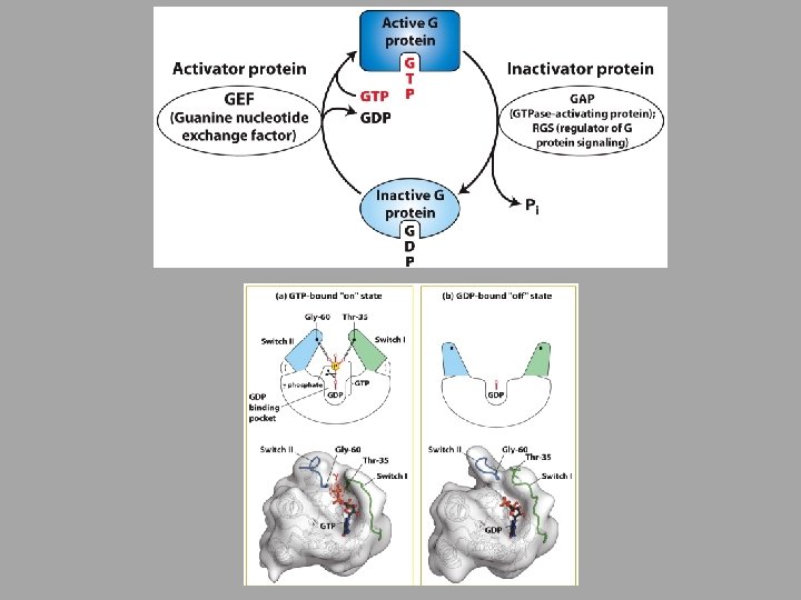
• Many intracellular signaling proteins behave as molecular switches and fall mainly into 2 categories: • Proteins activated or inactivated by phosphorylation – Protein kinase (serine/threonine kinases, tyrosine kinases) – Protein phosphatase • GTP-binding proteins – If GDP is bound, inactive – If GTP is bound, active

• GTP-binding proteins are frequently used in signal transduction pathways as on/off switches • Evolutionarily ancient • 2 large classes are used in signaling » Heterotrimeric G Proteins – directly bind to and are activated by cell-surface receptors » Monomeric G Proteins – play roles in cell division and motility (Ras, Ran and Sar are examples)

• Second messengers • One used in all metazoan cells is Ca 2+ ions » » Ca 2+ is usually low due to ATP-powered pumps Can be increased by release from ER In muscle cells this results in contraction In neurons, this results in excitation of neurotransmitter vesicles • c. AMP (cyclic adenosine monophosphate) » Can activate protein kinase A – cell metabolism » Can regulate ion channels

• Receptors are usually detected and quantified by their ability to bind radioactively or fluorescently labeled ligands

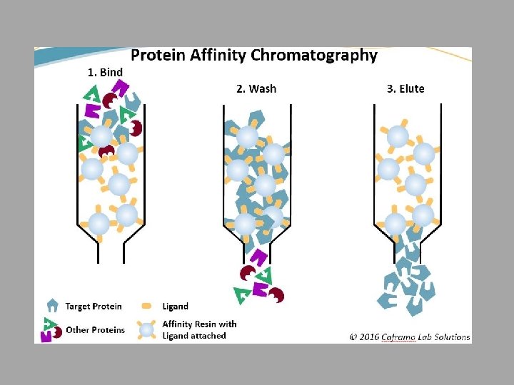
• Receptors can be purified by affinity chromatography techniques using antibodies specific for the receptor that is bound to the column • Need to purify in order to characterize their structures • Even though a mammalian cell can have 1, 000 – 50, 000 copies of a specific receptor, this only makes up 0. 1 -5% of plasma membranes

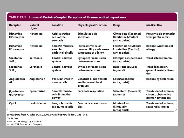
• G-Protein-Coupled Receptors • Largest family of cell-surface receptors – About one-third of all drugs today interact with GCPRs • Next 2 slides: – All GPCRs have similar structure – Stimulation of GPCRs activates G-Protein Subunits

• All G protein-coupled receptors have the same orientation in the membrane and contain 7 transmembrane α-helices, 4 extracellular segments, and 4 cytostolic fragments

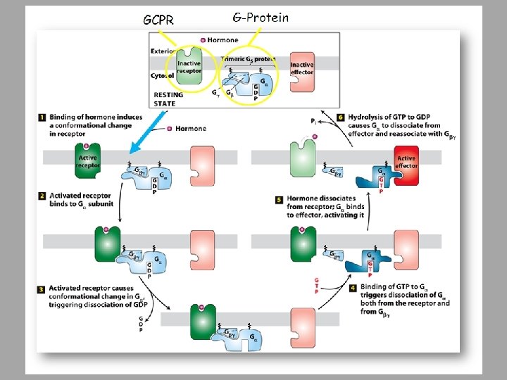
• Binding of ligand to a G-protein-coupled receptor results in • • Enabling the receptor to bind G subunit GDP is released GTP can bind Interacts and activates effector protein • See next figure

• Some bacterial toxins cause disease by altering the activity of G Proteins • Important to shut down a signal • Some toxins can keep the signal on


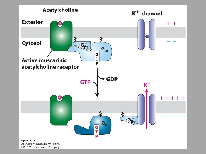
• Some G proteins directly regulate ion channels – Many neurotransmitter receptors are G proteincoupled receptors whose effector proteins are Na+ and K+ channels

• Many G proteins activate membrane-bound enzymes that produce small secondary messenger molecules

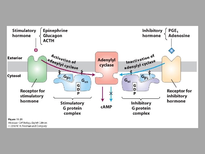
• Example of G-Protein activated membrane bound messenger: Adenylyl Cyclase – Activation of Adenylyl Cyclase activates the second messenger, cyclic AMP (c. AMP) – c. AMP then activates protein kinase (PKA)

• G protein-coupled receptors that use adenylyl cyclase as an effector protein and c. AMP as a second messenger are found in most mammalian cells – Example is the breakdown of glycogen • Release of epinephrine and glucagon during fight and flight response signals liver and muscle cells to depolymerize glycogen releasing glucose

• Some GPCRs activate G proteins that activate phospholipase C • Phospholipase C then cleaves membrane bound lipid molecule, inositol phospholipid • This generates inositol 1, 4, 5 -triphosphate (IP 3) and diacylglycerol (DAG) – IP 3 opens Ca 2+ channels – DAG stays in the membrane and activates PKC

• Step 1: GPCR activation of either the Gαo or Gαq subunit – activates phospholipase C (PLC). • Step 2: PLC cleaves PI(4, 5)P 2 – yields IP 3 and DAG • Step 3: IP 3 diffuses through the cytosol – IP 3 interacts with and opens IP 3 -gated Ca 2+ channels in the ER membrane • Step 4: Ca 2+ ions move down concentration gradient through the channel into the cytosol. • Step 5: Ca 2+ binding activates PKC and its recruitment to the plasma membrane. • Step 6: DAG activates membrane-associated PKC. • Step 7: Activated PKC-Ca 2+ leaves membrane to phosphorylate various cellular enzymes and transcription factors, activating proteins involved in cell growth and metabolism.

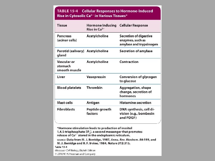
• Calcium ions play an essential role in regulating cellular responses to many signals • Small rise in cystolic calcium induces a variety of cellular responses • Hormone secretion by endocrine cells • Secretion of digestive enzymes by pancreatic cells • Muscle contraction • Triggers fusion of secretory vesicles with the plasma membrane and release of contents into extracellular space

• The Ca 2+ - Calmodulin complex mediates many cellular responses to external signals • Cystolic Ca 2+ binds to calmodulin and triggers a conformational change in the calmodulin • Allows calmodulin to bind and modulate the activity of many other enzymes and other proteins » Such as myosin and leads to contraction of smooth muscle

1. 2. 3. 4. 5. 6. 7. 8. Acetylcholine attaches to the GPCR receptor Activates and cleaves phospholipase C into DAG and IP 3 opens Ca 2+ channels Increase Ca 2+ activates nitric oxide (NO) synthase NO leaves cell and goes into muscle cell and attaches to GPCR receptor Activates G protein Activates PKG Muscle relaxation

• For cells to respond effectively in their environment, they must not only activate a signaling pathway, but they must be able to down-regulate or terminate the response • Feedback repression – end product blocks earlier step in the pathway • Enzymes also help in this process
- Slides: 42