Cell Biology and Physiology Culturing and Visualizing Cells

Cell Biology and Physiology Culturing and Visualizing Cells Chapter 4 Dr. Capers Molecular and Cell Biology, Lodish, 8 th edition

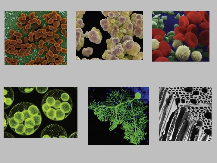
• First, let’s talk about some information from Chapter 1 • Biological organisms are very diverse – Cells come in astonishing variety of sizes and shapes – Some organisms are unicellular, some are multicellular – Oxygen kills some organisms • However, there is uniformity – all biological systems are composed of cells containing the same types of molecules

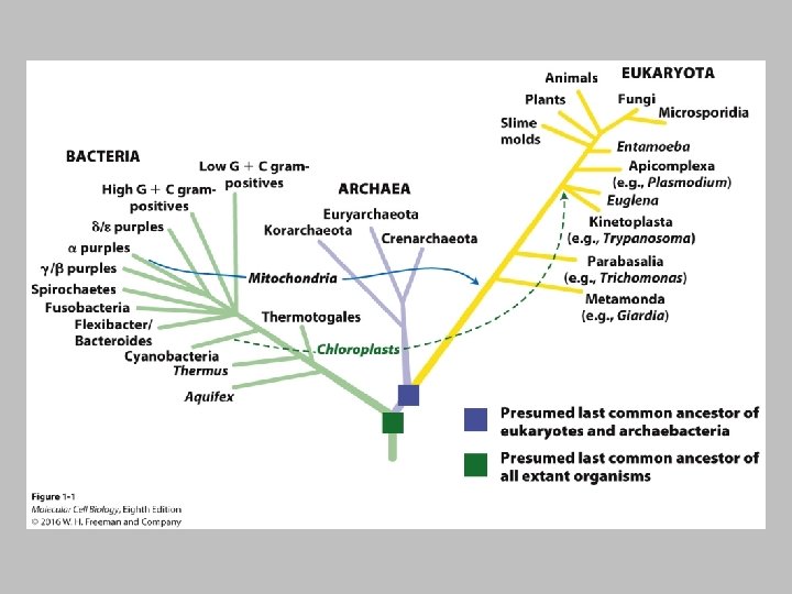
• Review: – 3 Domains of Life • Domain Archaea – prokaryotic • Domain Bacteria – prokaryotic • Domain Eukarya - eukaryotic

• Macromolecules • • Proteins Nucleic Acids Carbohydrates Lipids
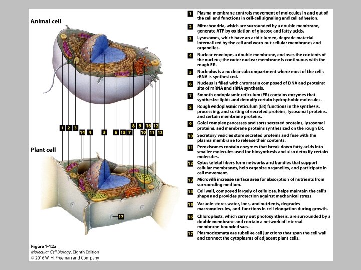

• The plasma membrane, mitochondria and chloroplasts we will talk about in more detail in other chapters

Lysosome • Acidic lumen • Degrade material phagocytized by the cell • Degrades old material found in the cell

Nucleus • Nucleus – has chromatin and proteins, double membrane, site of t. RNA and r. RNA synethesis • Nucleolus – most r. RNA is formed here

Rough Endoplasmic Reticulum • Synthesis and processing of proteins

Smooth Endoplasmic Reticulum • Lipid synthesis

Golgi Complex • Sorts secreted proteins, lysosomal proteins and membrane proteins

Peroxisomes • Peroxisomes hold enzymes that require oxygen • Breakdown fatty acids • Breakdown toxic chemicals

Vacuoles • Depending on cell, can be used to store water, food, minerals, ions, etc.

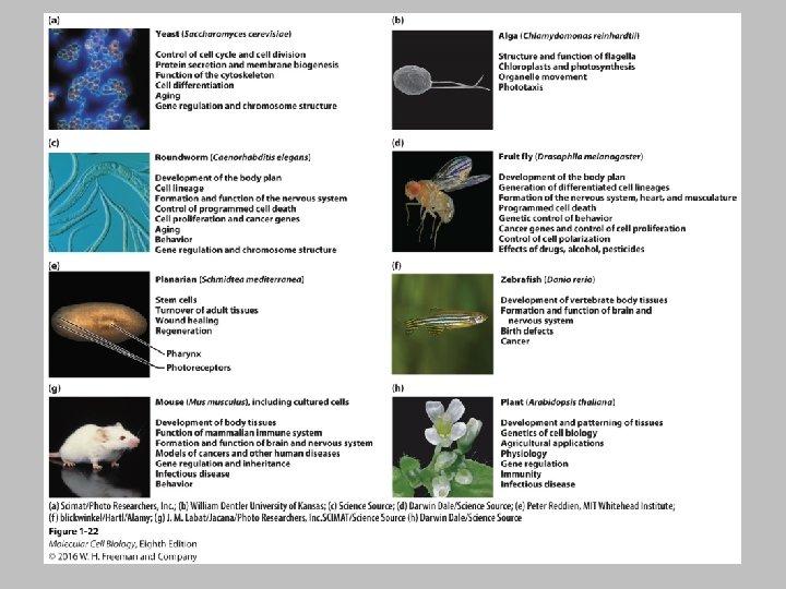
• Current understanding of the molecular functioning of eukaryotic cells largely rests on studies of a few types of model organisms • Because of the evolutionary conservation of genes, proteins, organelles, cell types, etc, discoveries about structures and functions with one organisms apply to another

Growing and Studying Cells in Culture • It is important to grow cells in culture • They can be examined microscopically • They can be subjected to treatments under normal conditions • It is generally quite easy to grow bacteria in culture • Animal and plant cells are a little more difficult • They are used to living in a multicellular environment, and are dependent on other cell types

• To permit survival and normal function of cultured tissues or cells several things have to be controlled: • • p. H Temperature Ionic strength Access to essential nutrients • Animal and plant cells are put into nutrientrich medium (culture medium) within specially coated plastic dishes or flasks

• Culture medium often contains antibiotics • Cultures are manipulated in sterile cabinets (hoods) to prevent contamination from airborne microorganisms

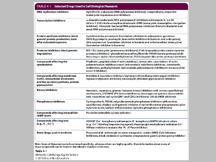
Growing and Studying Cells in Culture • Animal cells grow in culture when supplied with necessary nutrients and appropriate 2 D or 3 D substrates. • Primary cells have a finite life span; transformed tumor cells and cell line cells can grow indefinitely. • A fluorescence-activated cell sorter (FACS) sorts labeled cells. • Hybridoma cells produce monoclonal antibodies that bind one antigen epitope and are used for basic research and therapeutics. • Basic biological processes can be studied by using genetic manipulation or drugs to interfere with specific cell components. • Large chemical libraries can be screened for compounds that target specific processes.

• Media • Must supply 9 essential amino acids that cannot be synthesized by adult vertebrate cells • • • Phenylalanine Valine Threonine Tryptophan Isoleucine Methionine Leucine Lysine Histidine • Must also have 3 amino acids only produced by intact animal cells • Cysteine • Tyrosine • Arginine • • • Vitamins Salts Fatty acids Glucose Serum (proteins, insulin, growth factors, CAMs) Other components depending on cell type

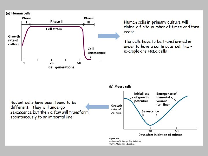
• Primary Cell Line • Isolated directly from tissues • Normal tissues (liver, kidney, skin) or whole embryos • These cells have a finite number of times they will divide » Cease to grow after a time, senescence » Exception to this is the embryonic stem cell (ES)
 • Pluripotent – differentiate into wide range of")
• Embryonic Stem Cells (ES) • Pluripotent – differentiate into wide range of cell types • Can be injected into the blastocoel of early mouse embryo and transplanted into uterus of pseudopregnant mouse • Induced Pluripotent Stem Cells (i. PS) • Adult somatic cells that can be induced into pluripotency by exposing to certain proteins • Stem cell niches in multicellular organisms • Multipotent – give rise to some but NOT all cell types • Germ-line stems cells produce sperm and oocytes • Intestinal stem cells • Hematopoietic stem cells • Meristems in plants

• Transformed Cell Line – Can grow indefinitely in culture – Often have abnormal DNA – Often have different chromosome #

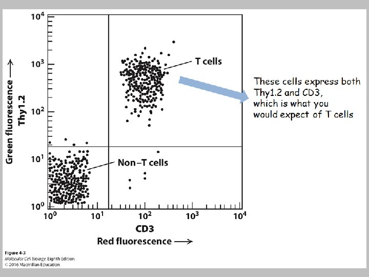
• Separating Cell Types • Some cells differ in density » Blood cells can be separated by centrifugation » Not all cells can be separated so easily so it must be done by other methods • Mark and sort » Flow cytometer • Fluorescence-activated cell sorter (FACS)

Differential labeling of cell types enables sorting from complex mixtures. Step 1: Labeled cells mixed with sheath fluid buffer solution pass single file through a laser light beam. Step 2: Both fluorescent light emitted and light scattered from each cell are measured and used to determine cell shape and size. Step 3: Forcing the cell suspension through a nozzle forms tiny droplets containing at most a single cell; each droplet obtains a negative electric charge proportional to fluorescence of that cell. Step 4: Passage through an electric field differentially deflects the droplets into different bins; as many as 10 million cells per hour can pass through the machine.

Growth of cells in Two-dimensional and threedimensional culture mimics the in vivo environment

• A wide variety of cell biological processes can be studied with cultured cells • • Easily subjected to a variety of manipulations Human diseases can be studied New drugs can be tested Old drugs – drugs that have been around for a long time but cell mechanism is not known; studying them can allow mapping out the exact effects on a particular cell type

• The invention of microscopes revolutionized the areas of microbiology and cell biology • Antony van Leeuwenhoek – 1600 s – Simple microscopes – single magnifying lens • Compound microscopes (2 -lens system) allow greater magnification – Technology has advanced to microscopes such as the electron microscope that allows even greater magnification

• Specimens can be visualized alive using microscopy. • However, often the specimens have to be fixed to a slide for microscopy.

• Often, specimens for light microscopy are fixed • Solution often contains chemicals that cross-link most proteins and nucleic acids – Example is formaldehyde • After fixation, one of 2 things happens: • the specimen is embedded in paraffin and cut into sections • 50µm thick • Has to be done with a microtome • Cultured cells can be grown on coverslip and fixed and are thin enough that they don’t have to be sliced Stain – Many chemical stains bind to molecules that have specific features – Examples: – Hematoxylin – binds to basic amino acids of certain proteins – Eosin – binds to acidic molecules (DNA, some amino acids) •
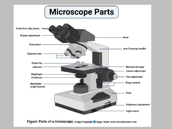

In bright-field light microscopy, light from a lamp is focused on the specimen (usually stained with dyes to enhance contrast) by a condenser lens below the stage. Condenser is used to collect and focus the light on a specimen. The diaphragm controls the amount of light.

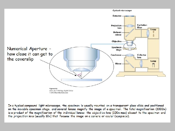
• Resolution – smallest distance that 2 neighboring points can be separated and still be discerned as 2 separate entities » Naked eye – 0. 1 mm » High-quality compound light microscope – 0. 2µm • Numerical aperture of the objective lenses – has to do with the ability for that objective to gather light and resolve fine specimen detail

Phase-contrast light microscopy - optical microscopy technique that converts phase shifts in light passing through a transparent specimen to brightness changes in the image. Helps to increase CONTRAST – difference between specimen and background.

• Inverted Microscopes • Light source and condenser on top of specimen • Objectives below specimen • Needed to visualize live cells that are in a dish of some sort as opposed to being on a thin slide

Fluorescence Microscopy

Fluorescence Microscopy – uses laser as light source 1. Specimen is treated with a fluorescent dye(s). The dye contains fluorophores (chemical that can re-emit light during excitation) that will attach to certain things in the specimen. 2. Laser travels through the excitation filter cube, only allowing certain wavelengths through (which cube is used depends on the dye used). 3. The light hits the specimen, if certain fluorophores are present, they will excite and fluoresce. 4. Excited light from specimen will travel through the emission filter cube, focusing the light to the detector. Click HERE to watch video

What can fluorescence microscopy do? • Localize and quantify specific molecules in live cells • Quantify ion concentrations with ion-sensitive fluorescent dyes • Detect specific proteins in fixed cells • Some fluorescent dyes attach directly to certain molecules, other fluorescent dyes are attached to monoclonal antibodies which will then attach to something present in the specimen

Immunofluorescence
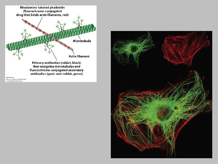

• Green Fluorescent Protein – GFP – Jellyfish Aequorea Victoria • Other fluorescent proteins expressed by bacteria can be isolated for use

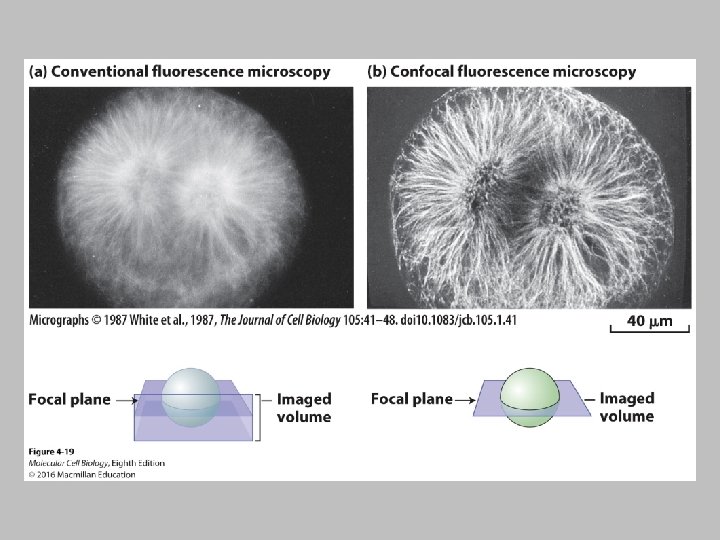
• Conventional fluorescent microscopy has limitations • Out of focus fluorescent light • Deconvolution microscopy and Confocal Microscopy help to resolve this

Electron Microscopy

• Electromagnetic lenses focus a high-velocity beam instead of visible light used by optical lenses • High resolution images • Single proteins, organelles, viruses can be imaged
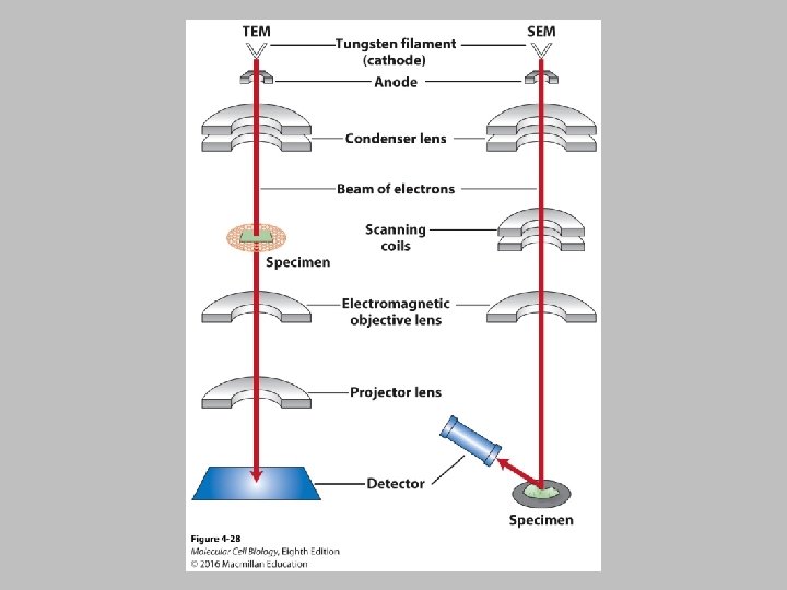

Transmission Electron Microscopy TEM samples are mounted on a small copper or gold grid that is covered with a very thin film of plastic and carbon to which a sample can adhere. Specimens are stained with heavy metal, such as uranyl acetate, and excess stain is removed. Negative stain of rotaviruses: The viruses exclude the stain and are seen in negative outline.

Transmission Electron Microscopy • Specimens such as cells and tissues that are too thick have to fixed and sliced

• Immunoelectron Microscopy

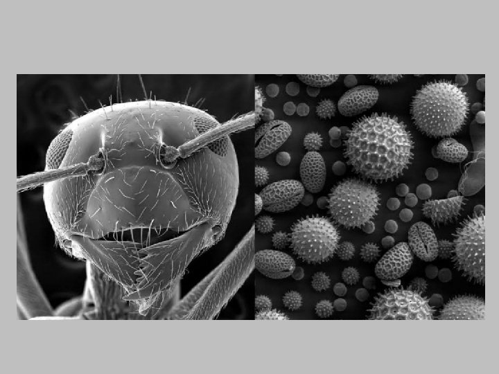
Scanning Electron Microscopy • SEM allows visualization of the surfaces of metal coated items

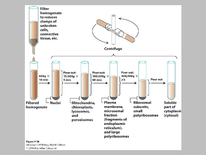
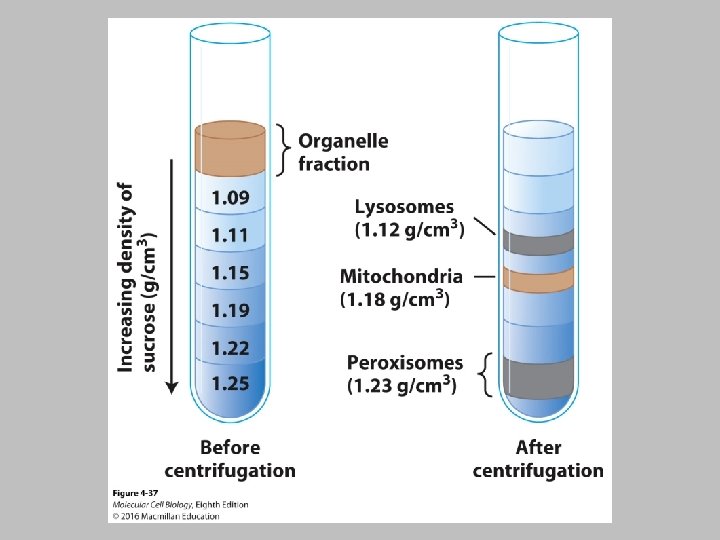
Isolation of Cell Organelles • It is necessary to isolate organelles in their native state and identify and dissect the function of each component • Lysosomes, for example, had been identified by light microscopy – But their function was only known after a method was developed to isolate them • Isolation techniques: – Disruption of cells by vigorous homogenization, sonication, or other techniques releases their organelles. Swelling cells in a hypotonic solution weakens the plasma membrane, making it easier to rupture. – Sequential differential centrifugation of a cell homogenate yields fractions of organelles that differ in mass and density. – Equilibrium density-gradient centrifugation separates cellular components by density for purification. – Immunological techniques using antibodies against organelle- specific membrane proteins purify organelles and vesicles of similar sizes and densities.

• What is the difference between a cell strain, a cell line and a clone? • A cell strain is a lineage of cells originating from a primary culture taken from an organism. Since these cells are not transformed, they have a limited lifespan in culture. • In contrast, a cell line is made of transformed cells and therefore these cells can divide indefinitely in culture. Such cells are said to be immortal. • A clone results when a single cell is cultured and gives rise to genetically identical progeny cells.

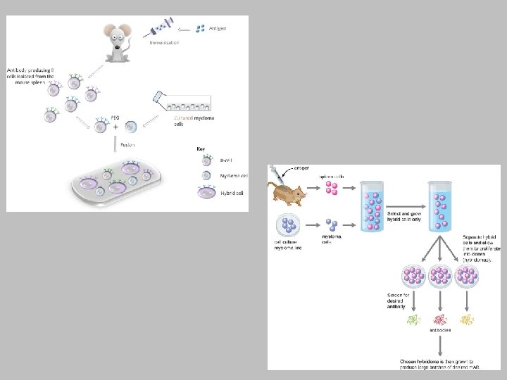
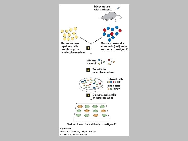
• Explain why the process of cell fusion is necessary to produce monoclonal antibodies used for research. – Normal B lymphocyte cells can produce a single type of antibody molecule. However, such cells have a finite lifespan in culture. Researchers use cell fusion of B lymphocytes and immortalized myeloma cells to create immortalized, antibody-secreting cells. Such cells, called hybridoma cells, retain characteristics of both parent cells, allowing for production of a singletype, or monoclonal, antibody.
- Slides: 65