Cell Biology 9 transport 4 th Sung Youn

Cell Biology 9 - transport 4 th Sung Youn Lee, Ph. D. Student Veterinary collage, Room 320 02 450 3719, 016 293 6059 leevet@paran. com

Cell membrane

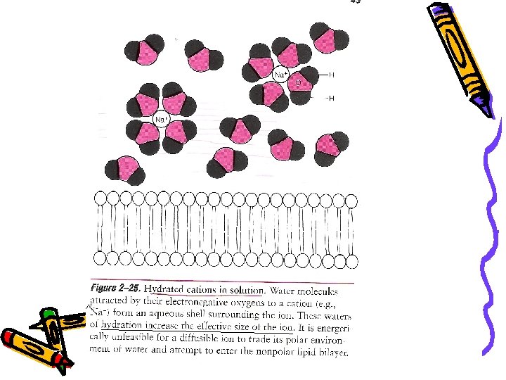
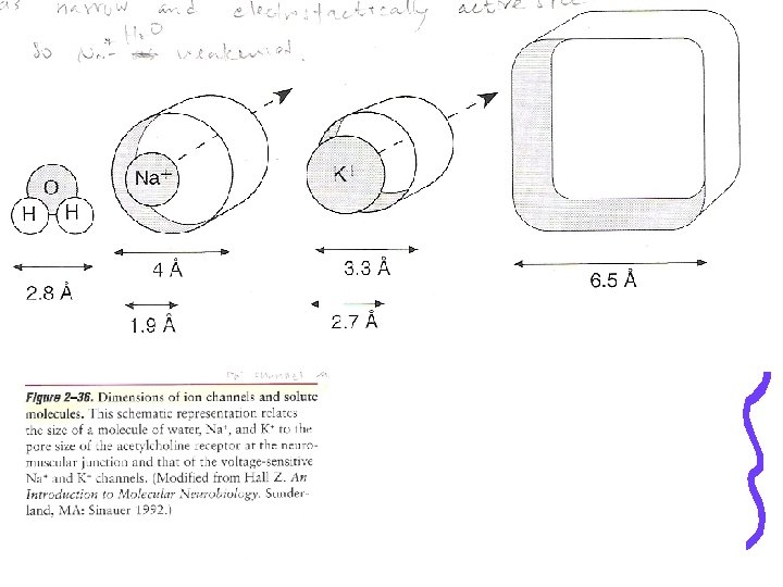
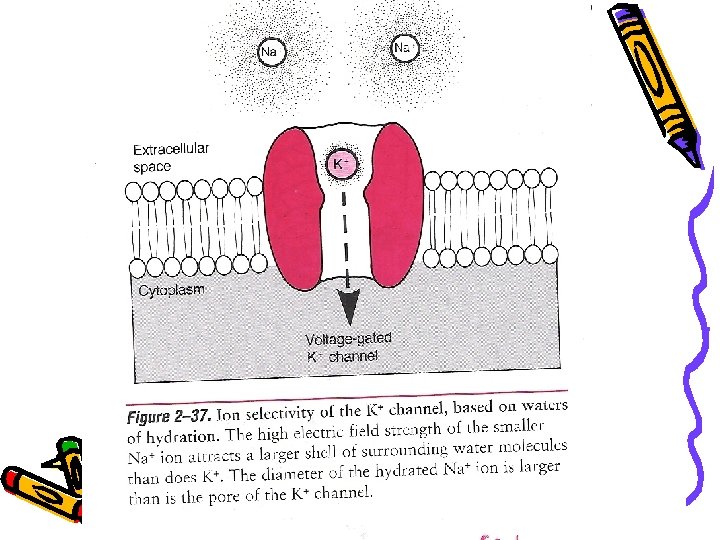
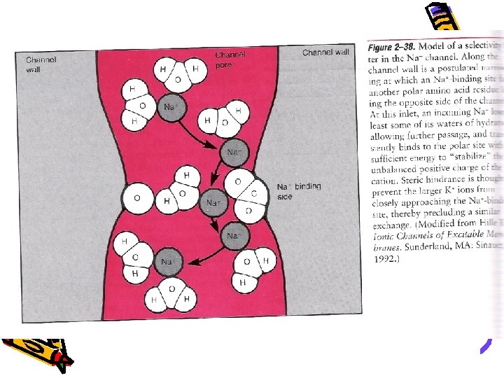
Ion channel and membrane potential • The hydrophobic environment of the phospholipid membrane bilayer is virtually impermeable to ions in aqueous solution. • Why? • Ion interact with water molecules, whether hydrated or dehydrated, ions can not cross the lipid bilayer by simple diffusion.

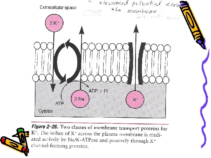
Protein channel permit the rapid flux of ions across membranes • Ion channels are distinguished physiologically from carrier proteins by the high velocity of ion flux they allow across the membrane, without expending energy. • Whereas permease-mediated transport operates at maximum of 105 ions/sec, ions flow at a rate 100~1, 000 times faster through channel. (nerve impulse and muscle contraction)

Ion channels have a common structural motif: The transmembrane α-helix • A major the breakthrough came with the discovery that ion channels could be artificially introduced into cell membranes treated with small hydrophobic proteins, termed ionophores. • Made primarily by different fungi, some ionophores have been used as antibiotics.


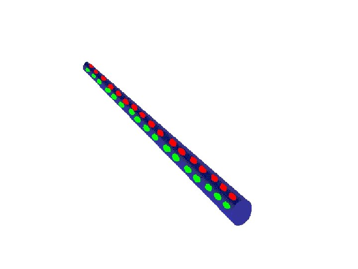
Gramicidin A • A linear polypeptide with 15 amino acids that, like all ionophores, is readily miscible with the phospholipid bilayer. • This ionophore is distinct because, then inserted into the membrane, it forms an unusual β-helix. (Fig 2 -27) • Alternating d- and l-amino acid residues in the molecule orient the polar (hydrophilic) carbonyl oxygen atoms and amide nitrogens of the peptide bonds toward the hollow center of the helix.

Two gamicidin. A peptides dimerize head to span the lipid bilayer Hydrophilic hollow center of the helix

Channel-forming protein in mammalian cells • Consist solely – L-amino acids • Carbonyl oxygens and nitrogens of polypeptide backbone do not behave as hydrophilic : α-helices are naturally hydrophobic

Hydrophilic residues point toward the aqueous channel and its hydrophobic residues face the lipid bilayer
 Resemble to neuromuscular junction by the acetylcholine receptor-5 subunit")
Synthetic channel (Leu & Ser) Resemble to neuromuscular junction by the acetylcholine receptor-5 subunit
")
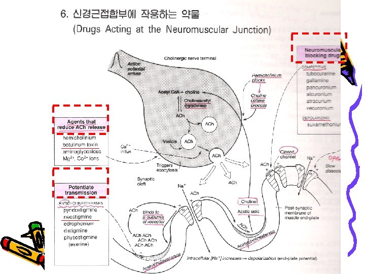
Ach receptor ion channel (ligand-gated ion channel)

Ion channels are allosteric proteins, the conformation of which is regulated by different stimuli • More than 75 different ion channels • All are glycoprotein • Two< conformation including stable open state and a stable closed state. • 4 types of ion channel : 3 -rapid, 1 slow
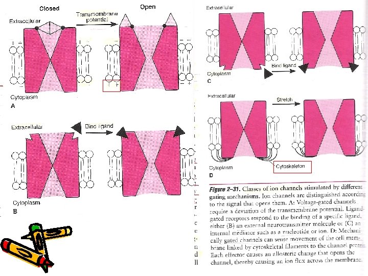

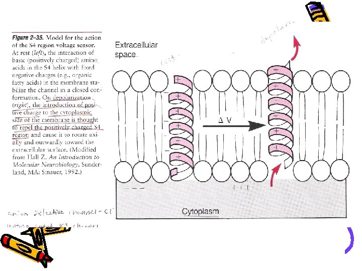
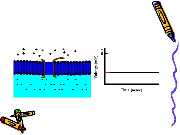
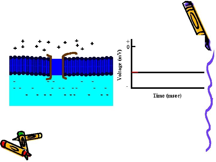
Voltage-gated channel • Propagation of electrical impulses over long distances in nerve and muscle. • They open specifically in response to a change in the electric field.

S 4 -voltage sensor P-ion selectivity
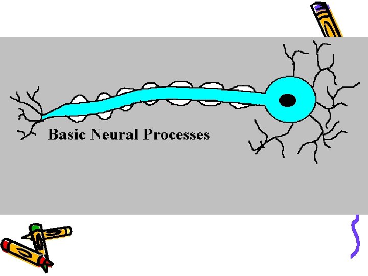

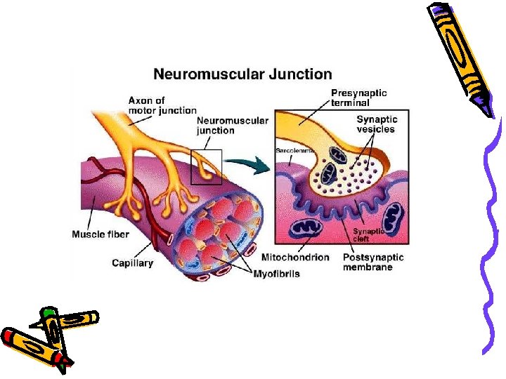
Ligand-gated ion channel • Insensitive to voltage change but opened by noncovalent, reversible binding of a chemical ligand. (including neurotransmitter) • Neuron-neuron, neuron-muscle, neuron-glandular

Nerve-Muscle Junction, Human Histology Photo. CD. Taken from the highest resolution PCD file that was ~18. 9 M, it was cropped & saved as a low compression JPG file, ~25 K.

Mechanically gated channel • The opening is controlled by cellular deformation.

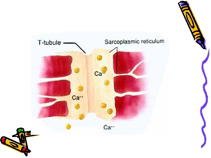
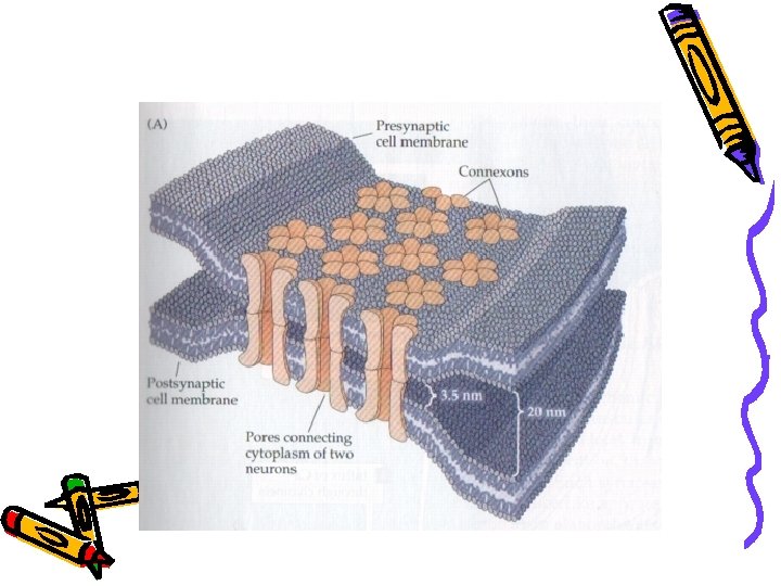
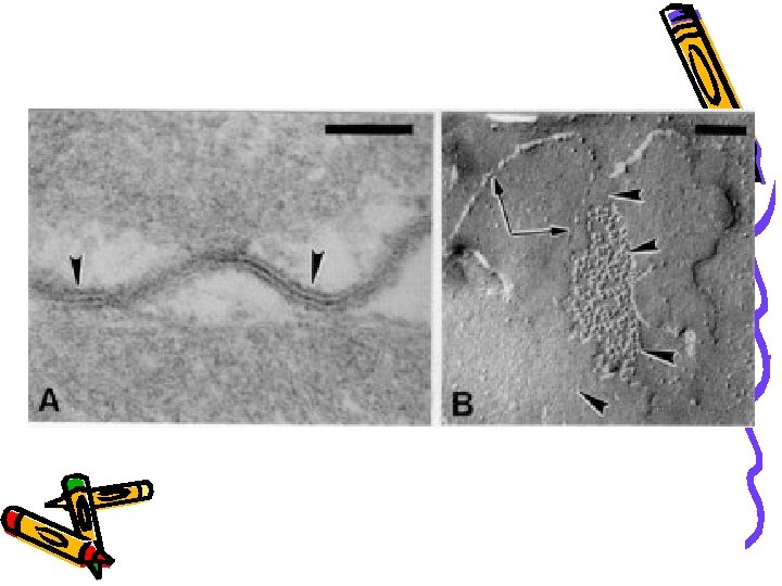
Gap junction • Enable ions to flow between adjacent cells without traversing the extracellular space. • Open/close in response to changes in the intracellular concentration of Ca 2+ and protons.

Thank you for your attention ~
- Slides: 38