Cell architecture structure and functions of cytoskeletal framework


- Slides: 15
Cell architecture; structure and functions of cytoskeletal framework Dr Mah Jabeen Muneera Assistant Professor Department of Anatomy KEMU
Microfilaments • Shape: double sranded helix • Diameter: 7 nm • Basic protein subunit: monomer of G-actin (globular or free actin) polymerizes to F- actin (filamentous), in presence of K & Mg. • Formin • Polarity: _ pointed end and + barbed end • Treadmilling • Stable- muscle cells and microvilli • Dissociate and reassemble • Profilin & Cofilin
Toxins that bind to actin • Prevent polymerization • Prevent depolymerization (phalloidin found in poisosnous mushrooms) • Disrupt the dynamic equilibrium between F-actin and G-actin causing cell death
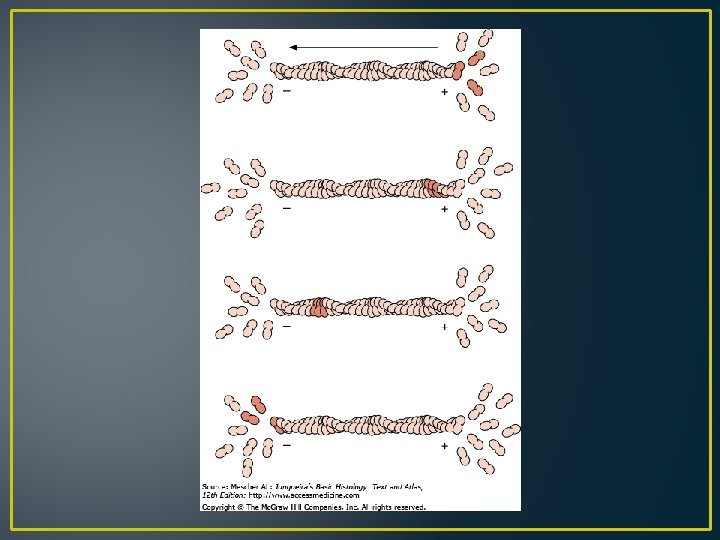
Cytokinesis
Microvillus Core of actin filaments ABPs- Actin Binding Proteins • Villin and fimbrin • Tropomyosin • Spectrin • Myosin 1 and 2
Cell cortex
Stress fibers
Intermediate filaments • • • 10 -12 nm Structural function Intercellular junctions Nuclear lamina Identification by means of immunocytochemical methods • Tissue specificity
Intercellular adhesions Desmosomes Zonula adherens
Tissue specificity Fibroblasts: Cells of mesenchymal origin- ? IF › 50 different types divided into 05 classes • 1. Keratins- epithelium • 2. Vimentin- mesenchymal cells and vascular smooth muscle • 3. Desmin- muscle except vascular smooth muscle • 4. Glial fibrillary acidic proteins- glial cells • 5. Neurofilamentsneurons
Information for diagnosis and treatment of cancer • Presence of a specific type of intermediate filament in tumors can reveal which cell originated the tumor • Identification of intermediate filament proteins by means of immunocytochemical methods is a routine procedure
Terminal Web • Microvilli bearing cells • Microfilaments & intermediate filaments • Desmosomes anchorage • ABP: spectrin & Myosin II
Alzheimers disease • Neurofibrillary tangles: presence of structurally abnormal but phosphorylated neurofilaments and microtubule associated proteins ? ?
Alexander disease • Disorder of CNS • Mutations in the coding region of GFAP gene • Altered GFAP prevents the assembly of intermediate filaments • Accumulation of intermediate filament protein GFAP leads to cytoplasmic inclusions in astrocytes (Rosenthal fibres)