CBD Stones Stricture Carcinoma Gall Bladder Cholangiocarcinoma Dr

CBD Stones, Stricture Carcinoma Gall Bladder Cholangiocarcinoma Dr Amit Gupta Associate Professor Dept Of Surgery
")
Common Bile Duct Stones (Choledocholithiasis)

• • May be small or large Single or multiple Found in 6 to 12% of patients with stones in the gallbladder Incidence increases with age • Secondary stones: – Formed within the gallbladder and migrate down the cystic duct to the common bile duct – Cholesterol stones • Primary stones : form in the bile ducts – Brown pigment type – Associated with biliary stasis and infection

Clinical Manifestations • Silent and often are discovered incidentally • May cause obstruction, complete or incomplete • Cholangitis or gallstone pancreatitis • Pain • Mild epigastric or right upper quadrant tenderness

Clinical Manifestations • Mild icterus • Symptoms may also be intermittent • Elevation of serum bilirubin, alkaline phosphatase, and transaminases are commonly seen in patients with bile duct stones • However, in about one third of patients with common bile duct stones, the liver chemistries are normal

Diagnosis Investigation Sensitivity Specificity US 25 -82% 56 -100% EUS 95 % 95 -98 % MRCP 95 % 97 % CT 87 % 97 %

Ultrasound shows a normal or mildly dilated common bile duct with a stone

ERCP shows multiple stones in the common bile duct
 on ultrasonography in a patient with")
• Dilated CBD (>8 mm in diameter) on ultrasonography in a patient with gallstones, jaundice, and biliary pain is highly suggestive • Magnetic resonance cholangiography (MRC) provides excellent anatomic detail • Endoscopic cholangiography is the gold standard for diagnosing common bile duct stones.

Management Options– CBD Stones • Open cholecystectomy + CBD exploration • ERCP + Endoscopic Sphincterotomy (followed by cholecystectomy – most frequently used). • Laparoscopic cholecystectomy + Laparoscopic CBD exploration – in specialized centers. • Choledochoscopy at laparoscopy or percutaneous choleydochoscopy or choleydochoscopy through T tube.

• ERCP has become a popular technique to clear CBD stones. • Currently in the laparoscopic era studies have shown that laparoscopic treatment of CBD stones is possible and is potentially as effective as ERCP. • This is most commonly done by a transcystic approach, though evidence of success in large volume cohorts with a more technically demanding laparoscopic Choledochotomy is emerging.

Common Bile Duct Stricture

Causes • operative injury MC by lap. cholecystectomy • fibrosis due to: Øchronic pancreatitis Øcommon bile duct stones Øacute cholangitis biliary obstruction: Ø cholecystolithiasis (Mirizzi's syndrome) Ø sclerosing cholangitis Ø Cholangiohepatitis Ø strictures of a biliary-enteric anastomosis

Clinical presentation • Episodes of cholangitis • Jaundice • Liver function tests usually show evidence of cholestasis

Diagnosis • Ultrasound / CT scan will show dilated bile ducts proximal to the stricture • MRC : anatomic information about the location and the degree of dilatation • Endoscopic cholangiogram will outline the distal bile duct

ERC showing stricture of the common hepatic duct

Management Depends on the location and the cause of the stricture • Percutaneous or endoscopic dilatation and/or stent placement give good results in more than one half of patients • Surgery with Roux-en-Y choledochojejunostomy or hepaticojejunostomy is the standard of care with good or excellent results in 80 to 90% of patients • Choledochoduodenostomy may be a choice for strictures in the distal-most part of the common bile duct

Carcinoma Gallbladder

Etiology • Accounts for 2 to 4% of malignant GI tumors • 2 -3 times more common in females than males • 90% of patients have gallstones • Larger stones (3 cm) are associated with tenfold increased risk of cancer • Polypoid lesions of the gallbladder (>10 mm) • Calcified "porcelain" gallbladder >20% incidence

• Choledochal cysts • Sclerosing cholangitis • Anomalous pancreaticobiliary duct junction • Exposure to carcinogens (azotoluene, nitrosamines)

Pathology • 80 and 90% of the tumors are adenocarcinomas – papillary, nodular, and tubular Squamous cell Adenosquamous Oat cell

Cancer of the gallbladder spreads through: • lymphatics • venous drainage • direct invasion into the liver parenchyma

Clinical Manifestation • • Abdominal discomfort Right upper quadrant pain Nausea & vomiting Jaundice Weight loss Anorexia Ascites Abdominal mass
 • Ultrasonography • CT scan • Percutaneous transhepatic or")
Diagnosis • FNAC /Biopsy (guided) • Ultrasonography • CT scan • Percutaneous transhepatic or endoscopic cholangiogram (in jaundiced pt) • MRCP
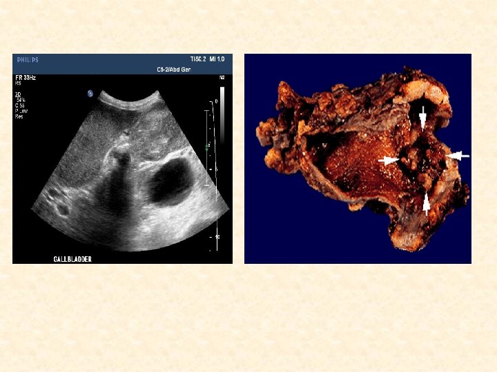

CT scan of a patient with gallbladder cancer
")
Staging: (AJCC 7 th Edition)

Treatment • Surgery : – Radical Cholecystectomy, Liver resection with regional lymphadenectomy • Radiotherapy – Adjuvant (p. T 1 b onwards) • Chemotherapy – Concurrent – Adjuvant – Palliative

Prognosis • 5 -year survival rate of all patients <less than 5% • Median survival: 6 months • T 1 disease treated with cholecystectomy have an excellent prognosis (85 - 100% 5 -year survival rate) • 5 -year survival rate for T 2 lesions treated with an extended cholecystectomy and lymphadenectomy compared with simple cholecystectomy is over 70% versus 25 to 40%, respectively • Patients with advanced but resectable gallbladder cancer are reported to have 5 -year survival rates of 20 to 50% • Median survival for patients with distant metastasis at the time of presentation is only 1 to 3 months

noma Cholangiocarci

• Rare tumor arising from the biliary epithelium • May occur anywhere along the biliary tree • About 2/3 rd are located at the hepatic duct bifurcation • Male to female ratio is 1. 3: 1 • Average of presentation is between 50 to 70 years

Etiology Ulcerative Colitis Thorotrast Exposure Sclerosing Cholangitis Typhoid Carrier Choledochal Cysts Adult Polycystic Kidney Disease Hepatolithiasis Liver Flukes Papillomatosis of Bile Ducts

Distribution • Right or left hepatic duct = 10% • Bifurcation = 20% • Proximal CBD = 30% • Distal CBD = 30%

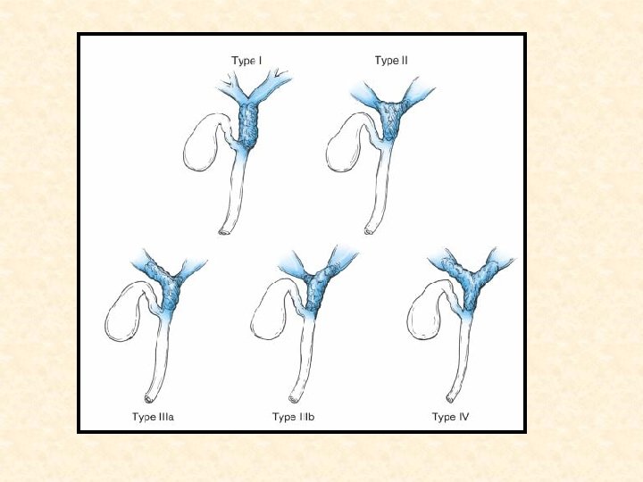
Pathology • Over 95% of bile duct cancers are adenocarcinomas. • Anatomically they are divided into distal, proximal, or perihilar tumors. • Intrahepatic cholangiocarcinomas are treated like hepatocellular carcinoma, with hepatectomy when possible. • About two-thirds of cholangiocarcinomas are located in the perihilar location • Perihilar cholangiocarcinomas, also referred to as Klatskin tumors, are further classified based on anatomic location by the Bismuth-Corlette classification

Bismuth-Corlette classification • Type I: confined to the common hepatic duct • Type II: involve the bifurcation without involvement of the secondary intrahepatic ducts • Type IIIA &IIIB: extend into the right and left secondary intrahepatic ducts, respectively • Type IV: involve both the right and left secondary intrahepatic ducts

Clinical Presentation • • Painless jaundice Pruritus Mild right upper quadrant pain Anorexia Fatigue Weight loss Cholangitis Elevated ALK PO 4 and GGT levels

Intra and Extra-hepatic Cholangiocarcinoma

Diagnosis • • Ultrasound abdomen CT scan Cholangiography : biliary anatomy is defined PTC Defines the proximal extent of the tumor, which is the most important factor in determining resectability. • ERC: evaluation of distal bile duct tumors • Celiac angiography: evaluation of vascular involvement • MRI: has the potential of evaluating the biliary anatomy, lymph nodes, vascular involvement, tumor growth

ERCP: Distal CBD Cancer

MRCP of Extra-hepatic Cholangiocarcinoma at the Bifurcation Klatskin tumor

Treatment • Surgical excision is the only potentially curative treatment • Location and local extension of the tumor dictates the extent of the resection

Bismuth-Corlette type I or II with no signs of vascular involvement: • local tumor excision with portal lymphadenectomy, cholecystectomy, common bile duct excision, and bilateral Roux-en-Y hepaticojejunostomies Bismuth-Corlette type IIIa or IIIb: • right or left hepatic lobectomy respectively should also be performed
 Unresectable distal bile duct cancer:")
Distal bile duct tumors: • pylorus-preserving pancreatoduodenectomy (Whipple procedure) Unresectable distal bile duct cancer: • Roux-en-Y hepaticojejunostomy, cholecystectomy and gastrojejunostomy

Roux-en-Y Hepaticojejunostomy

Cholangiocarcinoma Extra-hepatic Disease: Positive Margins or Unresectable • Stent and Chemo/Radiation Therapy • 5 -FU based or Gemcitabine or Clinical Trial • Survival with surgery and chemo/radiation is 24 to 36 m • With chemo/radiation alone survival is 12 to 18 m.

Cholangiocarcinoma Extra-hepatic Disease: Unstentable • Bypass if possible • If not use proximal decompression and feeding jejunostomy • Chemotherapy/Radiation Therapy/Brachy therapy as tolerated or clinical trial.

Prognosis • Best Result are with distal CBD tumors completely excised. Cure = 40% • Incomplete resection plus radiation gives a median survival of 30 m. • Stenting plus chemo/radiation gives a median survival of 17 to 27 m • Those stented alone live only a few months
- Slides: 48