
CASE REPORT GASTRIC NEUROENDOCRINE NEOPLASMA Dimitrios Thomas Endocrinologist




- Slides: 37
CASE REPORT: GASTRIC NEUROENDOCRINE NEOPLASMA Dimitrios Thomas, Endocrinologist Endocrine Department, Pathophysiology Clinic, “Laikon” Hospital, National and Kapodistrian University of Athena “Laikon” Hospital
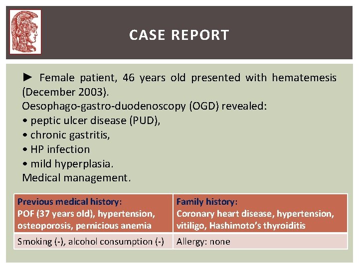
CASE REPORT ► Female patient, 46 years old presented with hematemesis (December 2003). Oesophago-gastro-duodenoscopy (OGD) revealed: • peptic ulcer disease (PUD), • chronic gastritis, • HP infection • mild hyperplasia. Medical management. Previous medical history: POF (37 years old), hypertension, osteoporosis, pernicious anemia Family history: Coronary heart disease, hypertension, vitiligo, Hashimoto’s thyroiditis Smoking (-), alcohol consumption (-) Allergy: none
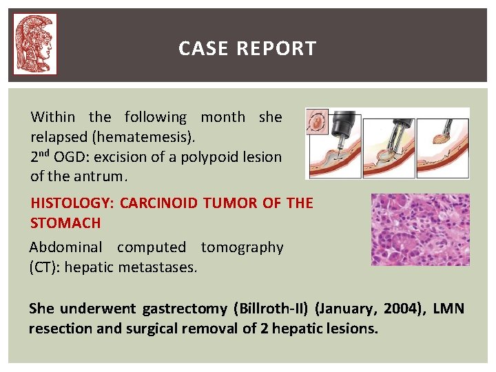
CASE REPORT Within the following month she relapsed (hematemesis). 2 nd OGD: excision of a polypoid lesion of the antrum. HISTOLOGY: CARCINOID TUMOR OF THE STOMACH Abdominal computed tomography (CT): hepatic metastases. She underwent gastrectomy (Billroth-II) (January, 2004), LMN resection and surgical removal of 2 hepatic lesions.
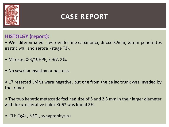
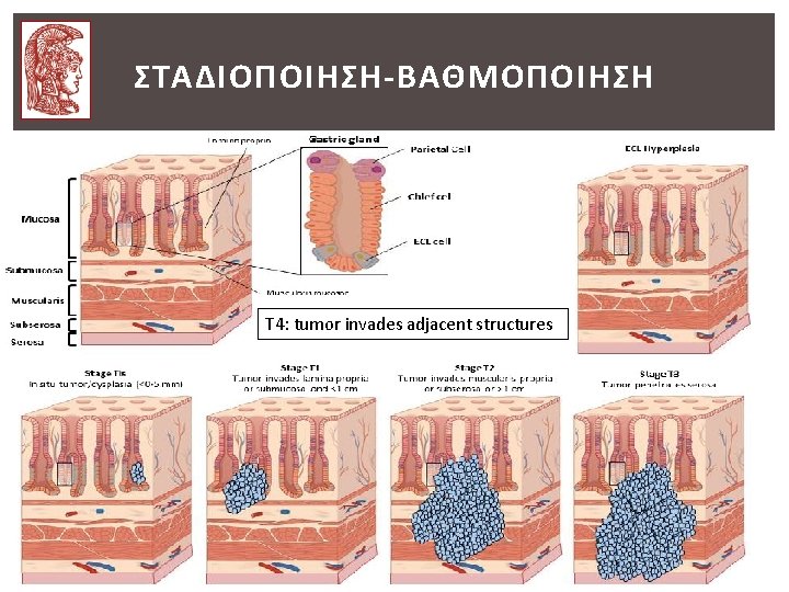
CASE REPORT HISTOLGY (report): • Well diferentiated neuroendocrine carcinoma, dmax=3, 5 cm, tumor penetrates gastric wall and serosa (stage T 3). • Mitoses: 0 -3/10 HPF, ki-67: 2%. • No vascular invasion or necrosis. • 17 resected LMNs were negative, but one from the celiac trunk was invaded by the tumor. • The two hepatic metastatic foci had size of 5 and 2. 3 mm in their larger diameter and the proliferative index Ki-67 was found 8%. • ICH: Cg. A+, NSE+, synaptophysin+
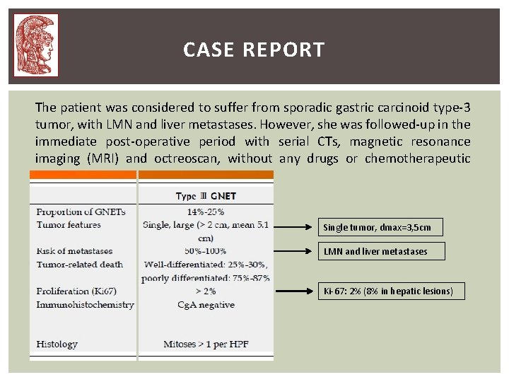
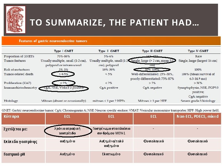
CASE REPORT The patient was considered to suffer from sporadic gastric carcinoid type-3 tumor, with LMN and liver metastases. However, she was followed-up in the immediate post-operative period with serial CTs, magnetic resonance imaging (MRI) and octreoscan, without any drugs or chemotherapeutic regimens. Single tumor, dmax=3, 5 cm LMN and liver metastases Ki-67: 2% (8% in hepatic lesions)
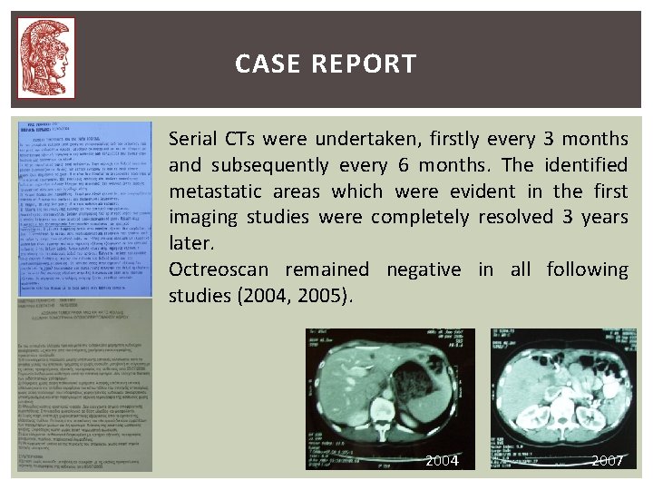
CASE REPORT Serial CTs were undertaken, firstly every 3 months and subsequently every 6 months. The identified metastatic areas which were evident in the first imaging studies were completely resolved 3 years later. Octreoscan remained negative in all following studies (2004, 2005). 2004 2007
CASE REPORT The patient visited our Department (January 2007) for further management. A full haematological and biochemical investigation, neuroendocrine markers and imaging study, at the time, did not show any evidence of disease, but she had… ► Increased thyroid peroxidase (anti-TPO) and thyroglobulin (anti-Tg) autoantibodies levels and ► Increased anti-parietal cell antibodies (APCA).
CASE REPORT Because of her indolent course and regression of hepatic lesions a revision of the pathological report was requested. Furthermore, the following had to be further investigated: 1. Gastrin and Cg. A levels were not measured before the initial operation. 2. APCA, anti-TPO and anti-Tg levels were increased post-surgically. 3. ICH was strongly positive for Cg. A. 4. The number of mitoses/HPF was relatively small.
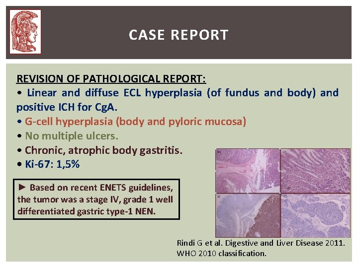
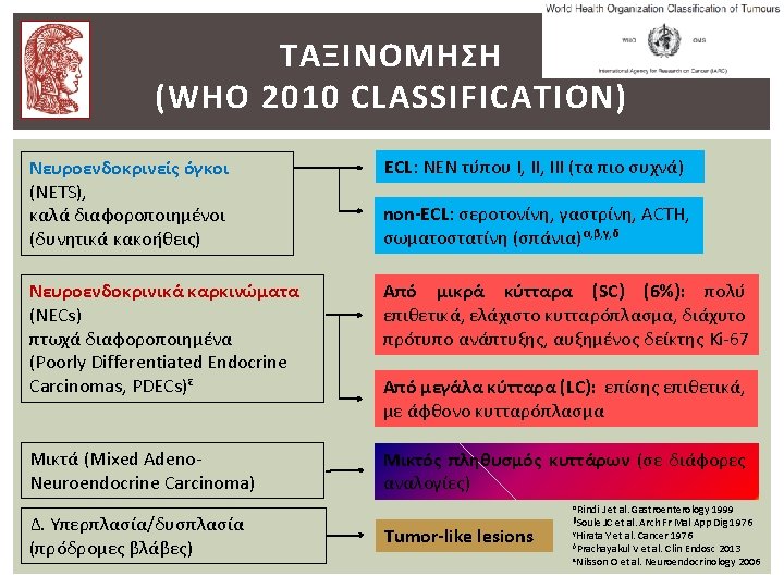
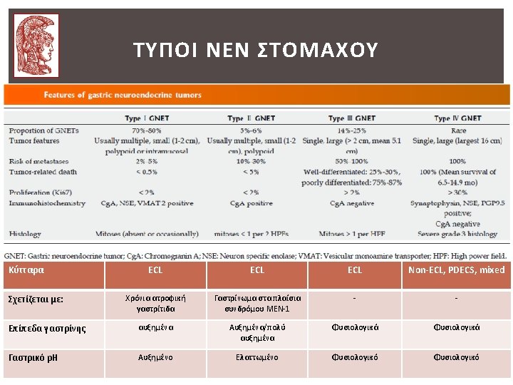
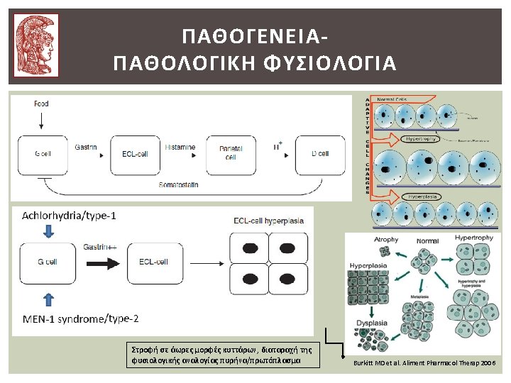
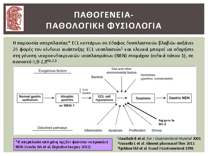
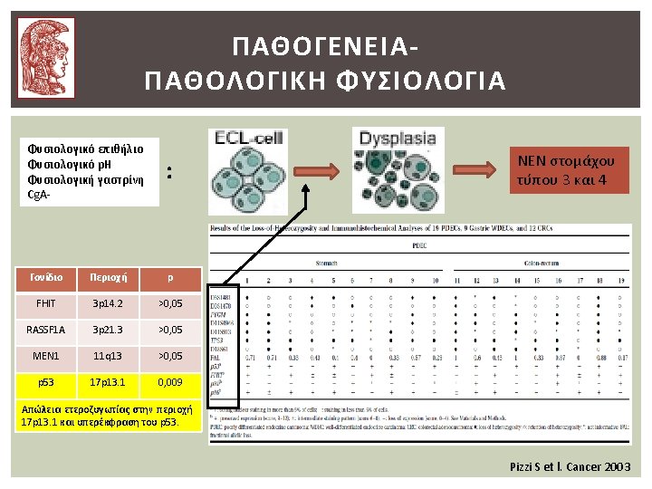
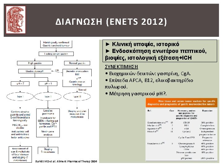
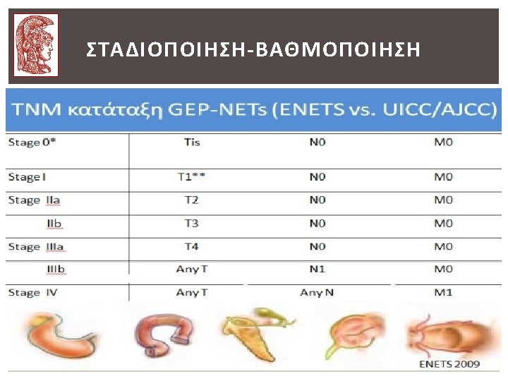
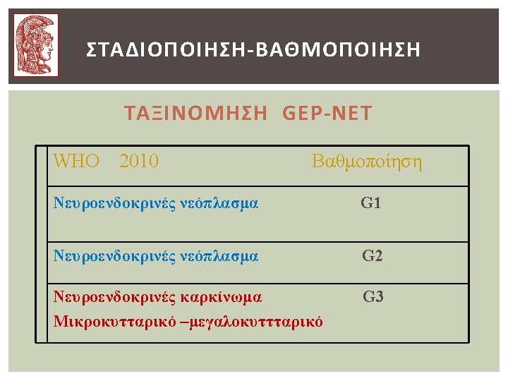
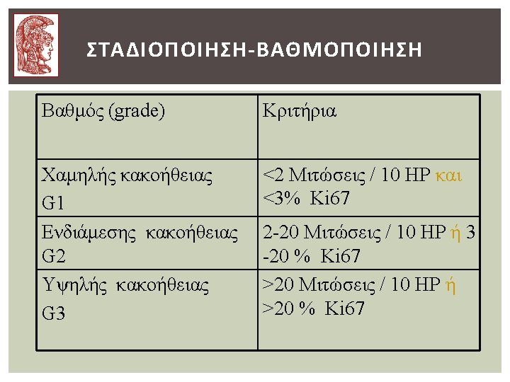
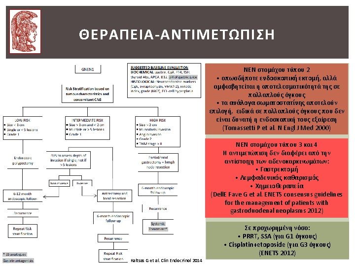
CASE REPORT REVISION OF PATHOLOGICAL REPORT: • Linear and diffuse ECL hyperplasia (of fundus and body) and positive ICH for Cg. A. • G-cell hyperplasia (body and pyloric mucosa) • No multiple ulcers. • Chronic, atrophic body gastritis. • Ki-67: 1, 5% ► Based on recent ENETS guidelines, the tumor was a stage IV, grade 1 well differentiated gastric type-1 NEN. Rindi G et al. Digestive and Liver Disease 2011. WHO 2010 classification.
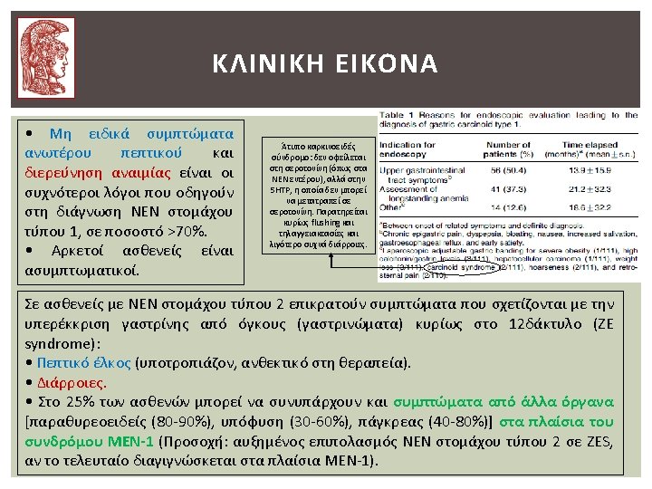
CASE REPORT The patient remains well and healthy 10 years after her primary surgery and she is followed-up yearly with haematological and imaging studies, without findings indicative of recurrence or distant metastasis. ► OGD (yearly): mild atrophy, HP infection ► Octreoscan (2008 και 2013): normal findings. ► abdominal MRI (/6 months) and us (yearly): no evidence of tumor relapse. ► Bone scan: no pathological findings ► Colonoscopy (2008, 2009, 2010, 2013): large bowel polyps and diverticulosis, haemorrhoids. ► Bone densitometry (yearly): βελτίωση του T-score (lumbar spine) -4, 1 (2008), -2, 9 (2014). ► Biochemical tests, neuroendocrine makers (/6 months): B 12 and iron deficiency.
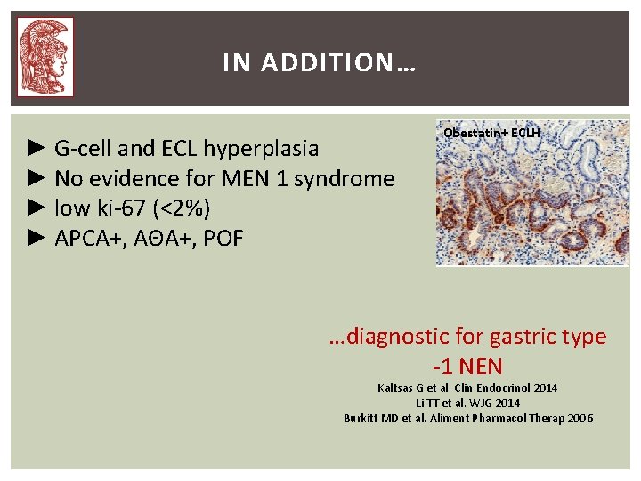
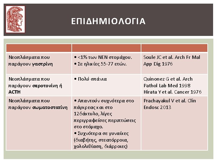
IN ADDITION… ► G-cell and ECL hyperplasia ► No evidence for MEN 1 syndrome ► low ki-67 (<2%) ► APCA+, ΑΘΑ+, POF Obestatin+ ECLH …diagnostic for gastric type -1 NEN Kaltsas G et al. Clin Endocrinol 2014 Li TT et al. WJG 2014 Burkitt MD et al. Aliment Pharmacol Therap 2006
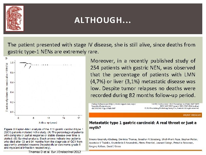
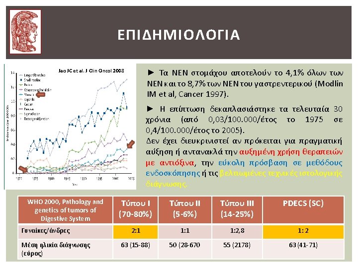
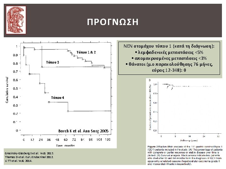
ALTHOUGH. . . The patient presented with stage IV disease, she is still alive, since deaths from gastric type-1 NENs are extremely rare. Moreover, in a recently published study of 254 patients with gastric NEN, was observed that the percentage of patients with LMN (4, 7%) or liver (3, 1%) metastatic disease was low. Despite tumor relapses no deaths were recorded during 82 months follow-up period. Thomas D et al. Eur J Endocrinol 2012
Thank you for your attention!
ΣΤΑΔΙΟΠΟΙΗΣΗ-ΒΑΘΜΟΠΟΙΗΣΗ T 4: tumor invades adjacent structures